The pituitary gland occupies a small, saddle-shaped depression of the sphenoid bone known as the sella tursica, and it hangs from the hypothalamus by a slender stalk. That anatomical arrangement is not incidental: it is the physical expression of a control relationship in which the hypothalamus governs the pituitary, and the pituitary in turn governs a remarkable range of distant tissues. Understanding this gland therefore means understanding a hierarchy of command, where neural signals from above are translated into chemical instructions that travel through the bloodstream to the thyroid, the adrenal cortex, the gonads, the mammary glands, the kidneys and the skeleton itself.
Anatomically, the gland divides into an adenohypophysis and a neurohypophysis, and this division is far more than a labelling convenience. The adenohypophysis, comprising the pars distalis and the pars intermedia, is true glandular tissue that synthesises its own hormones. The neurohypophysis, also called the pars nervosa or posterior pituitary, is nervous tissue that manufactures nothing; it merely stores and releases two hormones made in the hypothalamus and carried down to it along axons. Recognising which lobe makes a hormone and which merely warehouses one is the single most useful distinction a learner can carry into clinical reasoning about this gland.
This lesson works systematically through the pituitary: its location and attachment, its anatomical and functional subdivisions, the hormones of each region and their specific actions, and the disorders that arise when growth hormone or vasopressin goes wrong. It builds directly on the hypothalamic control discussed previously and prepares the ground for the pineal and thyroid glands that follow, since the thyroid's own activity depends on a pituitary signal introduced here.
On This Page
- Location, Attachment and the Architecture of the Pituitary
- The Hormones of the Pars Distalis and Their Functions
- The Pars Intermedia and Melanocyte Stimulating Hormone
- The Pars Nervosa: Storage and Release of Oxytocin and Vasopressin
- Disorders of Growth Hormone and Vasopressin
- Reading the Pituitary as a Coordinated System
Location, Attachment and the Architecture of the Pituitary
The pituitary gland sits within the sella tursica, a bony cavity that cradles it securely at the base of the skull, and it is attached to the hypothalamus by a stalk. This stalk is the conduit through which hypothalamic neurons send their axons into the posterior lobe, and through which the hypothalamic releasing and inhibiting factors reach the anterior lobe by way of the portal circulation. The diagrammatic representation of the pituitary and its relationship with the hypothalamus makes this dual connection visible: a neural bridge to one lobe and a vascular bridge to the other.
The Sella Tursica and the Hypothalamic Stalk
The bony cavity matters clinically as well as descriptively. Because the sella is a rigid compartment, any enlargement of the gland within it presses upon surrounding structures, which is why pituitary growths so often announce themselves through visual or neurological symptoms rather than through hormonal ones alone. The stalk, meanwhile, establishes the direction of control: the hypothalamus lies above and the pituitary below, and information flows downward. Every hormone discussed in this lesson is ultimately answerable to a hypothalamic signal, whether that signal is a releasing factor delivered by blood or an action potential delivered along an axon.
It is worth fixing the geography firmly, because the two lobes receive their hypothalamic instructions by entirely different routes. The anterior lobe is served by the portal circulation, a private vascular channel that carries hypothalamic factors directly to it. The posterior lobe is served by hypothalamic neurons whose axons run down the stalk and terminate within it. One lobe is commanded chemically through the blood; the other is commanded electrically through nerve fibres. This difference in wiring explains why the posterior lobe stores rather than synthesises its hormones.
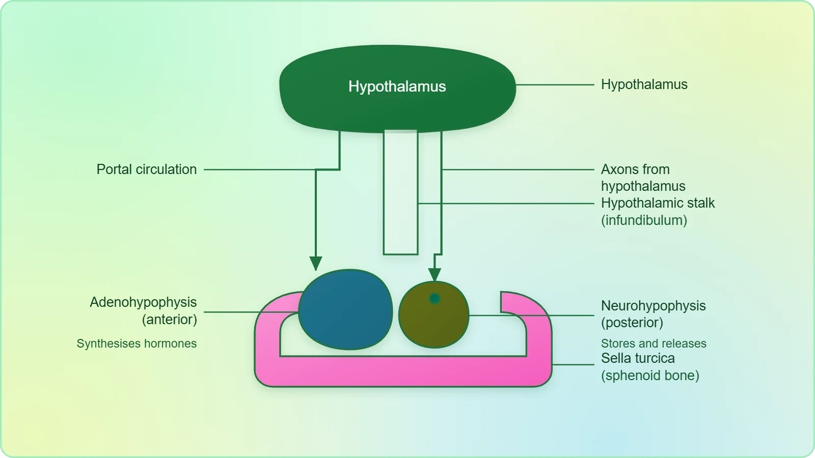
Adenohypophysis and Neurohypophysis: Two Lobes, Two Logics
The adenohypophysis consists of two portions, the pars distalis and the pars intermedia. The pars distalis, commonly called the anterior pituitary, is the great manufacturing centre of the gland, producing growth hormone, prolactin, thyroid stimulating hormone, adrenocorticotrophic hormone, luteinizing hormone and follicle stimulating hormone. The pars intermedia secretes only one hormone, melanocyte stimulating hormone, and in humans this region is almost merged with the pars distalis, a detail that explains why its contribution is comparatively modest.
The neurohypophysis, or pars nervosa, is known as the posterior pituitary and stores and releases two hormones, oxytocin and vasopressin. These are actually synthesised by the hypothalamus and transported axonally to the neurohypophysis. The wording deserves attention: the posterior pituitary is a release site, not a factory. When learners are asked to distinguish the lobes, the decisive question is not where a hormone acts but where it was made, and by that test oxytocin and vasopressin belong to the hypothalamus even though they enter the blood from the posterior lobe.
The Hormones of the Pars Distalis and Their Functions
The pars distalis produces six hormones, and they fall into recognisable families by target. Growth hormone and prolactin act on growth and lactation respectively; thyroid stimulating hormone and adrenocorticotrophic hormone drive other endocrine glands; and luteinizing hormone and follicle stimulating hormone, collectively called gonadotrophins, stimulate gonadal activity. Each hormone is named for its target or its effect, which makes the list easier to hold in memory than a bare recitation would suggest. The table below sets out each hormone against its principal action.
Growth Hormone and Prolactin
Growth hormone is the hormone most learners associate with the pituitary, and its effects are dramatic in both directions. Over-secretion stimulates abnormal growth of the body, leading to gigantism, while low secretion results in stunted growth, producing pituitary dwarfism. Prolactin has a narrower remit: it regulates the growth of the mammary glands and the formation of milk in them. The two hormones are chemically related but functionally distinct, and the contrast between a body-wide growth signal and a gland-specific milk signal illustrates how the same small gland can serve very different physiological agendas.
The timing of growth hormone excess matters enormously, and this is where the disorders diverge. In a growing individual, excess hormone lengthens the bones and produces the overall stature of gigantism. In adults, whose long bones can no longer extend, the same excess produces severe disfigurement, especially of the face, a condition called acromegaly. The underlying hormonal abnormality is identical; only the state of the skeleton differs. This is a clean illustration of a principle worth carrying forward: the effect of a hormone depends as much on the responsiveness of the tissue as on the quantity of hormone delivered.
Thyroid Stimulating Hormone, ACTH and the Gonadotrophins
Thyroid stimulating hormone stimulates the synthesis and secretion of thyroid hormones from the thyroid gland, and adrenocorticotrophic hormone stimulates the synthesis and secretion of steroid hormones called glucocorticoids from the adrenal cortex. Both are tropic hormones in the strict sense: they do not act on the body's working tissues directly but command another gland to do so. This is the mechanism behind the pituitary's reputation as a controller of controllers, and it explains why a pituitary fault can masquerade as a thyroid or adrenal disorder.
Luteinizing hormone and follicle stimulating hormone stimulate gonadal activity and are therefore called gonadotrophins. In males, LH stimulates the synthesis and secretion of hormones called androgens from the testis, while FSH and androgens together regulate spermatogenesis. In females, LH induces ovulation of fully mature follicles, the graafian follicles, and maintains the corpus luteum formed from the remnants of the graafian follicles after ovulation. FSH stimulates growth and development of the ovarian follicles. The same two hormones thus orchestrate quite different reproductive programmes in the two sexes.
The Pars Intermedia and Melanocyte Stimulating Hormone
The pars intermedia is the smallest functional division of the adenohypophysis and secretes only one hormone, melanocyte stimulating hormone. That hormone acts on the melanocytes, the melanin containing cells, and regulates pigmentation of the skin. In humans the pars intermedia is almost merged with the pars distalis, so its separate identity is more anatomical than practical. Even so, the hormone earns its place in the scheme because pigmentation is a visible endocrine readout, and changes in skin colour can occasionally signal disturbances in this region.
Melanocytes and the Regulation of Pigmentation
Melanin is the pigment that gives skin its colour, and the cells that hold it respond to melanocyte stimulating hormone by adjusting pigmentation. The relationship is a straightforward example of a hormone acting on a single, well-defined cell type rather than on a whole organ system, which makes it a useful teaching case. Where growth hormone reshapes the skeleton and gonadotrophins reorganise the reproductive cycle, melanocyte stimulating hormone simply modulates the output of pigment cells, and the contrast in scale is instructive.
Because the pars intermedia is nearly merged with the pars distalis in humans, learners should resist the temptation to treat melanocyte stimulating hormone as a major player in human physiology. Its inclusion in the pituitary scheme is a matter of completeness and comparative anatomy. The essential point to retain is that the adenohypophysis contains a region dedicated to pigmentation, that it secretes exactly one hormone, and that this hormone targets melanocytes specifically. Everything else about the region is detail that supports, rather than replaces, that core fact.
The Pars Nervosa: Storage and Release of Oxytocin and Vasopressin
The neurohypophysis, or pars nervosa, stores and releases two hormones, oxytocin and vasopressin, both synthesised by the hypothalamus and transported axonally to the posterior lobe. This arrangement is the clearest example in the endocrine system of a division of labour between manufacture and release. The hypothalamus writes the message; the posterior pituitary posts it. Because the hormones are peptides produced in nerve cell bodies and moved along axons, the posterior lobe behaves less like a gland and more like a terminal for neural cargo awaiting the right signal to discharge.
Oxytocin and Smooth Muscle Contraction
Oxytocin acts on the smooth muscles of the body and stimulates their contraction. In females, it stimulates a vigorous contraction of the uterus at the time of child birth, and milk ejection from the mammary gland. These two actions, though separated anatomically, share a common logic: both involve the contraction of smooth muscle in response to a neurohypophysial signal at a moment of reproductive importance. The uterine action assists delivery, while the mammary action releases milk already formed under the influence of prolactin, so the two pituitary lobes cooperate in the same physiological event.
The pairing of prolactin and oxytocin is worth pausing over, because it shows the anterior and posterior lobes working in sequence rather than in isolation. Prolactin, from the pars distalis, regulates the growth of the mammary glands and the formation of milk in them. Oxytocin, from the pars nervosa, then stimulates milk ejection from the mammary gland. Production and release are thus separate hormonal events, controlled from different parts of the gland, and a learner who confuses the two will misdescribe lactation at every stage.
Vasopressin as Antidiuretic Hormone
Vasopressin acts mainly at the kidney and stimulates resorption of water and electrolytes by the distal tubules, thereby reducing loss of water through urine, a process called diuresis. Because it opposes diuresis, it is also called antidiuretic hormone. The name captures the function precisely: the hormone conserves water by reclaiming it from the forming urine before it is lost. This is one of the clearest examples of endocrine regulation of fluid balance, and it explains why the hormone is discussed alongside kidney function rather than in isolation.
The logic of water conservation can be expressed compactly. If ##R## is the volume of water resorbed by the distal tubules and ##L## the volume lost as urine, then vasopressin raises ##R## and therefore lowers ##L##, so that the net conservation of body water increases. The relationship is directional rather than numerical, but it captures the essential mechanism:
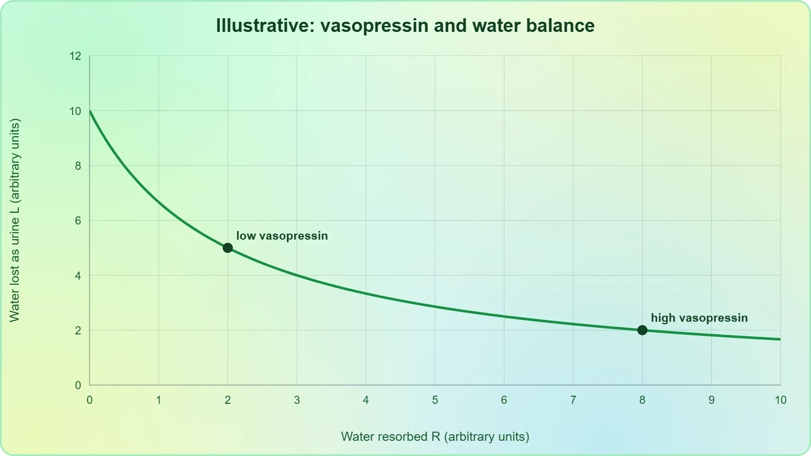
When vasopressin signalling is intact, the distal tubules reclaim water efficiently and urine remains concentrated. When the signal fails, the kidney loses its ability to conserve water, and the consequences are described in the next section. The formula above is a reminder that the two quantities move in opposite directions: any increase in resorption necessarily reduces the volume lost, which is precisely why the hormone is called antidiuretic.
Disorders of Growth Hormone and Vasopressin
The pituitary's disorders are unusually instructive because each one maps onto a specific hormone and a specific direction of error. Growth hormone excess or deficiency produces gigantism, pituitary dwarfism or acromegaly depending on the age of the individual, while impaired synthesis or release of vasopressin produces diabetes insipidus. Taken together, these conditions show how a single gland can generate a spectrum of disease through too much or too little of a single chemical messenger, and they reward careful attention to the timing and the target of the hormonal fault.
Gigantism, Pituitary Dwarfism and Acromegaly
Over-secretion of growth hormone stimulates abnormal growth of the body, leading to gigantism, while low secretion results in stunted growth, producing pituitary dwarfism. Excess secretion in adults, especially in middle age, can result in severe disfigurement, especially of the face, called acromegaly, which may lead to serious complications and premature death if unchecked. The distinction between gigantism and acromegaly rests entirely on the state of the skeleton at the time of excess: growing bones lengthen, mature bones thicken and distort.
Acromegaly is hard to diagnose in the early stages and often goes undetected for many years, until changes in external features become noticeable. This delayed recognition is a direct consequence of the disease's gradual nature: the hormonal excess accumulates silently while the visible alterations emerge only slowly. The clinical lesson is that a condition defined by external disfigurement may be well advanced before it is externally obvious, which is why the disorder is described as difficult to catch early rather than merely uncommon.
Diabetes Insipidus and the Failure of Water Conservation
An impairment affecting the synthesis or release of vasopressin results in a diminished ability of the kidney to conserve water, leading to water loss and dehydration. This condition is known as diabetes insipidus. The name points to the symptom that defines it: the production of large volumes of dilute urine, driven not by excess sugar but by the absence of the antidiuretic signal. The kidney itself may be perfectly capable of resorbing water; what fails is the hormonal instruction that tells it to do so.
Diabetes insipidus therefore belongs to the posterior pituitary in origin even though its symptoms appear at the kidney. This is a recurring pattern in endocrinology: the site of hormonal failure and the site of clinical signs are often different organs. A learner who traces the pathway from hypothalamic synthesis, through axonal transport, to release from the pars nervosa, and finally to the distal tubules of the kidney, will understand both why the condition arises and why it presents as it does.
Reading the Pituitary as a Coordinated System
Once the individual hormones are known, the gland can be read as a single coordinated system rather than a list of separate secretions. The hypothalamus issues releasing and inhibiting factors; the pars distalis translates them into tropic and direct-acting hormones; the pars intermedia contributes a pigmentation signal; and the pars nervosa releases two hypothalamic peptides that act on smooth muscle and kidney. The portal circulation and the axonal tract are the two channels of command, and every hormone in the lesson can be placed on one of them. This systems view is what makes the details memorable.
From Hypothalamic Signal to Peripheral Effect
A useful way to consolidate the lesson is to trace a single pathway from start to finish. A hypothalamic factor enters the portal circulation and reaches the pars distalis, which releases thyroid stimulating hormone; that hormone stimulates the synthesis and secretion of thyroid hormones from the thyroid gland; and those hormones then act on the body's tissues. At each step the signal changes form, from neural to hormonal to hormonal again, and the final effect depends on the integrity of every link. The same chain logic applies to ACTH and the glucocorticoids of the adrenal cortex.
The posterior lobe offers a contrasting pathway. A hypothalamic neuron synthesises vasopressin, transports it axonally to the neurohypophysis, and releases it into the blood, whence it reaches the distal tubules of the kidney and stimulates resorption of water and electrolytes. No intermediate gland is involved, and the effect is direct. Placing the two pathways side by side clarifies why the anterior lobe is described as glandular and the posterior lobe as neural, and why the posterior hormones are attributed to the hypothalamus despite their release from the pituitary.
Preparing for the Pineal and Thyroid Glands
The next lesson turns to the pineal gland and the thyroid gland, and the present material has already laid the groundwork for both. The thyroid's activity depends on thyroid stimulating hormone from the pars distalis, so the thyroid lesson will build directly on the tropic relationship introduced here. The pineal gland, which secretes melatonin and regulates a 24-hour diurnal rhythm, will extend the theme of hypothalamic control into the realm of biological timing. Learners who have mastered the pituitary's two lobes and their hormones will find both topics considerably easier to place.
A final consolidation is worth the effort. The pituitary is located in the sella tursica and attached to the hypothalamus by a stalk; it divides into adenohypophysis and neurohypophysis; the pars distalis produces six hormones, the pars intermedia one, and the pars nervosa stores and releases two. Growth hormone disorders produce gigantism, dwarfism and acromegaly; vasopressin failure produces diabetes insipidus. These facts form a compact framework, and every clinical detail in the lesson hangs upon it. Mastery here pays dividends in the lessons that follow.
Going further
The pituitary is often called the master gland because several of its hormones control the activity of other endocrine glands. Thyroid stimulating hormone commands the thyroid, adrenocorticotrophic hormone commands the adrenal cortex, and the gonadotrophins command the gonads, so a single small structure at the base of the brain exerts leverage over much of the endocrine system. The title is a convenience rather than a literal description, since the hypothalamus ultimately directs the pituitary, but it captures the gland's strategic position in the hierarchy of chemical coordination.
Acromegaly is described as hard to diagnose in its early stages because changes in external features become noticeable only later. This is a useful reminder that endocrine disease can progress substantially before it becomes visible, and that the absence of obvious signs is not evidence of the absence of hormonal excess. Recognising the insidious character of such conditions helps explain why they often go undetected for many years, and why an understanding of the underlying hormonal mechanism is more valuable than reliance on surface appearance alone.
Also Read
- No links available.
From our network :
- EV 2.0: The Solid-State Battery Breakthrough and Global Factory ExpansionDiscover the impact of the Solid-State Battery Breakthrough on the EV market. Learn about CATL and BYD stock growth, global factory expansion in Europe, and the future of EV 2.0 technology.
- Vite 6/7 'Cold Start' Regression in Massive Module GraphsIn-depth analysis of Vite 6/7 cold start regressions in massive module graphs. Learn about barrel file issues, pre-bundling optimizations, and how to improve Vite dev server performance.
- Mastering DB2 LUW v12 Tables: A Comprehensive Technical GuideComprehensive guide on DB2 LUW v12 tables. Explore CREATE TABLE syntax, BLU Acceleration, range partitioning, data types, and security features for IBM DB2 LUW v12.
- Mastering DB2 12.1 Instance Design: A Technical Deep Dive into Modern Database ArchitectureComprehensive guide to DB2 12.1 Instance Design. Explore architectural components, HADR setup, buffer pool tuning, and security policies for IBM DB2 12.1. Includes 10 functional SQL and Shell samples.
- 98% of Global MBA Programs Now Prefer GRE Over GMAT Focus EditionExplore why 98% of global MBA programs now prefer the GRE over GMAT Focus. Learn about the shorter GRE format, verbal logic importance, and strategic versatility for students.
- Analyzing Trump Deportation Numbers Insights Into The 2026 Immigration Crackdown
- Trump Political Strategy How Geopolitical Stunts Serve As Media Diversions
- AI-Powered 'Precision Diagnostic' Replaces Standard GRE Score ReportsDiscover the new GRE Precision Diagnostic. Learn how AI-powered behavioral analytics and time-to-solve tracking are replacing standard GRE score reports to empower students.
- 10 Physics Numerical Problems with Solutions for IIT JEEPrepare for IIT JEE with 10 physics numerical problems, complete with detailed solutions and conceptual explanations. Ideal for mastering kinematics, thermodynamics, electromagnetism, and optics.
RESOURCES
- Disorders of the Pituitary Gland | Johns Hopkins Medicinehopkinsmedicine.orgThe pituitary gland, sometimes called the “master gland,” produces hormones that enable other glands in the body to function. · Pituitary gland disorders include ...
- Pituitary Gland Disorders in women - Pacific Neuroscience Institutepacificneuroscienceinstitute.orgMay 26, 2025 ... What Are Pituitary Disorders and How Do They Affect Women? · Prolactinoma: A noncancerous tumor that causes excess prolactin production,…
- Understanding Pituitary Disorders | Brain Institute - OHSUohsu.eduTypes of pituitary disorders · Acromegaly · Craniopharyngioma · Cushing disease/syndrome · Growth hormone deficiency · Nonfunctioning pituitary adenoma · Prolactinoma ...
- Pituitary Gland: What It Is, Function & Anatomy - Cleveland Clinicmy.clevelandclinic.orgApr 4, 2022 ... Your pituitary gland also tells other endocrine system glands to release hormones. ... What conditions and disorders are related to…
- Pituitary Gland Disorders - University of Utah Healthhealthcare.utah.eduDisorders of Pituitary Hormone Under-Production · Adrenal Insufficiency · Arginine Vasopressin Deficiency · Growth Hormone Deficiency · Low Luteinizing Hormone (LH) ...
- Pituitary Gland Disorders: Symptoms, Diagnosis, Treatmentsuchicagomedicine.orgCommon Pituitary Conditions · Functional pituitary adenomas that result in overproduction of hormones · Prolactinoma · Cushing's disease · Acromegaly · Non- ...
- Overview of the Pituitary Gland - Hormonal and Metabolic Disordersmerckmanuals.comPituitary gland malfunction · Acromegaly or gigantism: Growth hormone · Cushing disease: Adrenocorticotropic hormone (ACTH) · Galactorrhea (the secretion of ...
- Pituitary Gland Disorders and Pituitary Tumors | Froedtert & MCWfroedtert.comTypes of Pituitary Tumors · Acromegaly – an excess in growth hormone production · Prolactinoma – an excess in the hormone prolactin, which stimulates…
- Pituitary Disorders - PubMedpubmed.ncbi.nlm.nih.govThere are 6 hormones secreted by the anterior pituitary gland: thyroid-stimulating hormone, adrenocorticotropic hormone, follicle-stimulating hormone, ...
- Pituitary tumors and adenomas - Symptoms and causes - Mayo Clinicmayoclinic.orgDec 24, 2025 ... Some pituitary tumors cause the pituitary gland to make too many hormones. ... Impulse control disorders in pituitary adenoma: What…
- Physiology, Pituitary Hormones - StatPearls - NCBI Bookshelfncbi.nlm.nih.gov... . Pituitary Gland Associated Diseases. Central diabetes insipidus (deficiency of vasopressin). Gigantism and acromegaly (excess of growth hormone).
- Adrenal Gland Disorders | Addison's Disease - MedlinePlusmedlineplus.govJan 19, 2024 ... The pituitary gland releases hormones that affect how the adrenal glands work. Changes in genes (mutations). These changes can cause…
- Pituitary Gland Functionpituitarysociety.orgLearn about pituitary gland hormones and how they regulate body systems with Pituitary Society. Discover hormone roles, anatomy, and common disorders.
- Pituitary Disorders - MedlinePlusmedlineplus.govDec 16, 2024 ... With pituitary disorders, you often have too much or too little of one of your hormones. Injuries can cause pituitary…
- Pituitary Disorders | Department of Medicine - Georgetown Universitydom.georgetown.eduPituitary tumor · Giantism or acromegaly (excess growth hormone) · Hyperthyroidism (excess thyroid stimulating hormone) · Cushing's syndrome (excess ...