The pineal gland and the thyroid gland occupy two very different positions in the architecture of human chemical coordination, yet both illustrate how a small cluster of cells can govern rhythms and metabolism across the entire body. The pineal gland sits on the dorsal side of the forebrain and secretes melatonin, a hormone that anchors the body's 24-hour diurnal rhythm, including the sleep-wake cycle and body temperature. The thyroid gland, by contrast, is a bilobed structure straddling the trachea, and its follicular cells synthesise the iodine-dependent hormones T4 and T3, which set the pace of the basal metabolic rate.
This lesson, the fourth in a sequence on chemical coordination and integration, builds directly on the pituitary gland discussed previously and prepares the ground for the parathyroid gland and thymus that follow. It works through the material in its own order of ideas: first the pineal gland and melatonin, then the structure of the thyroid gland, its hormones, the essential role of iodine, and the conditions that arise when thyroid output falters or surges, including goitre, cretinism, hyperthyroidism and exopthalmic goitre. The lesson closes with thyrocalcitonin, the thyroid's protein hormone that regulates blood calcium levels.
Learners at beginner to intermediate level will find the treatment deliberately sequential. Each gland is introduced by location, then by hormonal product, then by physiological action, and finally by the clinical consequences of dysfunction. That pattern mirrors the way endocrine physiology is actually examined, so the lesson doubles as preparation for structured answers. A short "Going further" block at the end adds well-established background on iodine chemistry and the anatomical scale of the thyroid, kept clearly separate from the core material so that the backbone of the lesson remains undistorted.
On This Page
The Pineal Gland and the Diurnal Rhythm
The pineal gland is a small endocrine structure located on the dorsal side of the forebrain, and its significance lies less in its size than in its timing. It secretes a hormone called melatonin, which plays a very important role in the regulation of a 24-hour, or diurnal, rhythm of the body. This rhythm is not a vague background hum; it coordinates predictable physiological cycles that recur with the turning of day and night. Melatonin is the chemical signal through which the pineal gland communicates the time of day to the rest of the organism.
Location and Hormonal Product
The anatomical description is brief but precise: the pineal gland is located on the dorsal side of the forebrain. From this position it secretes melatonin into the circulation, from which the hormone reaches its targets throughout the body. Learners should note that the material presents the pineal gland as a single-hormone gland, in contrast with the multi-hormone pituitary discussed in the previous lesson. That simplicity makes it an ideal entry point for understanding how a gland's position, its secretory product and its physiological role are described together in endocrine physiology.
Melatonin itself is the only hormone attributed to the pineal gland in this material, and its identity as the pineal secretion is the key fact to retain. The gland does not act through a cascade of releasing and stimulating factors; it responds to the diurnal cycle and issues a single chemical instruction. This makes the pineal gland a clean illustration of the general principle that endocrine glands release specific chemical messengers into the blood, where they act on distant target tissues. The next subsection turns to the specific rhythms melatonin governs.
Functions of Melatonin
Melatonin helps in maintaining the normal rhythms of the sleep-wake cycle and of body temperature. These two examples are given explicitly in the material and deserve emphasis, because they show melatonin acting on both behavioural and physiological cycles. The sleep-wake cycle is the most familiar expression of the diurnal rhythm, while body temperature follows its own daily oscillation. Together they demonstrate that the 24-hour rhythm regulated by melatonin is a coordinated property of the whole body rather than a feature of a single organ system.
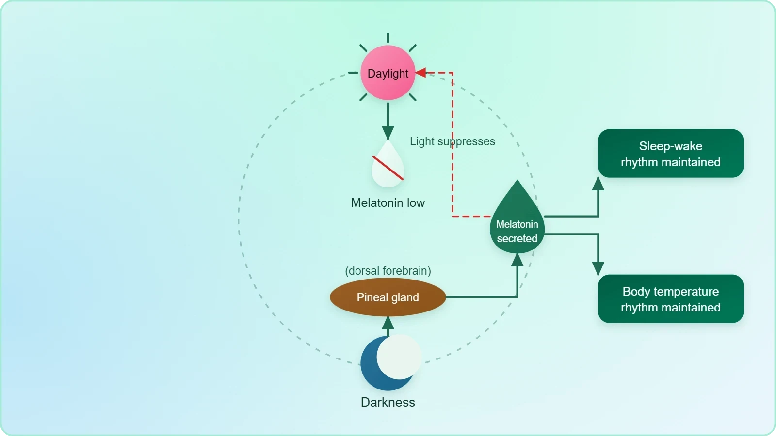
Beyond these rhythms, melatonin also influences metabolism, pigmentation, the menstrual cycle and the body's defense capability. This list widens the reach of the pineal gland considerably: a single hormone touches energy handling, skin colour, reproductive cycling and immune function. The material presents these as influences rather than as primary regulatory roles, and that distinction should be preserved. The pineal gland is therefore best understood as a timing device whose signal ripples outward into several physiological systems, adjusting their activity to the phase of the daily cycle.
The Structure of the Thyroid Gland
The thyroid gland is composed of two lobes located on either side of the trachea, and both lobes are interconnected with a thin flap of connective tissue called the isthmus. This bilobed arrangement, bridged at the midline, gives the gland its characteristic shape and explains why it is described as straddling the airway. The structural description matters because the gland's functional units, the follicles, are distributed throughout both lobes, so the whole organ participates in hormone synthesis rather than any single region acting alone.
Lobes, Isthmus and Follicles
The thyroid gland is composed of follicles and stromal tissues. Each thyroid follicle is composed of follicular cells, enclosing a cavity. This two-part architecture, with secretory cells arranged around a central space and supported by stroma, is the histological signature of the gland. The follicular cells are the functional units that synthesise hormones, while the stromal tissue provides the supporting framework in which the follicles are embedded. Recognising this arrangement is essential before the hormonal products themselves are introduced.
The isthmus deserves particular attention because it is the structure that unites the two lobes into a single gland. Without it, the thyroid would be two separate masses on either side of the trachea; with it, the lobes form one interconnected organ. The material names the isthmus explicitly as a thin flap of connective tissue, and this detail is frequently tested. Learners should be able to describe the gland in sequence: two lobes, one on each side of the trachea, joined by the isthmus, built of follicles and stroma, with follicular cells enclosing a cavity.
Follicular Cells and Hormone Synthesis
These follicular cells synthesise two hormones, tetraiodothyronine or thyroxine (T4) and triiodothyronine (T3). The nomenclature is instructive: the prefixes tetra and tri refer to the number of iodine atoms incorporated into each molecule, four in T4 and three in T3. Both hormones are produced by the same follicular cells, so the thyroid does not partition its output between separate cell types as some other glands do. The material presents T4 and T3 together as the paired products of follicular cell activity, and this pairing should be retained in any account of thyroid function.
Iodine is essential for the normal rate of hormone synthesis in the thyroid. This single sentence carries enormous physiological weight, because it links a dietary element directly to the gland's capacity to produce its hormones. The follicular cells can only assemble T4 and T3 when iodine is available in sufficient quantity, so the rate of synthesis is contingent on iodine supply. The structural account of the gland therefore culminates not in anatomy but in a nutritional requirement, which sets up the discussion of deficiency and disease that follows.
Thyroid Hormones and Their Physiological Actions
Thyroid hormones play an important role in the regulation of the basal metabolic rate, and this is the central fact around which their other actions cluster. The basal metabolic rate is the energy expenditure of the body at rest, so a hormone that regulates it effectively sets the idle speed of every tissue. From this primary role follow a series of supporting functions: the hormones support the process of red blood cell formation, control the metabolism of carbohydrates, proteins and fats, and influence the maintenance of water and electrolyte balance. Each of these actions extends the thyroid's reach into haematology, intermediary metabolism and fluid homeostasis.
Basal Metabolic Rate and Metabolism
The regulation of the basal metabolic rate is the headline function, and it explains why thyroid dysfunction produces such widespread effects. When thyroid hormone levels rise, the resting rate of energy expenditure climbs; when they fall, it declines. The material also states that thyroid hormones control the metabolism of carbohydrates, proteins and fats, which means the gland governs how the body's three major fuel classes are handled. This is a broader claim than metabolic rate alone, since it concerns the processing of specific substrates rather than the aggregate energy cost of living.
Support for red blood cell formation is a less obvious but equally important action. Because red cells carry oxygen, a hormone that assists their production indirectly influences the oxygen supply available to every tissue. The maintenance of water and electrolyte balance is also influenced by thyroid hormones, linking the gland to the same homeostatic territory that vasopressin governs at the kidney. Taken together, these actions show that the thyroid is not merely a metabolic throttle but a participant in blood formation and fluid balance, which is why its disorders present with such varied clinical features.
Thyrocalcitonin and Calcium Regulation
The thyroid gland also secretes a protein hormone called thyrocalcitonin (TCT), which regulates the blood calcium levels. This is a distinct product, different in chemical nature from T4 and T3, and it addresses a different physiological problem. Calcium regulation is a matter of precise balance, and thyrocalcitonin contributes to it from the thyroid side. The material introduces TCT here and notes its regulatory role without elaborating a mechanism, leaving the fuller treatment of calcium balance to the parathyroid gland in the next lesson.
Learners should appreciate that the thyroid therefore secretes two chemically distinct classes of hormone: the iodine-containing thyroid hormones T4 and T3 from the follicular cells, and the protein hormone thyrocalcitonin. The pairing of TCT with parathyroid hormone in the service of calcium balance is a theme that will be developed when the parathyroid gland is discussed. For now, the essential point is that the thyroid's endocrine output is not exhausted by its metabolic hormones; it also contributes a calcium-regulating signal that operates alongside the parathyroid glands.
Iodine Deficiency, Goitre and Cretinism
Deficiency of iodine in the diet results in hypothyroidism and enlargement of the thyroid gland, commonly called goitre. This is the pivotal clinical consequence of the iodine requirement established earlier: without adequate iodine, the normal rate of hormone synthesis cannot be sustained, and the gland enlarges in the process. The material treats goitre as the visible sign of an underlying hypothyroid state, so the two should be understood together rather than as separate conditions. The enlargement is the anatomical expression of a gland struggling to meet demand.
Hypothyroidism in Pregnancy and Cretinism
Hypothyroidism during pregnancy causes defective development and maturation of the growing baby, leading to stunted growth (cretinism), mental retardation, low intelligence quotient, abnormal skin and deaf-mutism. This is the most serious consequence described in the material, because it affects the developing child rather than the adult. The list of features is specific and should be retained: stunted growth, mental retardation, low intelligence quotient, abnormal skin and deaf-mutism. Cretinism is the name given to the stunted growth that results, and it stands as the clearest illustration of why maternal thyroid function matters.
The mechanism behind these effects is implicit in the material's logic: thyroid hormones regulate the basal metabolic rate and support normal development, so their absence during pregnancy deprives the growing baby of the signals needed for proper maturation. The phrase defective development and maturation captures this precisely. Learners should note that the material presents cretinism as a consequence of hypothyroidism during pregnancy specifically, not of adult-onset hypothyroidism, and that the features listed are developmental rather than merely metabolic.
Adult Hypothyroidism and Menstrual Irregularity
In adult women, hypothyroidism may cause the menstrual cycle to become irregular. This is a narrower but practically important consequence, and it connects back to the earlier statement that melatonin influences the menstrual cycle as well. The adult presentation of hypothyroidism is thus distinguished from the developmental presentation in the offspring of a hypothyroid pregnancy. The material does not elaborate further on adult symptoms, so the menstrual irregularity stands as the representative adult consequence to be remembered alongside the goitre that commonly accompanies iodine deficiency.
The distinction between the two settings is worth emphasising in revision. In pregnancy, hypothyroidism affects the growing baby and produces cretinism with its associated features. In adult women, it disturbs the menstrual cycle. Both arise from the same underlying deficiency of thyroid hormone, but the consequences differ according to the stage of life at which the deficiency operates. This developmental perspective is a recurring theme in endocrinology and prepares learners for the parathyroid and thymus material that follows.
Hyperthyroidism and Exopthalmic Goitre
Due to cancer of the thyroid gland or due to development of nodules of the thyroid glands, the rate of synthesis and secretion of the thyroid hormones is increased to abnormally high levels, leading to a condition called hyperthyroidism, which adversely affects the body physiology. The material identifies two causes, cancer of the thyroid gland and the development of nodules, and one consequence, an abnormally high rate of hormone synthesis and secretion. Hyperthyroidism is thus the mirror image of the deficiency state, and its adverse effects on body physiology follow from the same metabolic roles that thyroid hormones perform.
Causes and General Effects
The two named causes deserve separate attention. Cancer of the thyroid gland is a neoplastic process, while nodules are discrete growths within the gland; both can drive the rate of synthesis and secretion upward. The material does not distinguish further between them, presenting them as alternative routes to the same hyperthyroid state. The phrase adversely affects the body physiology is deliberately general, signalling that the consequences are systemic rather than confined to the gland. Because thyroid hormones regulate the basal metabolic rate and control the metabolism of carbohydrates, proteins and fats, an excess of them disturbs energy handling throughout the body.
Learners should connect hyperthyroidism back to the functional account given earlier. If thyroid hormones regulate the basal metabolic rate, then abnormally high levels should raise that rate; if they control the metabolism of carbohydrates, proteins and fats, then excess should disturb all three. The material's general statement that body physiology is adversely affected is best understood through these established roles. This is also why hyperthyroidism is described as a distinct condition rather than merely a laboratory finding: the elevated hormone levels translate into measurable physiological disturbance.
Exopthalmic Goitre (Graves' Disease)
Exopthalmic goitre is a form of hyperthyroidism, characterised by enlargement of the thyroid gland, protrusion of the eyeballs, increased basal metabolic rate and weight loss, also called Graves' disease. The four features form a compact clinical picture: the gland enlarges, the eyes protrude, the basal metabolic rate rises and body weight falls. The protrusion of the eyeballs is the feature that gives the condition its name, and it distinguishes exopthalmic goitre from other forms of hyperthyroidism. The alternative name, Graves' disease, should be memorised alongside the descriptive term.
The increased basal metabolic rate and weight loss are direct expressions of the hormonal excess described in the previous subsection. When the resting rate of energy expenditure climbs, the body burns fuel faster than it is replaced, and weight falls. The enlargement of the thyroid gland parallels the goitre of iodine deficiency, but the underlying hormonal state is opposite: here the gland is overactive rather than underactive. This contrast between goitre with hypothyroidism and exopthalmic goitre with hyperthyroidism is one of the most examinable distinctions in the lesson.
Consolidating the Lesson and Looking Ahead
The pineal and thyroid glands together demonstrate two contrasting modes of endocrine control. The pineal gland, on the dorsal side of the forebrain, issues a single hormone, melatonin, that regulates the 24-hour diurnal rhythm and thereby maintains the normal rhythms of the sleep-wake cycle and body temperature, while also influencing metabolism, pigmentation, the menstrual cycle and defense capability. The thyroid gland, bilobed and joined by the isthmus, builds follicles of follicular cells that synthesise T4 and T3 under the essential influence of iodine, and adds the protein hormone thyrocalcitonin for calcium regulation. One gland times the body; the other sets its metabolic pace.
Key Distinctions to Retain
Several distinctions are worth fixing firmly. Goitre accompanies iodine-deficiency hypothyroidism, whereas exopthalmic goitre is a form of hyperthyroidism marked by protrusion of the eyeballs, increased basal metabolic rate and weight loss, and is also called Graves' disease. Cretinism arises from hypothyroidism during pregnancy and carries stunted growth, mental retardation, low intelligence quotient, abnormal skin and deaf-mutism, while adult hypothyroidism may simply render the menstrual cycle irregular. Thyrocalcitonin regulates blood calcium levels and will be paired with parathyroid hormone in the next lesson, where calcium balance is treated more fully.
The material's order of ideas is itself a study aid: location, then hormonal product, then physiological action, then clinical consequence. Applied to the pineal gland, this yields forebrain position, melatonin, diurnal rhythm, and influences on metabolism and pigmentation. Applied to the thyroid, it yields the bilobed structure with isthmus, the follicular cells producing T4 and T3, the metabolic and haematological actions, and the deficiency and excess states. Learners who internalise this pattern will find the parathyroid gland and thymus easier to absorb, because the same sequence of description recurs there.
Going further
Iodine is a component of the thyroid hormones, which is why iodine deficiency in the diet affects hormone synthesis. The prefixes in the names tetraiodothyronine and triiodothyronine record the number of iodine atoms in each molecule, four and three respectively, so the element is not a catalyst but a structural constituent of the hormones themselves. This chemical fact explains why the material treats iodine as essential for the normal rate of hormone synthesis rather than as a mere accelerator, and why dietary deficiency translates so directly into hypothyroidism and goitre.
The thyroid gland is one of the largest endocrine glands and lies in the neck, wrapped around the trachea. This anatomical scale is consistent with the bilobed structure described in the material, with the two lobes flanking the airway and the isthmus bridging them across the midline. Because the gland is superficial and closely applied to the trachea, its enlargement in goitre is readily palpable, which is part of why goitre has long served as the visible marker of thyroid disease in clinical observation and public health surveys.
Also Read
- No links available.
From our network :
- AI-Powered 'Precision Diagnostic' Replaces Standard GRE Score ReportsDiscover the new GRE Precision Diagnostic. Learn how AI-powered behavioral analytics and time-to-solve tracking are replacing standard GRE score reports to empower students.
- 98% of Global MBA Programs Now Prefer GRE Over GMAT Focus EditionExplore why 98% of global MBA programs now prefer the GRE over GMAT Focus. Learn about the shorter GRE format, verbal logic importance, and strategic versatility for students.
- Vite 6/7 'Cold Start' Regression in Massive Module GraphsIn-depth analysis of Vite 6/7 cold start regressions in massive module graphs. Learn about barrel file issues, pre-bundling optimizations, and how to improve Vite dev server performance.
- Trump Political Strategy How Geopolitical Stunts Serve As Media Diversions
- 10 Physics Numerical Problems with Solutions for IIT JEEPrepare for IIT JEE with 10 physics numerical problems, complete with detailed solutions and conceptual explanations. Ideal for mastering kinematics, thermodynamics, electromagnetism, and optics.
- Mastering DB2 LUW v12 Tables: A Comprehensive Technical GuideComprehensive guide on DB2 LUW v12 tables. Explore CREATE TABLE syntax, BLU Acceleration, range partitioning, data types, and security features for IBM DB2 LUW v12.
- Mastering DB2 12.1 Instance Design: A Technical Deep Dive into Modern Database ArchitectureComprehensive guide to DB2 12.1 Instance Design. Explore architectural components, HADR setup, buffer pool tuning, and security policies for IBM DB2 12.1. Includes 10 functional SQL and Shell samples.
- EV 2.0: The Solid-State Battery Breakthrough and Global Factory ExpansionDiscover the impact of the Solid-State Battery Breakthrough on the EV market. Learn about CATL and BYD stock growth, global factory expansion in Europe, and the future of EV 2.0 technology.
- Analyzing Trump Deportation Numbers Insights Into The 2026 Immigration Crackdown
RESOURCES
- Changes in thyroid hormones, leptin, ghrelin and, galanin following ...pmc.ncbi.nlm.nih.govMay 14, 2019 ... Melatonin is secreted from pineal gland and thyroid c-cells under thyroid stimulating hormone ... T3 and T4 levels, melatonin may…
- Opposite effects of zinc and melatonin on thyroid hormones in ratssciencedirect.com... (T4) and thyroid-stimulating hormone (TSH) levels. The free T3, T4 and TSH levels were lower in the melatonin ... thyroid gland. Zinc has…
- Thyroid hormone and adrenergic signaling interact to control pineal ...pmc.ncbi.nlm.nih.govWe also tested the effects of thyroxine (T4), and triiodothyronine (T3) because of the large daily rhythm in the activity in the pineal gland…
- Physiology, Endocrine Hormones - StatPearls - NCBI Bookshelf - NIHncbi.nlm.nih.govMelatonin is a hormone synthesized within the pineal gland from the amino acid tryptophan. ... As mentioned in the TSH section, there are 2…
- Effects of a temperature rise on melatonin and thyroid hormones ...pubmed.ncbi.nlm.nih.gov... melatonin (MEL; the time-keeping hormone), throughout smoltification. We also studied the impact of temperature history on pineal gland MEL production in vitro.
- Melatonin and the thyroid - NeL.edunel.eduOct 18, 2001 ... melatonin; pineal gland; thyroid hormones; thyroid gland; growth ... T3,T4. TSH. Pineal. Pituitary. Thyroid. +. MNL? MBH-TRH. Fig. 1. General ...
- Leptin, neuropeptide Y (NPY), melatonin and zinc levels ... - PubMedpubmed.ncbi.nlm.nih.govMar 2, 2018 ... ... pineal gland, is generally accepted to exercise an inhibitor effect on the thyroid gland. Zinc mediates the effects of…
- The Role of Melatonin in Thyroid Function in Adult Rats. | EBSCOhostopenurl.ebsco.com... hormones, such as triiodothyronine (T3), thyroxine (T4), and calcitonin. The pineal gland produces and releases melatonin, which is stimulated in the dark ...
- The Role of Melatonin in Thyroid Function in Adult Ratsijvm.ut.ac.irBackground: The thyroid gland, a vital endocrine organ, the thyroid gland synthesizes and secretes hormones, such as triiodothyronine (T3), thyroxine (T4), ...
- Molecular economy of nature with two thyrotropins from different ...archivesofmedicalscience.comJan 5, 2021 ... ... pineal gland-gonadal hormones and T3 [60, 61]. Photoperiodic DIO-3 ... hormone glycoforms Photoperiodism Thyroid hormones, seasons and ...
- Chronothyroidology: Chronobiological Aspects in Thyroid Function ...mdpi.comMay 10, 2021 ... ... thyroid gland to produce T4(thyroxine) and T3(triiodotyronine). Thyroid hormones regulate, by a negative feedback, the secretion of TRH and ...
- Is Thyroxine a Peptide Hormone - CETAFcetaf.orgOct 12, 2026 ... * Thyroid Hormone (T4/T3): These are tw Amino acid-derived hormones ... thyroid gland) and melatonin (produced by the pineal gland).
- Hormones: What They Are, Function & Types - Cleveland Clinicmy.clevelandclinic.orgWhich body tissues make hormones? · Hypothalamus · Pituitary gland · Pineal gland · Thyroid gland · Parathyroid glands · Adrenal glands · Pancreas…
- Protective Effects of Melatonin against Carcinogen-Induced ... - MDPImdpi.comApr 25, 2024 ... Melatonin, primarily synthesized in the pineal gland, plays a crucial role in regulating circadian rhythms and possesses significant ...
- Are thyroid hormones peptide or steroid - CETAFcetaf.orgOct 13, 2026 ... ... thyroid gland) and melatonin (produced by the pineal gland). ... * Thyroid hormone (T3/T4): Derived from tyro Steroid vs…