Calcium is one of the body's most tightly policed ions, and the two small glands that sit almost invisibly against the back of the thyroid are central to that policing. This lesson examines the parathyroid glands and the thymus, two structures that share a region of the neck and chest yet serve entirely different physiological purposes. The parathyroid glands guard the calcium concentration of the blood with a peptide hormone that acts on bone, kidney and gut, while the thymus trains the immune system's T-lymphocytes through a family of peptide hormones called thymosins. Together they illustrate how chemical coordination extends from mineral homeostasis to immune competence.
The lesson follows a clear sequence. It begins with the anatomy of the four parathyroid glands and the regulation of parathyroid hormone by circulating calcium ions, then traces the three target tissues through which PTH raises blood calcium: bone resorption, renal tubular reabsorption and intestinal absorption. From there it considers the partnership between PTH and thyrocalcitonin, the thyroid hormone introduced in the previous lesson, in maintaining calcium balance. The second half turns to the thymus, its location and lobular structure, the role of thymosins in T-lymphocyte differentiation and antibody production, and the degeneration of the gland in old age that weakens immune responses.
Because this lesson builds directly on the thyroid, readers should keep thyrocalcitonin in view as the counterweight to parathyroid hormone. The material deliberately pairs a hypercalcemic hormone with a hypocalcemic one, and the balance between them is the conceptual heart of the first half. The second half shifts register entirely, moving from mineral chemistry to cellular immunology, and asks why a gland that is prominent in youth becomes a vestige in later life. By the end, the reader should be able to name each hormone, its source, its target and its effect, and explain why the thymus matters most when the body is young.
On This Page
- The Parathyroid Glands: Position, Number and Secretory Product
- Parathyroid Hormone and Its Hypercalcemic Action
- Calcium Balance: PTH and Thyrocalcitonin Together
- The Thymus Gland: Structure and Position
- Thymosins and the Differentiation of T-Lymphocytes
- Ageing of the Thymus and Weakened Immune Responses
- Going Further
The Parathyroid Glands: Position, Number and Secretory Product
In humans, four parathyroid glands are present on the back side of the thyroid gland, arranged as one pair in each of the two lobes of the thyroid. They are small structures, easily overlooked in a dissection of the neck, yet their removal or damage has immediate and serious consequences for calcium homeostasis. The glands secrete a peptide hormone called parathyroid hormone, usually abbreviated PTH. Because the hormone is a peptide, it belongs to the same broad chemical class as insulin and glucagon rather than to the steroid or amine hormones, a point worth carrying forward when the mechanism of hormone action is studied later in the course.
Anatomical arrangement and relationship to the thyroid
The description is precise and worth memorising exactly: four glands, on the back side of the thyroid, one pair in each lobe. This arrangement explains why thyroid surgery carries a risk to calcium balance, since the parathyroid glands are embedded in the capsule region of the thyroid rather than standing apart from it. The proximity is anatomical only; the parathyroids are developmentally and functionally distinct from the thyroid, and their hormone is not a thyroid hormone. Students often confuse the two because of the shared neighbourhood, so it helps to state the distinction plainly: the thyroid secretes thyroxine, triiodothyronine and thyrocalcitonin, while the parathyroids secrete PTH alone.
Regulation of PTH secretion by circulating calcium
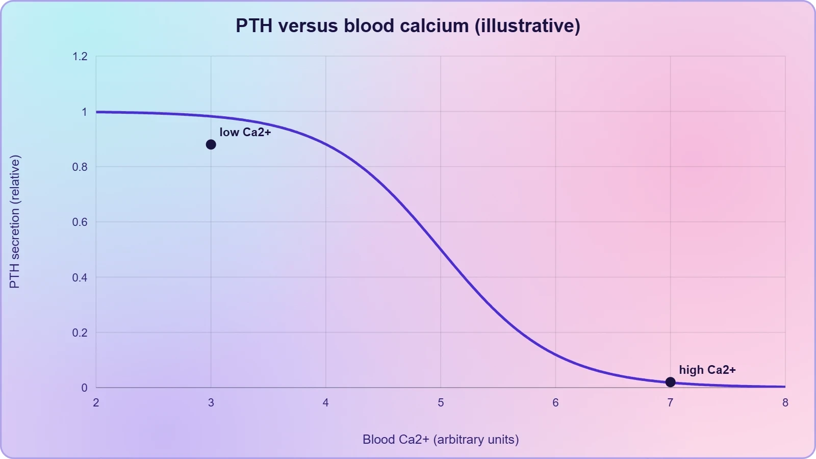
The secretion of PTH is regulated by the circulating levels of calcium ions. This is a direct negative feedback arrangement of the simplest kind: when blood calcium falls, PTH secretion rises, and when blood calcium rises, PTH secretion falls. No separate releasing hormone from the hypothalamus and no tropic hormone from the anterior pituitary intervenes, which makes the parathyroid an unusually autonomous endocrine gland. The sensor and the secretory cell are effectively the same cell, responding to the ion concentration in the fluid around it. This design keeps calcium within a narrow range with remarkable speed, and it explains why the parathyroid glands are described as the body's principal calcium-regulating tissue.
Parathyroid Hormone and Its Hypercalcemic Action
Parathyroid hormone increases the Ca²⁺ levels in the blood. The material states this as the defining action of the hormone and then explains it through three target tissues. PTH acts on bones and stimulates the process of bone resorption, that is, dissolution or demineralisation of bone tissue. It also stimulates reabsorption of Ca²⁺ by the renal tubules, so that calcium that would otherwise be lost in urine is returned to the blood. Finally, it increases Ca²⁺ absorption from the digested food. Through these three routes the hormone raises the calcium concentration of the blood, and it is therefore described as a hypercalcemic hormone.
Bone resorption: dissolution and demineralisation
Bone is not an inert scaffold but a mineral reservoir in constant exchange with the blood, and PTH draws on that reservoir when calcium is scarce. The material uses two words for the process, dissolution and demineralisation, and both are worth keeping. Dissolution suggests the chemical breakdown of the mineral phase, while demineralisation emphasises the loss of mineral from the matrix. The effect is a net release of calcium from bone into the circulation. This is a normal physiological response, not a disease, though sustained overactivity of the parathyroid would progressively weaken the skeleton, which is why the gland's feedback control matters so much.
Renal reabsorption and intestinal absorption of calcium
The kidney and the gut complete the picture. By stimulating reabsorption of Ca²⁺ by the renal tubules, PTH prevents calcium from being wasted in the urine and returns filtered calcium to the blood. By increasing Ca²⁺ absorption from the digested food, it raises the amount of calcium entering the body from the diet. The three actions are complementary rather than redundant: bone provides a rapid buffer, the kidney conserves what is already in the body, and the gut replenishes the body's supply from outside. Together they explain why a single peptide hormone can hold blood calcium steady across fasting, feeding and growth.
Calcium Balance: PTH and Thyrocalcitonin Together
Along with TCT, parathyroid hormone plays a significant role in calcium balance in the body. The previous lesson established that the thyroid gland secretes a protein hormone called thyrocalcitonin, which regulates the blood calcium levels. The two hormones pull in opposite directions, and that opposition is the point. PTH is hypercalcemic, raising blood Ca²⁺; TCT lowers it. The material does not elaborate the mechanism of TCT beyond its regulatory role, and this lesson respects that limit. What matters here is the principle of paired antagonistic hormones holding a single variable within a narrow band, a pattern the reader will meet again in the adrenal gland and in the pancreatic hormones.
Antagonistic hormones and the logic of balance
Endocrine control frequently works through opposing pairs rather than through a single regulator, and calcium is a textbook case. One hormone raises the variable, another lowers it, and the concentration settles wherever the two secretory rates balance. This arrangement gives fine control in both directions: a fall in calcium is corrected by more PTH, and a rise is corrected by more TCT. The value of such a design is stability. A single hormone could only push calcium upward or downward, and correcting an overshoot would require a separate mechanism anyway. Pairing the hormones makes the correction intrinsic to the system.
Why the calcium set-point is defended so tightly
The tight defence of blood calcium reflects how much depends on the ion. Calcium is required for the strength of bone, as the material's account of bone mineral implies, and it is also central to many cellular processes, including the contraction of muscle and the transmission of nerve impulses. A concentration that drifts too low or too high disturbs these processes quickly. The parathyroid glands therefore sit at the centre of a control loop whose importance far exceeds their modest size, and the partnership with thyrocalcitonin ensures that the loop can correct deviations in either direction rather than only one.
The Thymus Gland: Structure and Position
The thymus gland is a lobular structure located between the lungs, behind the sternum, on the ventral side of the aorta. The description is compact but sufficient to place the organ in the upper chest, in front of the great vessels and behind the breastbone. Its lobular construction, divided into discrete lobules, is characteristic of glands that organise their tissue into repeating functional units. Unlike the parathyroid glands, which are minute and hidden, the thymus occupies a substantial space in the chest, particularly in the young. Its position near the centre of the thorax reflects its role in the development of a system that circulates throughout the body.
Location and lobular organisation
Three landmarks fix the thymus in place: between the lungs, behind the sternum, and on the ventral side of the aorta. Each is worth noting because together they locate the gland unambiguously in the anterior mediastinum. The lobular structure matters functionally as well as descriptively, since the lobules provide the organised microenvironments in which developing lymphocytes are processed. The material does not describe the internal architecture of the lobule in detail, and this lesson does not add to it. What the reader should retain is the simple anatomical fact: a lobular gland in the chest, positioned in front of the aorta and behind the sternum.
A gland of the immune system rather than of metabolism
The thymus belongs to a different functional category from the endocrine glands studied so far. The thyroid, parathyroid and pineal glands regulate metabolism, calcium and daily rhythms; the thymus regulates the development of immune cells. It is an endocrine gland in the sense that it secretes hormones into the blood, but its hormones act chiefly on cells of the immune system rather than on distant metabolic targets. This dual identity, endocrine and immunological at once, is why the thymus is often treated separately in surveys of the endocrine system, and why its inclusion in this course is instructive.
Thymosins and the Differentiation of T-Lymphocytes
The thymus secretes peptide hormones called thymosins. These hormones play a major role in the differentiation of T-lymphocytes, the lymphocytes that provide cell-mediated immunity. In addition, thymosins promote the production of antibodies, and so contribute to humoral immunity as well. The two contributions are distinct: cell-mediated immunity depends on lymphocytes acting directly against infected or abnormal cells, while humoral immunity depends on antibodies circulating in the blood and other fluids. The material thus credits thymosins with supporting both arms of the adaptive immune response, with the differentiation of T-lymphocytes named as the primary role.
Cell-mediated immunity through T-lymphocyte differentiation
Differentiation is the process by which immature cells acquire the specialised features of a mature cell type, and for T-lymphocytes the thymus is the organ in which this happens. The name T-lymphocyte itself reflects the gland's role, since the T stands for thymus-derived. Thymosins are the peptide signals that drive this maturation. The outcome is a population of lymphocytes capable of recognising and acting against specific targets, which is the essence of cell-mediated immunity. Without the thymic signal, the maturation of these cells would be incomplete, and the body's ability to respond to intracellular pathogens and abnormal cells would be correspondingly weakened.
Humoral immunity and antibody production
Beyond cell-mediated immunity, thymosins also promote the production of antibodies, and so support humoral immunity. Antibodies are proteins that bind specifically to foreign substances and mark them for destruction, and their production is the defining feature of the humoral response. The material presents this as an additional action of thymosins rather than as their principal one, and the distinction is worth preserving. The thymus is therefore not solely a school for T-lymphocytes; its hormones also influence the antibody side of immunity, which means the gland's reach extends across the adaptive immune system as a whole.
Ageing of the Thymus and Weakened Immune Responses
The thymus is degenerated in old individuals, resulting in a decreased production of thymosins. As a result, the immune responses of old persons become weak. The material states this causal chain in three steps, and the sequence deserves attention: degeneration of the gland, decline in hormone output, weakening of immune responses. It is a clean example of an endocrine explanation for an age-related change, and it links the gland's structure to its function in a way that is easy to test in the mind. The thymus is thus unusual among endocrine glands in that its activity declines markedly with age rather than remaining roughly constant.
Degeneration of the gland and falling thymosin output
Degeneration here means the progressive replacement of functional thymic tissue, so that the gland produces less thymosin as the years pass. The consequence is a reduced supply of the peptide signal that drives T-lymphocyte differentiation. Because the hormone is the link between the gland and the immune cells, the decline in hormone output translates directly into a decline in the maturation of new lymphocytes. The material does not specify the rate of decline or the age at which it begins, and this lesson does not invent such figures. What it does establish is the direction of the change and its immunological consequence.
Consequences for immunity in later life
The weakening of immune responses in old persons follows from the reduced production of thymosins. With less hormonal drive for T-lymphocyte differentiation, the supply of mature, newly trained lymphocytes falls, and the cell-mediated arm of immunity becomes less vigorous. The material states the outcome plainly and does not elaborate further. The broader implication is that the immune system is not static but has an endocrine dependency that fades with age, which helps explain why older individuals are often more susceptible to infections. This is a physiological observation, not a clinical prescription, and it closes the lesson on a note of change over the lifespan.
Going Further
Two pieces of background help place this material in a wider frame without altering it. The first concerns why calcium balance matters so much. Calcium is needed for bone strength, as the account of bone mineral implies, and it is also required for many cellular processes, including muscle contraction, nerve impulse transmission and the release of neurotransmitters. A hormone that raises blood calcium therefore protects not only the skeleton but the signalling systems that depend on a stable calcium concentration. The second concerns the thymus and age. The gland is largest in childhood and shrinks thereafter, which fits the statement that it degenerates in old individuals and produces less thymosin.
Calcium as a shared currency of bone and cell
Bone stores calcium, but the same ion also serves as a signal inside and between cells. This dual role explains why the body tolerates only a narrow range of blood calcium and why the parathyroid glands respond so promptly to small deviations. The material's three target tissues, bone, kidney and gut, are best understood as the three routes by which the body can adjust its calcium economy: releasing stored mineral, conserving what is filtered, and absorbing what is eaten. The hypercalcemic action of PTH is the coordinated use of all three routes in the service of one concentration.
The shrinking thymus and the arc of immune life
The observation that the thymus is largest in childhood and shrinks with age is a familiar anatomical fact that aligns neatly with the material's account of degeneration in old age. Early life is when the immune repertoire is built and when thymosin output is highest; later life is when the gland's functional tissue declines and immune responses weaken. Reading the two statements together gives a coherent picture of an organ whose importance is front-loaded in the lifespan. The lesson does not claim that immunity depends on the thymus alone, only that the gland's hormonal contribution fades with time.
Also Read
- No links available.
From our network :
- Vite 6/7 'Cold Start' Regression in Massive Module GraphsIn-depth analysis of Vite 6/7 cold start regressions in massive module graphs. Learn about barrel file issues, pre-bundling optimizations, and how to improve Vite dev server performance.
- Trump Political Strategy How Geopolitical Stunts Serve As Media Diversions
- EV 2.0: The Solid-State Battery Breakthrough and Global Factory ExpansionDiscover the impact of the Solid-State Battery Breakthrough on the EV market. Learn about CATL and BYD stock growth, global factory expansion in Europe, and the future of EV 2.0 technology.
- Mastering DB2 LUW v12 Tables: A Comprehensive Technical GuideComprehensive guide on DB2 LUW v12 tables. Explore CREATE TABLE syntax, BLU Acceleration, range partitioning, data types, and security features for IBM DB2 LUW v12.
- Analyzing Trump Deportation Numbers Insights Into The 2026 Immigration Crackdown
- Mastering DB2 12.1 Instance Design: A Technical Deep Dive into Modern Database ArchitectureComprehensive guide to DB2 12.1 Instance Design. Explore architectural components, HADR setup, buffer pool tuning, and security policies for IBM DB2 12.1. Includes 10 functional SQL and Shell samples.
- 98% of Global MBA Programs Now Prefer GRE Over GMAT Focus EditionExplore why 98% of global MBA programs now prefer the GRE over GMAT Focus. Learn about the shorter GRE format, verbal logic importance, and strategic versatility for students.
- 10 Physics Numerical Problems with Solutions for IIT JEEPrepare for IIT JEE with 10 physics numerical problems, complete with detailed solutions and conceptual explanations. Ideal for mastering kinematics, thermodynamics, electromagnetism, and optics.
- AI-Powered 'Precision Diagnostic' Replaces Standard GRE Score ReportsDiscover the new GRE Precision Diagnostic. Learn how AI-powered behavioral analytics and time-to-solve tracking are replacing standard GRE score reports to empower students.
RESOURCES
- Thymus-Associated Parathyroid Hormone Has Two Cellular Origins ...pmc.ncbi.nlm.nih.govDec 23, 2010 ... Despite their distinct primary functions, the parathyroid and thymus organs have a close relationship during organogenesis, initially developing ...
- Thymus-associated parathyroid hormone has two cellular ... - PubMedpubmed.ncbi.nlm.nih.govDec 23, 2010 ... In mammals, parathyroid hormone (PTH) is a key regulator of extracellular calcium and inorganic phosphorus homeostasis.
- Perspectives on the Parathyroid–Thymus Interconnection—A ... - PMCpmc.ncbi.nlm.nih.govJun 23, 2025 ... Recent studies suggest that parathyroid hormone (PTH) may influence thymic function, including T-cell maturation and immune regulation, while ...
- Congenital aplasia of parathyroid glands and thymus - PubMedpubmed.ncbi.nlm.nih.govTetralogy of Fallot / complications*; Thymus Gland / abnormalities*; Vitamin D / therapeutic use. Substances. Parathyroid Hormone; Vitamin D; Phosphorus ...
- Parathyroid hormone and premature thymus ageing in patients with ...nature.comJan 28, 2019 ... Premature immune ageing, including thymic atrophy, is observed in patients with chronic kidney disease (CKD). Parathyroid hormone (PTH) and ...
- Embryology, Parathyroid - StatPearls - NCBI Bookshelf - NIHncbi.nlm.nih.govThe initial synthesis of parathyroid hormone takes place within the parathyroid gland ... thymus and parathyroid glands. Poor thymic development leads to abnormal ...
- Cross-Expression of Thymic and Parathyroid Hormone Receptors ...mdpi.comNov 28, 2025 ... The thymus and parathyroid glands share a common embryological origin from the third pharyngeal pouch, yet their potential morphological and ...
- Anatomy, Head and Neck, Parathyroid - StatPearls - NCBI Bookshelfncbi.nlm.nih.govAug 8, 2023 ... ... parathyroid gland releases a hormone called parathermone or parathyroid hormone (PTH). ... Thymus and Parathyroid Glands Organogenesis Paradigm. J ...
- Limits to in vivo fate changes of epithelia in thymus and parathyroid ...nature.comAug 8, 2022 ... Gcm2-mediated fate changes were assessed by the expression of the parathyroid hormone (Pth) gene, one of Gcm2´s key down-stream targets…
- Ectopic PTH-producing parathyroid cyst inside the thymus: a case ...link.springer.comDec 21, 2022 ... The hallmark of hyperparathyroidism is hypersecretion of parathyroid hormone (PTH) which results in hypercalcemia and hypophosphatemia.
- MHS 209 Thyroid-Parathyroid-Thymus - Histology Guidehistologyguide.comHistology of a parathyroid gland embedded within the thyroid which itself is fused with the thymus ... Secrete thyroid hormones when active.
- Thymus: The Function of the Gland & Why it is Importantmy.clevelandclinic.orgMay 15, 2022 ... What does the thymus do? · Thymopoietin: fuels the production of T-cells and tells the pituitary gland to release hormones.…
- Ectopic parathyroid adenoma in thymus - MedCrave onlinemedcraveonline.comJul 13, 2017 ... Three months before the adenoma of right parathyroid gland was discovered by USG and the increased level of parathyroid hormone-615,8pg/ml, ...
- Parathyroid Gland Anatomy: Overview, Natural Variants - Medscapeemedicine.medscape.comMar 7, 2025 ... These four glands produce parathyroid hormone (PTH), a single-chain polypeptide of 84 amino-acid residues that regulates calcium and phosphorus ...
- Anatomy of the Endocrine System | Johns Hopkins Medicinehopkinsmedicine.orgThe hypothalamus secretes hormones that stimulate or suppress the release of hormones ... The thyroid gland and parathyroid glands are located in front of…