The heart is the only organ in the body that can, quite literally, run itself. Strip away every nerve connecting it to the brain, and a strip of its own tissue will keep contracting in a dish, beating out a rhythm that no external command supplied. This intrinsic self-regulation is the foundation of cardiac physiology, and it is the starting point for understanding how the body then layers external control on top of that internal rhythm. The heart is not a passive pump waiting for instructions; it is a self-starting engine that the nervous system merely tunes.
This lesson examines the regulation of cardiac activity as the body actually performs it: first through the specialised nodal tissue that generates the heartbeat from within, and then through the autonomic nervous system that speeds it up or slows it down from the medulla oblongata. We will trace how sympathetic and parasympathetic signals exert opposing effects on heart rate, conduction and contractile strength, and how hormones from the adrenal medulla add a further layer of control over cardiac output. Each mechanism is treated in the order the material presents it, building directly on the double circulation pathways already covered.
By the end, you should be able to explain why the heart is described as myogenic, identify the neural centre that moderates cardiac function, and distinguish precisely between the effects of the two divisions of the autonomic nervous system. This prepares the ground for the next lesson on circulatory disorders, where the consequences of a regulatory system pushed beyond its limits become the central concern.
On This Page
- The Myogenic Heart: Intrinsic Autoregulation by Nodal Tissue
- Sympathetic Neural Signals: Raising Rate and Contractile Force
- Parasympathetic Neural Signals: Slowing the Heart
- Integrating Neural and Hormonal Regulation of Cardiac Output
- Going Further: The Physiological Logic of Autonomic Balance
- From Regulation to Disorder: Setting Up the Next Lesson
- Working with the Regulatory Model: A Computational Illustration
- Review and Consolidation: The Regulatory Framework in Full
The Myogenic Heart: Intrinsic Autoregulation by Nodal Tissue
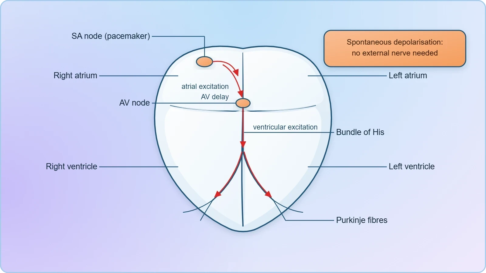
Normal activities of the heart are regulated intrinsically, meaning they are auto regulated by specialised muscles known as nodal tissue. This is the single most important idea in the lesson: the heartbeat originates within the heart muscle itself, not from a command issued by the brain. Because the heart generates its own rhythm, it is described as myogenic. The term distinguishes cardiac muscle from skeletal muscle, which contracts only when a motor neuron delivers a signal, and it explains why a heart can continue beating even when its nerve supply is severed.
The nodal tissue responsible for this automaticity is a specialised cardiac muscle that has lost most of its contractile function and gained the ability to depolarise spontaneously. These cells do not wait for an external stimulus; their membrane potential drifts steadily until it reaches a threshold and fires. This self-generated impulse then spreads through the cardiac musculature, setting the pace for the entire organ. The result is a rhythmic, coordinated contraction that persists independently of any neural input, which is precisely what the term myogenic captures.
Why Automaticity Matters for the Whole Organ
The practical consequence of myogenic regulation is that the heart possesses a built-in safety margin. Even if autonomic nerves were completely silenced, the organ would continue to beat at its intrinsic rate. This intrinsic rate is not arbitrary; it is set by the firing frequency of the nodal tissue itself, which acts as the natural pacemaker of the heart. Every beat you have ever felt, from before birth to this moment, traces back to this spontaneous electrical activity generated within the cardiac muscle.
This arrangement also means that external regulation is a matter of modulation rather than initiation. The nervous system does not start the heart; it adjusts a rhythm that is already running. Understanding this distinction is essential before examining the autonomic control mechanisms, because it clarifies that sympathetic and parasympathetic signals act as accelerators and brakes on an engine that is always idling, never as the ignition key itself.
The Medulla Oblongata as the Moderating Centre
A special neural centre in the medulla oblongata can moderate cardiac function through the autonomic nervous system. The word moderate is carefully chosen: this centre does not generate the heartbeat but adjusts its rate and force. Located in the brainstem, the medulla oblongata houses the cardiovascular centres that receive input from higher brain regions and from sensory receptors throughout the body, then issue the neural commands that travel down the autonomic pathways to the heart.
This central moderation allows cardiac output to be matched to the demands of the moment. During rest, the medulla permits the heart to settle toward its slower, energy-conserving rhythm; during exertion or stress, it adjusts the balance of autonomic outflow to raise output. The medulla thus functions as a control hub, continuously integrating signals and fine-tuning the myogenic rhythm to suit the body's circumstances without ever taking over the generation of the beat itself.
Sympathetic Neural Signals: Raising Rate and Contractile Force
Neural signals through the sympathetic nerves, which form part of the autonomic nervous system, can increase the rate of heartbeat, the strength of ventricular contraction, and thereby the cardiac output. This is the excitatory arm of cardiac regulation, and its effects are threefold: faster rhythm, stronger contraction, and greater volume of blood expelled per minute. Each of these changes compounds the others, so a modest increase in rate combined with a stronger contraction produces a substantial rise in cardiac output.
The sympathetic pathway achieves this by releasing neurotransmitters that act on the nodal tissue and the ventricular muscle alike. At the nodes, the effect is to accelerate the spontaneous depolarisation, shortening the interval between beats. At the ventricles, the effect is to recruit more contractile force from the muscle fibres. The combination means the heart not only beats more often but also empties more completely with each beat, delivering a greater volume of blood to the systemic circulation.
Cardiac Output and Its Components
Cardiac output is the volume of blood pumped by the heart per unit time, and it is the product of heart rate and stroke volume, the amount ejected per beat. Sympathetic stimulation raises both factors simultaneously, which is why its effect on cardiac output is so pronounced. A faster rate multiplies the number of ejection events, while a stronger ventricular contraction increases the volume released at each event. The two effects reinforce each other rather than merely adding together.
This relationship can be expressed compactly. Cardiac output is given by:
where ##CO## is cardiac output, ##HR## is heart rate in beats per minute, and ##SV## is stroke volume in millilitres per beat. Sympathetic signals raise ##HR## and ##SV## together, so ##CO## climbs steeply. This is why the sympathetic effect on cardiac output is described as an increase in both rate and strength of contraction, working in concert rather than in isolation.
Adrenal Medullary Hormones as a Parallel Pathway
Adrenal medullary hormones can also increase the cardiac output. This hormonal route operates alongside the neural one, providing a slower but longer-lasting amplification of the sympathetic effect. The hormones released from the adrenal medulla circulate in the blood and reach the heart directly, where they act on the same cardiac tissue that sympathetic nerves influence. The result is a sustained rise in cardiac output that outlasts the momentary burst of neural activity.
The existence of this hormonal pathway means the heart can be driven to higher output even when neural signals have subsided. During prolonged stress or sustained physical effort, the adrenal medullary contribution helps maintain elevated cardiac performance. Together, the sympathetic nerves and adrenal medullary hormones form a coordinated excitatory system, one fast and precisely targeted, the other slower and more diffuse, both converging on the same goal of increased cardiac output.
Parasympathetic Neural Signals: Slowing the Heart
Parasympathetic neural signals, the other component of the autonomic nervous system, decrease the rate of heartbeat, the speed of conduction of action potential, and thereby the cardiac output. This is the inhibitory arm of cardiac regulation, and its effects mirror the sympathetic ones in reverse. Where sympathetic signals accelerate, parasympathetic signals decelerate; where sympathetic signals strengthen, parasympathetic signals restrain. The result is a reduction in the volume of blood pumped per minute, achieved through a slower rhythm and a slower spread of the electrical impulse.
The parasympathetic effect on conduction speed is particularly significant. By slowing the conduction of action potentials through the nodal tissue and the conducting pathways, parasympathetic signals lengthen the interval before the next beat is triggered. This is not merely a matter of rate; it is a change in how quickly the electrical command travels through the heart. The combined result is a heart that beats less often and conducts its impulses more slowly, lowering cardiac output accordingly.
The Antagonistic Balance of Autonomic Control
The heart is therefore subject to two opposing streams of autonomic input, and its actual performance at any moment reflects the balance between them. Sympathetic signals push rate, contractile strength and conduction speed upward; parasympathetic signals pull rate and conduction speed downward. The medulla oblongata adjusts this balance continuously, so that cardiac output can be raised during exertion and lowered during rest without any change in the intrinsic myogenic rhythm itself.
This antagonistic arrangement provides fine control. Rather than switching the heart on and off, the autonomic system shifts the balance point between acceleration and deceleration. A slight predominance of parasympathetic outflow keeps the resting heart slow and economical; a shift toward sympathetic predominance raises output rapidly when demand increases. The myogenic heart supplies the rhythm, and the autonomic balance determines the tempo at which that rhythm is played.
Conduction Speed and the Spread of Excitation
The speed at which action potentials conduct through the heart determines how quickly the chambers are activated and how tightly the contraction is coordinated. Sympathetic signals accelerate this conduction, while parasympathetic signals reduce it. Because conduction speed governs the timing of chamber activation, its modulation directly affects the efficiency of each beat. A faster spread means quicker, more synchronized activation; a slower spread means a more drawn-out sequence of excitation.
This is why the material lists conduction speed as a distinct variable alongside heart rate. The two are related but not identical: rate concerns how often a beat occurs, while conduction speed concerns how rapidly the electrical signal travels to trigger that beat. Parasympathetic signals reduce both, and sympathetic signals raise both, so the two variables move together under autonomic control, jointly shaping the cardiac output that the body ultimately receives.
Integrating Neural and Hormonal Regulation of Cardiac Output
Cardiac output emerges from the interplay of three regulatory inputs: the intrinsic myogenic rhythm of the nodal tissue, the opposing autonomic signals from the medulla oblongata, and the circulating hormones of the adrenal medulla. The myogenic rhythm sets the baseline; the autonomic nerves adjust it moment to moment; the adrenal hormones provide a slower, sustained amplification. Together they allow the heart to match its output to the body's needs across a wide range of conditions, from deep rest to intense exertion.
The elegance of this system lies in its hierarchy. The heart does not depend on any single input for its basic function, because the myogenic mechanism guarantees a beat even in the absence of neural or hormonal signals. External regulation then refines that beat, raising or lowering it as circumstances demand. This layered design ensures both reliability and flexibility, a combination that no single control mechanism could provide on its own.
A Summary of the Regulatory Inputs
The table below consolidates the regulatory inputs described in the material, pairing each source with its effect on cardiac function. It serves as a reference for the distinctions that matter most: which inputs are intrinsic and which are extrinsic, which raise cardiac output and which lower it, and which act through nerves and which through hormones. Keeping these categories clear prevents the common error of conflating the myogenic origin of the beat with its autonomic modulation.
Why the Myogenic Design Confers Reliability
A heart that depended on external nerves for every beat would be vulnerable to any interruption in that supply. The myogenic design removes this vulnerability by making the beat self-generating. The nodal tissue fires on its own, and the autonomic system merely adjusts the tempo. This means that even under conditions of neural damage or pharmacological blockade, the heart continues to beat, preserving circulation at its intrinsic rate. Reliability is built into the tissue itself.
This principle also clarifies the clinical significance of the regulatory system. Because the heart can function without external input, disorders of autonomic regulation tend to manifest as inappropriate rates rather than as complete cessation. The heart keeps beating, but it may beat too fast, too slow, or with insufficient force for the body's needs. Understanding the myogenic baseline is therefore essential for interpreting what goes wrong when regulation fails, a theme that leads directly into the next lesson.
Going Further: The Physiological Logic of Autonomic Balance
The material establishes the mechanisms of cardiac regulation; a few sentences of well-established background can illuminate why these mechanisms are arranged as they are. Sympathetic signals prepare the body for activity by raising heart rate and contraction strength, mobilising the cardiovascular system to deliver more oxygen and nutrients to working tissues. This is the physiological logic of the excitatory arm: when demand rises, output must rise with it, and the sympathetic pathway delivers that increase rapidly and precisely.
Parasympathetic signals, by contrast, slow the heart and the conduction of action potentials, conserving energy and allowing the organ to recover during rest. The two divisions thus serve complementary purposes, one geared toward action and the other toward restoration. The medulla oblongata's role is to arbitrate between them, shifting the balance in response to the body's changing state. This arrangement explains why the resting heart beats slowly and why it accelerates so readily when activity begins.
Reading the Balance Through Observable Signs
Because the autonomic balance determines cardiac performance, it can be inferred from observable signs. A rising heart rate during exertion reflects increasing sympathetic outflow; a slowing rate during rest reflects parasympathetic predominance. The strength of the pulse and the speed of recovery after activity likewise carry information about the regulatory balance. These signs are not merely descriptive; they are windows into the autonomic control that the material describes, allowing the state of the regulatory system to be assessed from the outside.
This connection between mechanism and observation is what makes the study of cardiac regulation practically meaningful. The concepts of myogenic rhythm, autonomic moderation and hormonal amplification are not abstract; they manifest in every pulse you can feel and every rate you can count. Recognising them in action reinforces the material's central claim: the heart regulates itself intrinsically, and the nervous system moderates that self-regulation to meet the demands of the moment.
From Regulation to Disorder: Setting Up the Next Lesson
The regulatory system described here maintains cardiac output within a narrow, healthy range under ordinary conditions. When that system is overwhelmed, or when the vessels themselves are compromised, the result is disorder. The next lesson turns to the disorders of the circulatory system, beginning with high blood pressure, defined as blood pressure higher than normal, with 120/80 mm Hg as the reference measurement. The regulatory concepts developed here provide the vocabulary needed to understand what goes wrong when control fails.
Understanding regulation first is essential because disorders of circulation are, in large part, disorders of regulation. A heart driven too hard by sympathetic signals, or a vasculature subjected to persistently elevated pressure, illustrates the consequences of a control system pushed beyond its design limits. The transition from normal regulation to pathological disorder is therefore a natural progression, and the mechanisms covered in this lesson form the foundation for interpreting the clinical material that follows.
Consolidating the Core Distinctions
Before moving on, consolidate the distinctions that define this lesson. The heart is myogenic because its rhythm arises from nodal tissue within the cardiac muscle. The medulla oblongata moderates that rhythm through the autonomic nervous system. Sympathetic signals increase rate, contraction strength and conduction speed, raising cardiac output; parasympathetic signals decrease rate and conduction speed, lowering it. Adrenal medullary hormones provide an additional route to increased cardiac output. These five statements capture the material's essential content.
Each distinction matters because it separates mechanisms that are easily confused. The myogenic origin of the beat is not the same as its autonomic modulation; the neural pathway is not the same as the hormonal one; and the sympathetic effect is not the same as the parasympathetic one. Holding these categories apart allows the regulatory system to be analysed accurately, whether in the context of normal physiology or in the disordered states that the next lesson will examine.
Working with the Regulatory Model: A Computational Illustration
The regulatory relationships described in this lesson can be expressed as a simple computational model, which helps clarify how the components interact. The model treats cardiac output as the product of heart rate and stroke volume, then applies separate multipliers for sympathetic, parasympathetic and hormonal influences. This is not a clinical tool but a teaching device: it makes explicit the arithmetic that underlies the qualitative statements in the material, and it shows how small changes in each input combine to shift the final output.
Such a model reinforces the lesson's central point about hierarchy. The myogenic baseline provides the starting values of rate and stroke volume; the autonomic and hormonal inputs then modify those values. By adjusting the multipliers, you can simulate the effect of sympathetic activation, parasympathetic dominance, or adrenal hormone release, and observe how each changes cardiac output. The exercise makes the antagonistic balance of autonomic control tangible and prepares the ground for the disorders that arise when regulation is disturbed.
Expressing the Model in Code
The following snippet implements the model in Python. It defines a baseline heart rate and stroke volume, then applies the regulatory multipliers to compute cardiac output under different conditions. The code is deliberately simple, mirroring the conceptual structure of the lesson rather than any clinical complexity. Running it with different multiplier values illustrates how the sympathetic and parasympathetic pathways push cardiac output in opposite directions, and how the hormonal input adds a further increment.
def cardiac_output(heart_rate, stroke_volume, sympathetic=1.0, parasympathetic=1.0, adrenal=1.0):
    """Estimate cardiac output from rate, stroke volume and regulatory multipliers."""
    effective_rate = heart_rate * sympathetic * parasympathetic
    effective_stroke = stroke_volume * sympathetic * adrenal
    return effective_rate * effective_stroke

baseline_hr = 72      # beats per minute at rest
baseline_sv = 70      # millilitres per beat

resting = cardiac_output(baseline_hr, baseline_sv)
sympathetic_drive = cardiac_output(baseline_hr, baseline_sv, sympathetic=1.3)
parasympathetic_drive = cardiac_output(baseline_hr, baseline_sv, parasympathetic=0.8)
adrenal_boost = cardiac_output(baseline_hr, baseline_sv, adrenal=1.2)

print("Resting cardiac output:", resting)
print("With sympathetic drive:", sympathetic_drive)
print("With parasympathetic drive:", parasympathetic_drive)
print("With adrenal boost:", adrenal_boost)
Interpreting the Output
When the snippet is run, the resting cardiac output serves as the reference value. Applying a sympathetic multiplier above one raises both the effective rate and the effective stroke volume, so the output climbs by more than the multiplier alone would suggest. Applying a parasympathetic multiplier below one lowers the effective rate, reducing output. The adrenal multiplier raises the effective stroke volume, producing an increase that operates through a different route than the neural one, illustrating the parallel hormonal pathway described in the material.
The model also exposes a subtlety worth noting: because cardiac output is a product, changes in rate and stroke volume compound. A modest sympathetic increase in both variables yields a disproportionately large rise in output, which is precisely why the sympathetic effect on cardiac output is so powerful. Conversely, a parasympathetic reduction in rate alone can meaningfully lower output even without a change in contractile strength. These arithmetic relationships clarify the physiological statements and make the regulatory logic easier to retain.
Review and Consolidation: The Regulatory Framework in Full
The regulatory framework presented in this lesson can now be assembled into a coherent whole. The heart's normal activities are auto regulated by specialised nodal tissue, making the organ myogenic. A special neural centre in the medulla oblongata moderates cardiac function through the autonomic nervous system. Sympathetic signals increase the rate of heartbeat, the strength of ventricular contraction and the cardiac output; parasympathetic signals decrease the rate of heartbeat, the speed of conduction of action potential and the cardiac output; and adrenal medullary hormones can also increase cardiac output. These statements, taken together, define the regulatory system.
What unites them is the principle of layered control. The myogenic mechanism guarantees a beat; the autonomic nerves adjust its tempo and force; the adrenal hormones extend and amplify the excitatory effect. No single layer is sufficient on its own, and no single layer is redundant. The system achieves both reliability and responsiveness, allowing the heart to serve the body's needs across the full range of conditions it encounters, from sleep to strenuous exertion, without ever losing its intrinsic rhythm.
Preparing for Circulatory Disorders
The next lesson examines what happens when this regulatory framework is stressed beyond its limits. High blood pressure, defined as blood pressure higher than normal, with 120/80 mm Hg as the reference and 140/90 or above on repeated checks indicating hypertension, is the first disorder to be considered. The regulatory concepts developed here, particularly the relationship between cardiac output, vascular resistance and pressure, provide the tools needed to understand how persistent elevation of pressure arises and why it matters.
Carrying the regulatory vocabulary forward is therefore essential. Terms such as cardiac output, heart rate, stroke volume, sympathetic and parasympathetic will recur in the discussion of disorders, and their meanings must remain precise. The myogenic heart, moderated by the medulla oblongata and modulated by autonomic and hormonal signals, is the baseline against which disorder is defined. With that baseline firmly established, the study of circulatory pathology can proceed on a secure foundation.
Also Read
- No links available.
From our network :
- Mastering DB2 LUW v12 Tables: A Comprehensive Technical GuideComprehensive guide on DB2 LUW v12 tables. Explore CREATE TABLE syntax, BLU Acceleration, range partitioning, data types, and security features for IBM DB2 LUW v12.
- 10 Physics Numerical Problems with Solutions for IIT JEEPrepare for IIT JEE with 10 physics numerical problems, complete with detailed solutions and conceptual explanations. Ideal for mastering kinematics, thermodynamics, electromagnetism, and optics.
- Mastering DB2 12.1 Instance Design: A Technical Deep Dive into Modern Database ArchitectureComprehensive guide to DB2 12.1 Instance Design. Explore architectural components, HADR setup, buffer pool tuning, and security policies for IBM DB2 12.1. Includes 10 functional SQL and Shell samples.
- Analyzing Trump Deportation Numbers Insights Into The 2026 Immigration Crackdown
- AI-Powered 'Precision Diagnostic' Replaces Standard GRE Score ReportsDiscover the new GRE Precision Diagnostic. Learn how AI-powered behavioral analytics and time-to-solve tracking are replacing standard GRE score reports to empower students.
- 98% of Global MBA Programs Now Prefer GRE Over GMAT Focus EditionExplore why 98% of global MBA programs now prefer the GRE over GMAT Focus. Learn about the shorter GRE format, verbal logic importance, and strategic versatility for students.
- Vite 6/7 'Cold Start' Regression in Massive Module GraphsIn-depth analysis of Vite 6/7 cold start regressions in massive module graphs. Learn about barrel file issues, pre-bundling optimizations, and how to improve Vite dev server performance.
- EV 2.0: The Solid-State Battery Breakthrough and Global Factory ExpansionDiscover the impact of the Solid-State Battery Breakthrough on the EV market. Learn about CATL and BYD stock growth, global factory expansion in Europe, and the future of EV 2.0 technology.
- Trump Political Strategy How Geopolitical Stunts Serve As Media Diversions
RESOURCES
- Autonomic and endocrine control of cardiovascular function - PMCpmc.ncbi.nlm.nih.gov... cardiac output and blood pressure. Parasympathetic nervous system control and heart function: As mentioned earlier, parasympathetic activity produces ...
- Muscle reflex control of sympathetic nerve activity in heart failurepubmed.ncbi.nlm.nih.gov... cardiac output. The activation of sympathetic nervous system and engagement of the myogenic reflex serve as the controlling influence between the heart and ...
- Effects of body position on autonomic regulation of cardiovascular ...pmc.ncbi.nlm.nih.gov... myogenic activity, which is not associated with the cardiac autonomic nervous system. ... autonomic nervous activity to the heart did not differ between ...
- A view of the cardiac rhythm control: Intrinsic regulationlink.springer.comIntrinsic regulation of the heart rate (HR) includes the myogenic sublevel and the sublevels of cell-to-cell communication, the cardiac nervous system, and ...
- Autonomic Nervous System - Gabella - Wiley Online Libraryonlinelibrary.wiley.comAug 15, 2012 ... Autonomic nerves control the motility of viscera, but there is also myogenic activity, generated spontaneously by the muscle itself. Myogenic ...
- Neural Mechanisms in Cardiovascular Health and Diseaseahajournals.orgMay 23, 2025 ... This review examines the bidirectional interplay between the autonomic nervous system and the heart, highlighting the role of this interplay…
- Physiology, Baroreceptors - StatPearls - NCBI Bookshelf - NIHncbi.nlm.nih.govAug 7, 2026 ... ... regulation of sympathetic and parasympathetic activity ... cardiac vagal activity and withdraws sympathetic tone from the heart and vasculature.
- Identifying Cardiovascular Risk by Nonlinear Heart Rate Dynamics ...mdpi.comMar 14, 2025 ... This study demonstrates that HR dynamics under physiological conditions are determined by ANS control depending on tonic vagal activity (neither ...
- Neural Mechanisms in Cardiovascular Health and Diseaseahajournals.orgMay 22, 2025 ... Autonomic Control of Cardiac Function. Autonomic Regulation of Cardiac Rhythm and Heart Rate Variability. The autonomic innervation of the heart ...
- Effects of oral glucose tolerance test on microvascular and ... - Naturenature.comSep 25, 2025 ... ... autonomic nervous system (ANS) regulation. Available studies in both ... cardiac ANS activity toward sympathetic predominance. The ...
- Interplay Between Systemic Metabolic Cues and Autonomic Outputfrontiersin.orgElevated sympathetic nervous system activity has been linked to ... ). The autonomic nervous system and cardiac GLP-1 receptors control heart rate in mice.
- The phylogeny and ontogeny of autonomic control of the heart and ...journals.biologists.comMar 1, 2014 ... Heart rate in vertebrates is controlled by activity in the autonomic nervous system. In spontaneously active or experimentally prepared ...
- Vascular physiology - Knowledge @ AMBOSSamboss.comFeb 12, 2026 ... Blood flow is driven by cardiac activity ... regulate blood pressure in systemic circulation via signaling to the autonomic nervous…
- Endurance Exercise Ameliorates Aging-Related Bradyarrhythmia in ...elifesciences.orgApr 11, 2025 ... regulation in the aging heart may act through the autonomic nervous system, but ... autonomic nervous system in modulating cardiac.
- The Sympathetic Nervous System in Heart Failure - JACCjacc.orgOct 27, 2009 ... Azevedo E.R. and Parker J.D.: Parasympathetic control of cardiac sympathetic activity: normal ventricular function versus congestive heart ...