Circulatory systems are, at their core, solutions to a single engineering problem: how to move a transport fluid through a body large enough that diffusion alone can no longer serve the cells. The answer evolution settled on is a pump paired with a network of conduits, but the architecture of that network varies enormously across the animal kingdom. In some animals the fluid stays inside vessels for the whole circuit; in others it spills into open cavities and bathes the tissues directly. This lesson examines both designs, then traces how the vertebrate heart itself was rebuilt across fishes, amphibians, reptiles, birds and mammals, gaining chambers and separating bloodstreams along the way.
The comparison matters because it links structure to performance. A closed network permits precise control of pressure and flow to individual organs, while an open system trades that precision for lower resistance and a more diffuse mode of delivery. The vertebrate heart tells a parallel story: each additional chamber and each new septum is a step toward keeping oxygen-rich and oxygen-poor blood apart. By the end of this lesson you should be able to classify any named animal's circulation, count its heart chambers, and explain what its circulatory pattern achieves physiologically.
This is Lesson 8 of 15 in the course on body fluids and circulation. It builds directly on the previous lesson on lymph and tissue fluid, which established how interstitial fluid is collected and returned to the major veins, and it prepares the ground for the next lesson on the human heart's structure, valves and nodal tissue. The material below follows the chapter's own order of ideas, with terminology, definitions and numbers preserved, and a short "Going further" block adds background where it sharpens understanding.
On This Page
- Two Circulatory Patterns: Open and Closed Systems
- The Vertebrate Heart: A Muscular Chambered Pump
- Single Circulation in Fishes
- Incomplete Double Circulation in Amphibians and Reptiles
- Double Circulation in Crocodiles, Birds and Mammals
- Going Further: Reading the Designs Functionally
- Consolidating the Lesson: A Structured Review
Two Circulatory Patterns: Open and Closed Systems
The circulatory patterns found in animals fall into two types, open or closed, and the distinction rests on whether the blood pumped by the heart remains inside vessels throughout its circuit. In an open circulatory system, the blood leaves the heart through large vessels and then passes into open spaces or body cavities called sinuses, where it directly surrounds the tissues before returning. In a closed circulatory system, the blood pumped by the heart is always circulated through a closed network of blood vessels, never leaving the vascular compartment. This single structural difference shapes everything else about how each system performs.
Open Systems in Arthropods and Molluscs
The open circulatory system is present in arthropods and molluscs. Blood pumped by the heart passes through large vessels into the open spaces or body cavities known as sinuses. Because the fluid is not confined to narrow vessels for most of its journey, it moves at comparatively low pressure and comes into direct contact with the cells of the body. The heart or hearts generate the driving force, and the return of fluid to the heart depends on body movements and the arrangement of the sinuses rather than on a continuous return network of veins.
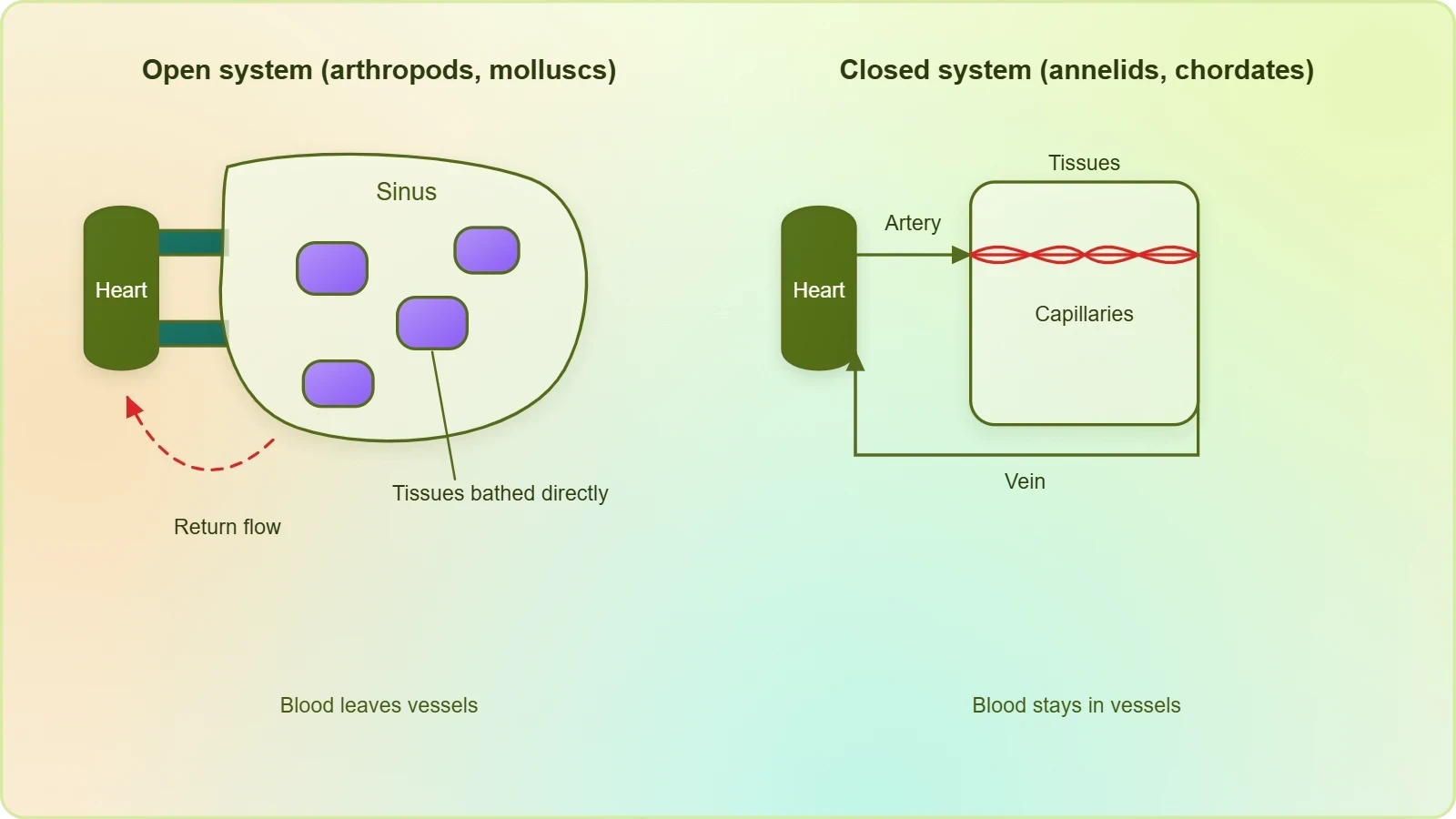
This design is economical and well suited to animals whose metabolic demands and body plans differ from those of vertebrates. The fluid in an open system serves both transport and, in a sense, a bathing function, since it washes the tissues directly. The trade-off is that the system offers limited ability to redirect flow to one organ and hold it there under sustained pressure. For many arthropods and molluscs, that limitation is entirely compatible with their way of life, and the open plan has persisted successfully across vast numbers of species.
Closed Systems in Annelids and Chordates
Annelids and chordates have a closed circulatory system, in which the blood pumped by the heart is always circulated through a closed network of blood vessels. This pattern is considered to be more advantageous because the flow of fluid can be more precisely regulated. Because blood stays within vessels, the system can vary the diameter of those vessels, direct blood preferentially to active tissues, and maintain higher pressures without the fluid simply dispersing into a cavity. The result is finer control over delivery of nutrients and removal of wastes.
The closed plan is the one that all vertebrates inherited, and it is the plan on which the rest of this lesson builds. Its precision comes from the vessel network itself: arteries carry blood away from the heart, capillaries bring it close to the cells, and veins return it. Regulation of flow is possible at each stage. When you later study the human heart and its nodal tissue, the value of this closed arrangement becomes concrete, because the heart's ability to adjust output only matters if the vessels can respond in kind.
The Vertebrate Heart: A Muscular Chambered Pump
All vertebrates possess a muscular chambered heart, and the number of chambers is one of the clearest structural markers of a species' circulatory pattern. The chambers are atria, which receive blood, and ventricles, which pump it out. Across the vertebrate classes the count rises from two to three to four, and each increase accompanies a change in how oxygenated and deoxygenated blood are handled. The heart is therefore not merely a pump of variable size but a record of how a lineage has solved the problem of separating two bloodstreams.
From Two Chambers to Four
Fishes have a 2-chambered heart with an atrium and a ventricle. Amphibians and the reptiles, except crocodiles, have a 3-chambered heart with two atria and a single ventricle. Crocodiles, birds and mammals possess a 4-chambered heart with two atria and two ventricles. The progression is not a ladder of improvement so much as a set of working solutions, each matched to the metabolic style and habitat of the animals that carry it. The chamber count directly determines whether the two bloodstreams can be kept apart.
Notice the precise wording of the exception: among reptiles, only crocodiles have four chambers. This detail is worth holding onto, because it shows that the boundary between three-chambered and four-chambered hearts does not fall neatly along the line between reptiles and birds. Crocodiles share the four-chambered condition with birds and mammals, while other reptiles retain the single ventricle. The table below sets out the full comparison so the pattern is easy to recall.
Why Chamber Number Matters
The number of chambers determines whether the two bloodstreams can be kept separate. With a single ventricle, oxygenated and deoxygenated blood necessarily share a pumping chamber and therefore mix. With two ventricles, each bloodstream has its own pump and the two never meet. This is why the chamber count is not a trivial anatomical detail but the structural basis of the circulatory pattern itself. When you read that an animal has double circulation, the four-chambered heart is the reason it can.
It also explains why the three-chambered heart is described as incomplete rather than simply intermediate. The two atria do their job of receiving separate bloodstreams, but the single ventricle cannot keep them apart on the way out. The inefficiency is built into the anatomy, not a failure of the animal. For amphibians and most reptiles, the arrangement supports their metabolic needs perfectly well, and the mixed blood leaving the ventricle is simply what their circulation delivers.
Single Circulation in Fishes
In fishes the heart pumps out deoxygenated blood, which is oxygenated by the gills and supplied to the body parts, from where deoxygenated blood is returned to the heart. This is single circulation: the blood passes through the heart once in each complete circuit of the body. The two-chambered heart, with its atrium and ventricle, is exactly matched to this route. Blood is received into the atrium, pumped by the ventricle to the gills, and then travels onward to the tissues before returning.
Tracing the Fish Circuit
Follow the path in order. Deoxygenated blood enters the heart, is pumped out by the ventricle, and travels to the gills, where it is oxygenated. From the gills the now oxygenated blood is supplied to the body parts. The tissues take up oxygen and release carbon dioxide, and the resulting deoxygenated blood is returned to the heart to begin the circuit again. At no point does the blood pass through the heart twice, which is precisely what the term single circulation denotes.
The elegance of this arrangement lies in its directness. The heart handles only deoxygenated blood, so there is no possibility of mixing and no need for a second ventricle. The gills sit between the heart and the body, acting as the oxygenation station through which all blood must pass. The pressure delivered to the tissues is therefore whatever remains after the blood has traversed the gill capillaries, a constraint that suits the generally lower metabolic demands of fishes compared with birds and mammals.
What Single Circulation Achieves
Single circulation keeps the design simple: one pump, one oxygenation station, one continuous loop. Because the heart receives only deoxygenated blood, the question of separating bloodstreams never arises. The system is efficient for its purpose and has served fishes across an enormous range of aquatic habitats. Its limitation is that blood loses pressure as it crosses the gills, so the driving force reaching the body tissues is lower than the heart's output alone would suggest.
This is a useful point of contrast for the lessons that follow. When you study the human heart, you will see two pumps in series, one serving the lungs and one serving the body, each able to generate the pressure its circuit requires. The fish heart, with its single ventricle, cannot do this. Recognising what single circulation cannot achieve makes the functional logic of double circulation much easier to grasp when it appears in the next sections.
Incomplete Double Circulation in Amphibians and Reptiles
In amphibians and reptiles, the left atrium receives oxygenated blood from the gills, lungs or skin, and the right atrium gets the deoxygenated blood from other body parts. However, they get mixed up in the single ventricle, which pumps out mixed blood. This is incomplete double circulation. The two atria perform their separate receiving roles correctly, but the shared ventricle means the two bloodstreams cannot remain distinct once they are pumped out to the body.
Two Atria, One Ventricle
The three-chambered heart has two atria and a single ventricle. The left atrium is the receiving chamber for oxygenated blood, which in amphibians and reptiles arrives from the gills, lungs or skin. The right atrium receives deoxygenated blood returning from the other body parts. Both atria then deliver their contents to the same ventricle. The ventricle therefore holds a mixture, and it is this mixture that is pumped out to the body. The separation achieved at the atrial level is lost at the ventricular level.
The phrase "gills, lungs or skin" is worth pausing over, because it reflects the diversity of respiratory surfaces across these groups. Amphibians in particular may rely on skin as well as lungs, and the circulatory description accommodates that. The constant across the group is the destination of oxygenated blood: it enters the left atrium. The constant across the group is also the limitation: a single ventricle cannot keep the two streams apart.
Why the Circulation Is Called Incomplete
The circulation is called incomplete because the separation of oxygenated and deoxygenated blood is not maintained throughout the circuit. The atria separate the streams on arrival, but the ventricle merges them on departure. Double circulation, by contrast, requires two separate pathways in which the two bloodstreams never mix. The three-chambered heart achieves half of that separation, which is precisely why the term incomplete is used rather than absent or partial in a vague sense.
This is a definition worth committing to memory, because examiners frequently test the distinction between incomplete and complete double circulation. The criterion is mixing. If oxygenated and deoxygenated blood mix in the ventricle, the double circulation is incomplete. If the ventricles pump the two bloodstreams out without any mixing, two separate circulatory pathways are present and the animal has double circulation. The next section shows how the four-chambered heart satisfies that criterion.
Double Circulation in Crocodiles, Birds and Mammals
In birds and mammals, oxygenated and deoxygenated blood received by the left and right atria respectively passes on to the ventricles of the same sides. The ventricles pump it out without any mixing up; that is, two separate circulatory pathways are present in these organisms, and hence these animals have double circulation. Crocodiles share this four-chambered condition with birds and mammals. The key structural feature is the complete separation of the two ventricles, which makes mixing impossible.
Two Separate Pathways
The four-chambered heart has two atria and two ventricles. Oxygenated blood received by the left atrium passes to the left ventricle, and deoxygenated blood received by the right atrium passes to the right ventricle. Each ventricle then pumps its own bloodstream out along its own pathway. Because the ventricles are separate, the two streams never meet. This is what the phrase "two separate circulatory pathways" describes, and it is the defining feature of double circulation as the term is used here.
The practical consequence is that the body receives fully oxygenated blood and the respiratory surfaces receive fully deoxygenated blood, with no dilution of either stream. This supports the higher metabolic rates characteristic of birds and mammals, which depend on a reliable supply of well-oxygenated blood to their tissues. The four-chambered heart is the structural foundation of that reliability, and it is why the condition is shared by the most metabolically demanding vertebrate groups.
Reading the Pattern Across Vertebrates
Set the three patterns side by side and the logic becomes clear. Fishes have a two-chambered heart and single circulation, with blood passing through the heart once per circuit. Amphibians and most reptiles have a three-chambered heart and incomplete double circulation, with mixing in the single ventricle. Crocodiles, birds and mammals have a four-chambered heart and double circulation, with no mixing at all. Each pattern is internally consistent, and each is matched to the animals that possess it.
This progression also prepares you for the human heart. The next lesson examines its structure, its valves and its nodal tissue in detail, and everything there presupposes the four-chambered, double-circulation plan described here. When you meet the left and right ventricles as separate pumps, you will already know why their separation matters and what it achieves. The vocabulary established in this lesson, from sinus to single circulation to incomplete double circulation, will recur throughout the remainder of the course.
Going Further: Reading the Designs Functionally
Two observations sharpen the material above without altering it. The first concerns what an open system actually does with its fluid. In an open system, blood is not always confined to vessels but bathes the cells in sinuses. This means the transport fluid and the interstitial environment are in direct continuity for much of the circuit, which is a fundamentally different arrangement from the closed systems of annelids and chordates, where the vascular wall intervenes between blood and tissue throughout.
The Cost of Mixing
The second observation concerns efficiency. Mixing of oxygenated and deoxygenated blood in a single ventricle makes circulation less efficient, because the blood delivered to the tissues carries less oxygen than it otherwise could. This is the functional reason the four-chambered heart is associated with the metabolically demanding birds and mammals. The structural separation of the ventricles is not an aesthetic refinement but a way of ensuring that the body receives the fullest possible oxygen load on every pass.
These two points are background rather than part of the core definitions, and they should be kept distinct from the material itself. The definitions, the chamber counts and the classification of groups remain exactly as stated: open systems in arthropods and molluscs, closed systems in annelids and chordates, two chambers in fishes, three in amphibians and most reptiles, four in crocodiles, birds and mammals. The functional reading simply explains why those arrangements work as they do.
Consolidating the Lesson: A Structured Review
A structured review ties the lesson together. Begin with the two circulatory patterns: open systems in arthropods and molluscs, where blood passes through large vessels into sinuses, and closed systems in annelids and chordates, where blood always circulates through a closed network of vessels and flow can be more precisely regulated. Then move to the vertebrate heart, noting that all vertebrates possess a muscular chambered heart and that the chamber count rises from two to three to four across the groups.
Checking Your Understanding
Test yourself against the definitions. Can you state which animals have an open system and which have a closed one? Can you name the chamber count for fishes, for amphibians and reptiles, and for crocodiles, birds and mammals? Can you explain why the circulation of amphibians and reptiles is called incomplete, and why that of birds and mammals is called double? If each answer comes readily, the core of the lesson is secure and you are ready for the human heart in the next lesson.
Pay particular attention to the exceptions and the precise wording. Crocodiles are the reptile exception with four chambers. The left atrium receives oxygenated blood from the gills, lungs or skin, while the right atrium receives deoxygenated blood from other body parts. In fishes the heart pumps out deoxygenated blood, which the gills oxygenate before it is supplied to the body. These details are exactly the kind that distinguish a precise answer from an approximate one.
Preparing for the Human Heart
The next lesson turns to the human heart's structure, valves and nodal tissue. Everything you have learned here about four chambers, two separate pathways and the absence of mixing applies directly. The human heart is a four-chambered heart operating on the double circulation plan, and its valves and nodal tissue are the mechanisms that keep that plan working in practice. The vocabulary of this lesson, from atrium and ventricle to single and double circulation, is the foundation on which that discussion rests.
Before moving on, return to the tables above and use them as a revision aid. The comparison of open and closed systems, the chamber counts by group, the step-by-step fish circuit, the summary of circulatory patterns and the glossary together cover the whole of this lesson's material. Reading them in sequence reconstructs the argument from structure to function, and that sequence is the one the course itself follows as it moves from the general principles of circulation to the specific anatomy of the human heart.
Also Read
- No links available.
From our network :
- 98% of Global MBA Programs Now Prefer GRE Over GMAT Focus EditionExplore why 98% of global MBA programs now prefer the GRE over GMAT Focus. Learn about the shorter GRE format, verbal logic importance, and strategic versatility for students.
- Mastering DB2 12.1 Instance Design: A Technical Deep Dive into Modern Database ArchitectureComprehensive guide to DB2 12.1 Instance Design. Explore architectural components, HADR setup, buffer pool tuning, and security policies for IBM DB2 12.1. Includes 10 functional SQL and Shell samples.
- Vite 6/7 'Cold Start' Regression in Massive Module GraphsIn-depth analysis of Vite 6/7 cold start regressions in massive module graphs. Learn about barrel file issues, pre-bundling optimizations, and how to improve Vite dev server performance.
- Analyzing Trump Deportation Numbers Insights Into The 2026 Immigration Crackdown
- AI-Powered 'Precision Diagnostic' Replaces Standard GRE Score ReportsDiscover the new GRE Precision Diagnostic. Learn how AI-powered behavioral analytics and time-to-solve tracking are replacing standard GRE score reports to empower students.
- EV 2.0: The Solid-State Battery Breakthrough and Global Factory ExpansionDiscover the impact of the Solid-State Battery Breakthrough on the EV market. Learn about CATL and BYD stock growth, global factory expansion in Europe, and the future of EV 2.0 technology.
- 10 Physics Numerical Problems with Solutions for IIT JEEPrepare for IIT JEE with 10 physics numerical problems, complete with detailed solutions and conceptual explanations. Ideal for mastering kinematics, thermodynamics, electromagnetism, and optics.
- Mastering DB2 LUW v12 Tables: A Comprehensive Technical GuideComprehensive guide on DB2 LUW v12 tables. Explore CREATE TABLE syntax, BLU Acceleration, range partitioning, data types, and security features for IBM DB2 LUW v12.
- Trump Political Strategy How Geopolitical Stunts Serve As Media Diversions
RESOURCES
- Evolutionary origins of the blood vascular system and endotheliumpmc.ncbi.nlm.nih.govWhy are certain blood circulatory systems open, while others are closed? Why ... system, highly efficient hearts and active lifestyles is seen in the…
- A Darwinian approach to the development of the vascular system in ...link.springer.comMar 27, 2025 ... In vertebrates, different closed circulatory systems are recognizable. In detail, fish have two-chambered hearts with unidirectional circulation ...
- 40.1 Overview of the Circulatory System - Biology 2e | OpenStaxopenstax.orgMar 28, 2018 ... In a closed circulatory system, blood is contained inside blood vessels and circulates unidirectionally from the heart around the systemic ...
- Overview of the Circulatory System | Biology II - Lumen Learningcourses.lumenlearning.comIn (a) closed circulatory systems, the heart pumps blood through vessels that are separate from the interstitial fluid of the body. Most vertebrates and…
- Circulatory system - Wikipediaen.wikipedia.orgIn vertebrates, the circulatory system is a system of organs that includes the heart, blood vessels, and blood which is circulated throughout the body.
- Circulatory System Architecture | Biology for Majors IIcourses.lumenlearning.comFigure 1. In (a) closed circulatory systems, the heart pumps blood through vessels that are separate from the interstitial fluid of the body. Most…
- Conservation and divergence of protein pathways in the vertebrate ...journals.plos.orgSep 6, 2019 ... Here, we present the cardiac proteomes of 4 model vertebrates with dual circulatory systems: the pig (Sus scrofa), the mouse…
- A Review of the “Open” and “Closed” Circulatory Systems: New ...onlinelibrary.wiley.comJan 26, 2009 ... Blood is driven at high pressures by the heart through a cell-lined (closed circulatory system) complex circulatory system that is…
- Circulatory systems of invertebrates | Health and Medicine - EBSCOebsco.com... circulatory system designs, typically categorized into open and closed systems. ... closed circulatory system of octopuses, squids, and cuttlefish, three hearts ...
- Evolution and development of the conduction system in the ...journals.biologists.comSep 2, 2024 ... ... open circulatory system, as opposed to the closed system seen in vertebrates. In embryonic cephalochordates (hagfish), the heart also…
- Circulatory System – ENT 425 – General Entomologygenent.cals.ncsu.eduIn a closed system, blood is always contained within vessels (arteries, veins, capillaries, or the heart itself). In an open system, blood (usually called ...
- Overview of the Circulatory System - OpenEd CUNYopened.cuny.edu... heart. In all vertebrate organisms, as well as some invertebrates, this is a closed-loop system, in which the blood is not free in…
- 6.1. Overview of the Circulatory System – Animal Physiologyopentext.uoregon.eduIn (a) closed circulatory systems, the heart pumps blood through vessels that are separate from the interstitial fluid of the body. Most vertebrates and…
- Circulatory system | Anatomy, Functions, Parts ... - Britannicabritannica.comNov 6, 2026 ... ... heart, respectively. The closed circulatory system found in vertebrates is not universal; a number of invertebrate phyla have an…
- 7.1 | Overview of the Circulatory System – Human Biologypressbooks-dev.oer.hawaii.eduFigure 7.2 In (a) closed circulatory systems, the heart pumps blood through vessels that are separate from the interstitial fluid of the body. Most…