Blood appears uniform to the naked eye, yet roughly 45 per cent of its volume is a busy cellular population suspended in plasma. These cells and cell fragments — erythrocytes, leucocytes and platelets — are collectively called the formed elements, and each family carries out a distinct assignment that keeps the body alive. Red cells ferry respiratory gases, white cells defend the organism against foreign invaders, and platelets stand ready to seal a breach in a vessel wall. Understanding how these three populations differ in number, structure and lifespan is the foundation for everything that follows in circulatory physiology.
This lesson examines each formed element in turn, following the order in which the material presents them. It begins with erythrocytes: their abundance, their site of formation, their biconcave architecture, the haemoglobin they carry and the 120-day lifespan that ends in the spleen. It then turns to leucocytes, distinguishing the granulocytes from the agranulocytes and assigning each subtype its percentage share and function. Finally, it treats platelets as fragments derived from megakaryocytes and links their number to the coagulation process. A short "Going further" block adds well-established background without disturbing the core sequence.
Because the preceding lesson established plasma and its proteins, and the next lesson takes up ABO grouping and donor compatibility, the discussion here stays deliberately within the cellular compartment. The numbers matter as much as the descriptions: five to five and a half million red cells per cubic millimetre, six to eight thousand white cells, and one and a half to three and a half lakh platelets. Hold these figures in mind, because they are the reference values against which clinical deviations are judged.
On This Page
The Formed Elements as a Whole
Erythrocytes, leucocytes and platelets are collectively called formed elements, and together they constitute nearly 45 per cent of the blood. The remaining portion is plasma, the fluid medium whose proteins, minerals and nutrients were examined in the previous lesson. This division of whole blood into a fluid phase and a cellular phase is more than a convenient description; it reflects two different systems of transport operating in parallel. Plasma carries dissolved substances in transit, while the formed elements carry oxygen, mount immune responses and repair vessel walls. The proportion itself is a useful diagnostic anchor, since a shift in the cellular fraction alters the consistency and behaviour of blood.
What unites the three populations is their origin. All are produced in the bone marrow, though the details differ: red cells form in the red bone marrow of adults, megakaryocytes reside in the marrow and shed platelet fragments there, and leucocytes arise from marrow precursors before entering the circulation. What separates them is equally instructive. Erythrocytes are the most abundant and the longest lived; leucocytes are nucleated, relatively scarce and generally short lived; platelets are not whole cells at all but fragments. The table below sets out this three-way comparison as a reference frame for the sections that follow.
Why the Cellular Fraction Matters
The 45 per cent figure is not a static constant so much as a regulated balance. Because red cells dominate the cellular fraction numerically, the overall proportion is governed chiefly by red cell number and by plasma volume. A fall in red cell production, an increase in destruction, or a dilution of plasma all shift the ratio, and each shift carries consequences for oxygen delivery and for the workload of the heart. Recognising that the formed elements are a population in dynamic turnover, rather than a fixed structural component, prepares the ground for understanding why the body invests so heavily in their continuous replacement.
Each population also has its own tempo. Red cells persist for months, leucocytes for hours to days, and platelets for a short span before being cleared. This means the marrow must run three production lines at different rates, each responsive to different signals. The sections that follow take these lines one at a time, beginning with the most numerous and ending with the smallest, mirroring the sequence in which the material presents them.
Reading the Numbers Correctly
Throughout this lesson, cell counts are expressed per cubic millimetre of blood, written as ##\text{mm}^{-3}##. This convention allows direct comparison between populations whose true numbers differ by orders of magnitude. A count of five million red cells beside eight thousand white cells is not a contradiction but a statement about relative abundance. When you meet a figure such as 1,500,00–3,500,00 platelets ##\text{mm}^{-3}##, read it as the lakh-scale range given in the material, and keep the unit attached to the number so that comparisons remain honest.
Precision in units is a habit worth forming early. The same numerical value means something entirely different depending on whether it is quoted per millilitre or per cubic millimetre, and clinical thresholds are always tied to a stated unit. For the purposes of this lesson, the counts given are the reference values for a healthy adult, and any deviation discussed later should be understood as a departure from these baselines rather than an absolute rule.
Erythrocytes: Structure, Haemoglobin and Lifespan
Erythrocytes, or red blood cells, are the most abundant of all the cells in blood. A healthy adult man has, on average, five million to five and a half million RBCs per cubic millimetre of blood. They are formed in the red bone marrow in adults, and in most mammals they are devoid of a nucleus. Their shape is biconcave, a geometry that increases surface area relative to volume and thereby favours the exchange of gases across the cell membrane. This combination of abundance, origin and architecture makes the erythrocyte the principal vehicle of respiratory transport in the body.
The red colour that gives these cells their name comes from haemoglobin, a red-coloured, iron-containing complex protein. A healthy individual has 12–16 grams of haemoglobin in every 100 millilitres of blood, and these molecules play a significant role in the transport of respiratory gases. The relationship between cell number, haemoglobin content and oxygen-carrying capacity is direct and worth stating explicitly: the cell is essentially a container, the pigment is the cargo, and the biconcave shape is the packaging that lets the cargo load and unload efficiently.
Formation and the Red Bone Marrow
In adults, red blood cells are formed in the red bone marrow. This is a point of practical importance because it locates the erythrocyte production line in a specific tissue rather than in the circulation itself. The marrow supplies the precursor cells and the raw materials, and the mature cells are released into the bloodstream once they have acquired their haemoglobin. Because the mature cell lacks a nucleus in most mammals, it cannot divide or repair itself; the burden of replacement therefore falls entirely on the marrow, which must sustain a continuous output to match the continuous loss of aged cells.
The steady-state logic is simple arithmetic. If a cell lives 120 days and the population is to remain constant, roughly one hundred and twentieth of the total must be replaced each day. This is why the marrow is among the most active tissues in the body, and why any interruption to its function shows up relatively quickly in the circulating count. The material does not elaborate on the intermediate stages of maturation, and this lesson does not invent them; what matters here is the site of formation and the fact that production is continuous.
Haemoglobin, Shape and the 120-Day Lifespan
Haemoglobin is the functional heart of the erythrocyte. Being iron-containing, it binds respiratory gases, and its presence in high concentration within each cell explains both the colour of blood and its capacity to carry oxygen far in excess of what plasma alone could dissolve. The biconcave shape complements this chemistry by presenting a large membrane surface for diffusion while keeping the diffusion distance to the cell interior short. Structure and function are thus matched: a pigment optimised for binding, enclosed in a shape optimised for exchange, distributed in numbers large enough to meet systemic demand.
RBCs have an average life span of 120 days, after which they are destroyed in the spleen, often described as the graveyard of RBCs. This single sentence carries two ideas. The first is that the erythrocyte is a terminally differentiated cell with a fixed tenure; the second is that clearance is an organised process localised in a particular organ. The spleen therefore sits at the end of the erythrocyte's story just as the marrow sits at its beginning, and the two organs together define the cell's complete arc.
Leucocytes: The Two Categories and Their Subtypes
Leucocytes are also known as white blood cells because they are colourless, a consequence of lacking haemoglobin. Unlike erythrocytes, they are nucleated, and they are relatively fewer in number, averaging 6,000–8,000 per cubic millimetre of blood. They are generally short lived. These four features — colourlessness, a nucleus, low abundance and brief tenure — distinguish the white cell population sharply from the red. Where the erythrocyte is a long-lived, nucleus-free carrier, the leucocyte is a short-lived, nucleated defender that circulates in comparatively small numbers but acts decisively when called upon.
Leucocytes fall into two main categories: granulocytes and agranulocytes. Neutrophils, eosinophils and basophils are the different types of granulocytes, while lymphocytes and monocytes are the agranulocytes. Within this classification, each subtype occupies a characteristic share of the total white cell count and performs a characteristic function. The percentages are not incidental detail; they describe a division of labour in which the most abundant cell type handles the most common threat, and the rarest handles a specialised inflammatory role.
Granulocytes: Neutrophils, Eosinophils and Basophils
Neutrophils are the most abundant cells of the total white blood cells, accounting for 60–65 per cent, and basophils are the least, at 0.5–1 per cent. Neutrophils are phagocytic cells which destroy foreign organisms entering the body. Basophils secrete histamine, serotonin, heparin and similar substances, and are involved in inflammatory reactions. Eosinophils, at 2–3 per cent, resist infections and are also associated with allergic reactions. The three granulocytes thus span the range from the numerically dominant phagocyte to the rarest secretory cell, and their functions map neatly onto that spread.
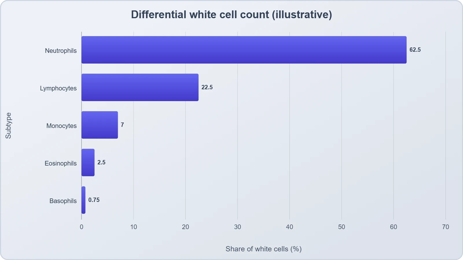
The pairing of neutrophils with monocytes in the material is worth noting: both are described as phagocytic cells which destroy foreign organisms entering the body. This shared function across a granulocyte and an agranulocyte shows that the granulocyte–agranulocyte division is a structural classification, not a functional one. A learner should therefore resist the temptation to assume that all granulocytes do one thing and all agranulocytes another. The categories describe appearance and lineage; the functions cut across them.
Agranulocytes: Lymphocytes and Monocytes
Monocytes account for 6–8 per cent of white cells and, as noted, are phagocytic. Lymphocytes account for 20–25 per cent and are of two major types, the B and T forms. Both B and T lymphocytes are responsible for immune responses of the body. This is the second most abundant white cell population after the neutrophils, and its division into B and T forms anticipates the detailed treatment of immunity that belongs to a later stage of study. For the present lesson, the essential point is that lymphocytes are the cells through which the body mounts specific immune responses.
Monocytes, though far fewer, complement the neutrophils in the phagocytic role. The arrangement is one of redundancy with specialisation: several cell types can engulf foreign organisms, but each does so in its own context and at its own tempo. The table below consolidates the five subtypes, their percentage shares and their functions, so that the classification and the division of labour can be read together rather than separately.
Platelets: Fragments from Megakaryocytes
Platelets, also called thrombocytes, are cell fragments produced from megakaryocytes, which are special cells in the bone marrow. Blood normally contains 1,500,00–3,500,00 platelets per cubic millimetre. Platelets can release a variety of substances, most of which are involved in the coagulation or clotting of blood. A reduction in their number can lead to clotting disorders, which will lead to excessive loss of blood from the body. The platelet is therefore the smallest of the formed elements in size and the most narrowly specialised in function, yet its role is indispensable to the integrity of the circulation.
The description of platelets as fragments rather than whole cells is deliberate and consequential. Because they are not complete cells, they do not carry a nucleus in the manner of leucocytes, and their functional repertoire is limited to the release of stored substances. That limitation is precisely their strength: the substances they release are the ones needed to initiate clotting, and the fragment is essentially a mobile packet of clotting-related material held in readiness until a vessel is damaged.
Origin, Number and the Coagulation Role
The megakaryocyte is the parent structure, a special cell of the bone marrow that produces platelet fragments. The normal range of 1,500,00–3,500,00 per cubic millimetre defines the healthy band, and the material links a fall below this band directly to clotting disorders. The logic is causal rather than merely correlational: fewer platelets mean less released clotting material, which means impaired coagulation, which in turn means excessive blood loss from the body. This chain from cell fragment to released substance to clot to haemostasis is the essential narrative of the platelet.
It is worth pausing on the phrase "most of which are involved in the coagulation or clotting of blood." The material does not claim that every released substance serves clotting, only that most do. This leaves room for other roles without contradicting the central function, and it is a reminder that biological descriptions are often stated with careful qualification. For examination purposes, the coagulation role is the one to retain, with the number range as its quantitative anchor.
When Platelet Numbers Fall
A reduction in platelet number leads to clotting disorders, and these in turn lead to excessive loss of blood from the body. The clinical significance of the platelet is thus inverse to that of the erythrocyte: too few red cells compromise oxygen transport, while too few platelets compromise the mechanical integrity of the vessel system. Both are deficiencies of supply, but they fail in different currencies — one in gas exchange, the other in haemostasis. Recognising this distinction helps organise the disorders of the circulatory system that a later lesson will address.
The platelet also illustrates a broader principle about the formed elements: function follows form. A fragment cannot divide or repair itself, but it can be produced in large numbers and consumed rapidly at a site of injury. The body's solution to the problem of vessel repair is therefore not a permanent cell but a disposable packet, manufactured continuously and deployed where needed.
Comparing the Three Populations
Placing the three populations side by side reveals a coherent design. The erythrocyte is numerous, nucleus-free, haemoglobin-bearing and long lived; the leucocyte is scarce, nucleated, colourless and short lived; the platelet is a fragment, produced in lakh-scale numbers and consumed at sites of injury. Each design choice serves a function: abundance serves gas transport, nucleation serves immune versatility, and fragmentation serves rapid, localised clotting. The comparison is not merely a revision aid but a demonstration of how structure, number and lifespan are tuned to physiological purpose.
The counts themselves span a remarkable range. Red cells number in millions per cubic millimetre, white cells in thousands, and platelets in lakhs. These orders of magnitude are not arbitrary; they reflect the frequency and urgency of each task. Oxygen must be carried continuously and in bulk, defence must be mounted rapidly but in smaller numbers, and clotting must be available instantly at any point in the vasculature. The table below arranges the three populations by count and lifespan to make these contrasts explicit.
Structure Matched to Function
The biconcave erythrocyte, the nucleated leucocyte and the anucleate platelet each embody a different solution to a different problem. The red cell maximises surface for gas exchange and fills its interior with haemoglobin; the white cell retains a nucleus so that it can respond flexibly to foreign organisms; the platelet abandons cellular completeness in favour of a concentrated packet of clotting substances. Reading the three together, one sees that the formed elements are not variations on a single theme but three distinct designs, each optimised for its own task and each produced on its own schedule.
This structural logic also explains the lifespans. A cell that cannot repair itself, like the mature erythrocyte, must be replaced on a fixed schedule, and 120 days is its allotted tenure. A cell that must respond to unpredictable threats, like the leucocyte, is deployed briefly and replaced as needed. A fragment that is consumed at the point of injury is produced continuously and in quantity. Lifespan, in each case, is a consequence of design rather than an arbitrary number.
What Comes Next
The formed elements described here circulate within a fluid whose proteins and minerals were treated in the previous lesson, and they carry on their surfaces the antigens that determine blood group. The next lesson takes up ABO grouping and donor compatibility, where the presence or absence of the A and B antigens on the red cell surface, and the corresponding antibodies in plasma, decide whether a transfusion will succeed or cause clumping. The red cell, in other words, is not only a gas carrier but also an immunological identity card, and that second role is the subject of the lesson to follow.
For now, the essential inventory is complete: erythrocytes in their millions, leucocytes in their thousands with five named subtypes, and platelets in their lakhs as fragments of megakaryocytes. Each has a number, a structure, a lifespan and a function, and each contributes to the 45 per cent of blood that constitutes the formed elements. Mastery of these particulars is the necessary preparation for the antigenic and compatibility questions that lie ahead.
Going further
Two points of well-established background help consolidate the material without altering it. First, the absence of a nucleus in mature mammalian erythrocytes is not a trivial detail but a design economy: by dispensing with the nucleus and most organelles, the cell frees internal volume for haemoglobin, so that a single red cell can carry far more oxygen-carrying pigment than a nucleated cell of the same size could. The trade-off is that the cell forfeits the capacity for repair and division, which is why its tenure is finite and why the marrow must replace it continuously.
Second, platelets are best understood as cell fragments rather than whole cells. They are shed from megakaryocytes, the large marrow cells named in the material, and they lack the complete cellular machinery of a leucocyte. This is why their functional repertoire centres on the release of stored substances rather than on phagocytosis or immune recognition. Recognising the platelet as a fragment clarifies both what it can do and what it cannot, and it explains why the body produces them in such large numbers: they are disposable units, manufactured to be spent at the site of injury.
Consolidating the Lesson
The formed elements form a coherent system in which number, structure, lifespan and function are interlocked. Erythrocytes dominate numerically and carry haemoglobin for respiratory transport; leucocytes are nucleated defenders divided into granulocytes and agranulocytes, each subtype with its own share and role; platelets are megakaryocyte fragments that release clotting substances. Together they account for nearly 45 per cent of blood, and together they illustrate how the body allocates different tasks to differently designed cells. The percentages and counts given here are the reference values against which any deviation is measured.
A useful way to test understanding is to ask, for each population, three questions: how many, how long, and what for? Red cells: millions, 120 days, gas transport. White cells: thousands, short lived, defence. Platelets: lakhs, fragments, clotting. If these three answers can be produced without hesitation, the core of the lesson has been absorbed. The remaining detail — the five white cell subtypes and their percentages — can then be layered on top as a finer-grained classification within the defence category.
Common Points of Confusion
Several distinctions are easily blurred. The first is between granulocytes and agranulocytes as structural categories and phagocytic versus non-phagocytic as functional ones; neutrophils and monocytes are both phagocytic despite belonging to different categories. The second is between the erythrocyte's lack of a nucleus and the leucocyte's possession of one, which is why only the latter can be described as nucleated. The third is between platelets as fragments and leucocytes as whole cells. Keeping these three distinctions clear prevents most of the errors that arise when the populations are compared.
A fourth point concerns the spleen. It is described as the graveyard of RBCs, which is a statement about clearance, not about production. Production occurs in the red bone marrow. Confusing the two organs would misplace the beginning and the end of the erythrocyte's life. A fifth point concerns the platelet range, which is quoted in lakhs and should not be mistaken for the thousands used for leucocytes. Unit discipline, as noted earlier, is the simplest safeguard against such slips.
Preparing for Blood Groups
The red cell carries not only haemoglobin but also surface antigens, and it is these antigens that will occupy the next lesson. The presence or absence of the A and B antigens on the red cell surface, together with the corresponding antibodies in plasma, defines the ABO groups and determines donor compatibility. Nothing in the present lesson needs to be revised to accommodate that material; the erythrocyte described here is simply the same cell viewed from a different angle, first as a gas carrier and next as an antigenic surface.
Similarly, the plasma proteins discussed in the previous lesson — fibrinogens for clotting, globulins for defence, albumins for osmotic balance — connect forward to the platelet's clotting role and to the leucocyte's defensive role. The three lessons together form a continuous account: plasma provides the medium and its proteins, the formed elements provide the cellular actors, and blood groups provide the compatibility rules that govern transfusion. Each lesson builds on the last, and the present one supplies the cellular inventory on which the next depends.
Also Read
- No links available.
From our network :
- Analyzing Trump Deportation Numbers Insights Into The 2026 Immigration Crackdown
- Mastering DB2 LUW v12 Tables: A Comprehensive Technical GuideComprehensive guide on DB2 LUW v12 tables. Explore CREATE TABLE syntax, BLU Acceleration, range partitioning, data types, and security features for IBM DB2 LUW v12.
- 10 Physics Numerical Problems with Solutions for IIT JEEPrepare for IIT JEE with 10 physics numerical problems, complete with detailed solutions and conceptual explanations. Ideal for mastering kinematics, thermodynamics, electromagnetism, and optics.
- 98% of Global MBA Programs Now Prefer GRE Over GMAT Focus EditionExplore why 98% of global MBA programs now prefer the GRE over GMAT Focus. Learn about the shorter GRE format, verbal logic importance, and strategic versatility for students.
- Vite 6/7 'Cold Start' Regression in Massive Module GraphsIn-depth analysis of Vite 6/7 cold start regressions in massive module graphs. Learn about barrel file issues, pre-bundling optimizations, and how to improve Vite dev server performance.
- AI-Powered 'Precision Diagnostic' Replaces Standard GRE Score ReportsDiscover the new GRE Precision Diagnostic. Learn how AI-powered behavioral analytics and time-to-solve tracking are replacing standard GRE score reports to empower students.
- EV 2.0: The Solid-State Battery Breakthrough and Global Factory ExpansionDiscover the impact of the Solid-State Battery Breakthrough on the EV market. Learn about CATL and BYD stock growth, global factory expansion in Europe, and the future of EV 2.0 technology.
- Trump Political Strategy How Geopolitical Stunts Serve As Media Diversions
- Mastering DB2 12.1 Instance Design: A Technical Deep Dive into Modern Database ArchitectureComprehensive guide to DB2 12.1 Instance Design. Explore architectural components, HADR setup, buffer pool tuning, and security policies for IBM DB2 12.1. Includes 10 functional SQL and Shell samples.
RESOURCES
- Introduction to Blood - SEER Training Modulestraining.seer.cancer.govDec 2, 2025 ... Three classes of formed elements: erythrocytes (red blood cells), leukocytes (white blood cells), and the thrombocytes (platelets).
- Blood Basics - Hematology.orghematology.orgIt has four main components: plasma, red blood cells, white blood cells, and platelets. ... Forming blood clots to prevent excess blood loss. Carrying ...
- Binding of 99Tcm sulphur colloid to blood components - PubMedpubmed.ncbi.nlm.nih.govIn humans, 68% of the total blood counts were bound to the formed elements (erythrocytes, leucocytes and platelets). The binding pattern (as percentage of ...
- Anatomy 163 Lab: Blood Exercise 29 Flashcards - Quizletquizlet.comThree types of formed elements found in blood include erythrocytes, leukocytes, and: platelets. The least numerous but largest of all agranulocytes is the ...
- Chapter 10 Blood Terminology - NCBI - NIHncbi.nlm.nih.gov... Blood components consist of erythrocytes (red blood cells), leukocytes (white blood cells), thrombocytes (platelets), and clotting factors.
- Blood cells and formed elements of the blood - GetBodySmartgetbodysmart.comMay 15, 2023 ... Plasma; Red blood cells (erythrocytes); White blood cells (leukocytes); Platelets (thrombocytes). These cellular components, also referred to as ...
- An Overview of Blood | Anatomy and Physiology II - Lumen Learningcourses.lumenlearning.comBlood is composed of formed elements—erythrocytes, leukocytes, and cell fragments called platelets—and a fluid extracellular matrix called plasma. More than ...
- The Oxidation of Long-Chain Fatty Acids by the Formed Elements of ...sciencedirect.comIn five experiments red blood cells, free of white blood cells and platelets, were incubated with phosphate-NaCl buffer, palmitate-l-C14, and glucose (5.55 mM).
- Production of the Formed Elements | Anatomy and Physiology IIcourses.lumenlearning.comThus, the body must form new blood cells and platelets quickly and continuously. ... erythrocytes, leukocytes, and platelets. Hemopoiesis begins in the red bone ...
- Production of the Formed Elements – Anatomy & Physiologypressbooks-dev.oer.hawaii.eduThus, the body must form new blood cells and platelets quickly and continuously. ... erythrocytes, leukocytes, and platelets. Hemopoiesis begins in the red bone ...
- Formed elements of blood: MedlinePlus Medical Encyclopedia Imagemedlineplus.govFeb 27, 2026 ... Blood transports oxygen and nutrients to body tissues and returns waste and carbon dioxide. Blood distributes nearly everything that is ...
- Viscosity of Blood - Cardiovascular Physiology Conceptscvphysiology.comThe addition of formed elements to plasma (red cells, white cells, and platelets) further increases the viscosity. Of these formed elements, red cells have…
- Comparative Effects of the Different Formed Elements on Normal ...journals.physiology.orgThe difference is due to the other formed elements: platelets, reticulocytes and leucocytes. ... They centrifuged blood and compared red cell suspensions low in ...
- 17.3: Cellular Defenses - Biology LibreTextsbio.libretexts.orgThe formed elements of the blood include red blood cells (erythrocytes), white blood cells (leukocytes), and platelets (thrombocytes).
- Microcirculation and Hemorheology - PMC - NIHpmc.ncbi.nlm.nih.gov... blood flow and the vascular wall. Flow of the blood formed elements (red blood cells (RBCs), white blood cells or leukocytes (WBCs) and…