Double circulation is the architectural principle that gives the human cardiovascular system its remarkable efficiency: two separate pumps, two distinct circuits, and a single continuous route that blood must follow without deviation. Every drop of blood leaving the right ventricle is destined for the lungs, and every drop leaving the left ventricle is bound for the body's tissues. This division of labour means oxygen-poor and oxygen-rich blood never mix in the healthy heart, and it allows the systemic circuit to operate at far higher pressure than the delicate pulmonary capillaries could tolerate. Understanding this dual arrangement is essential for anyone studying cardiac physiology, because it explains why the heart is effectively two pumps working in series rather than one pump serving the whole body.
This lesson, the twelfth in a fifteen-part course on body fluids and circulation, builds directly on the electrocardiogram discussion that preceded it. Having established how electrical activity is recorded, we now turn to the physical plumbing that electrical signal drives: the vessels themselves and the two great circulatory pathways they serve. We will examine the three-layered structure common to every artery and vein, trace the pulmonary circuit from right ventricle to left atrium, follow the systemic circuit from left ventricle to right atrium, and give proper attention to two specialised routes — the hepatic portal system and the coronary circulation — that deserve separate treatment because they deviate from the simple out-and-back pattern.
By the end of this lesson you should be able to describe the tunica intima, tunica media and tunica externa, explain why the tunica media is comparatively thin in veins, and sketch both circulatory pathways with their correct chambers and vessel types. You should also be able to articulate what the systemic circulation delivers to tissues and what it removes, and to explain why the hepatic portal vein and the coronary arteries are exceptions worth remembering. This foundation prepares you for the next lesson on regulation of cardiac activity, where the nervous and hormonal control of these pathways becomes the central concern.
On This Page
- The Three-Layered Architecture of Blood Vessels
- Pulmonary Circulation: Right Ventricle to Left Atrium
- Systemic Circulation: Left Ventricle to Right Atrium
- The Hepatic Portal System: A Detour Through the Liver
- The Coronary System: Supply to the Cardiac Musculature
- Reading the Pathways Together: A Unified View
- Going Further
The Three-Layered Architecture of Blood Vessels
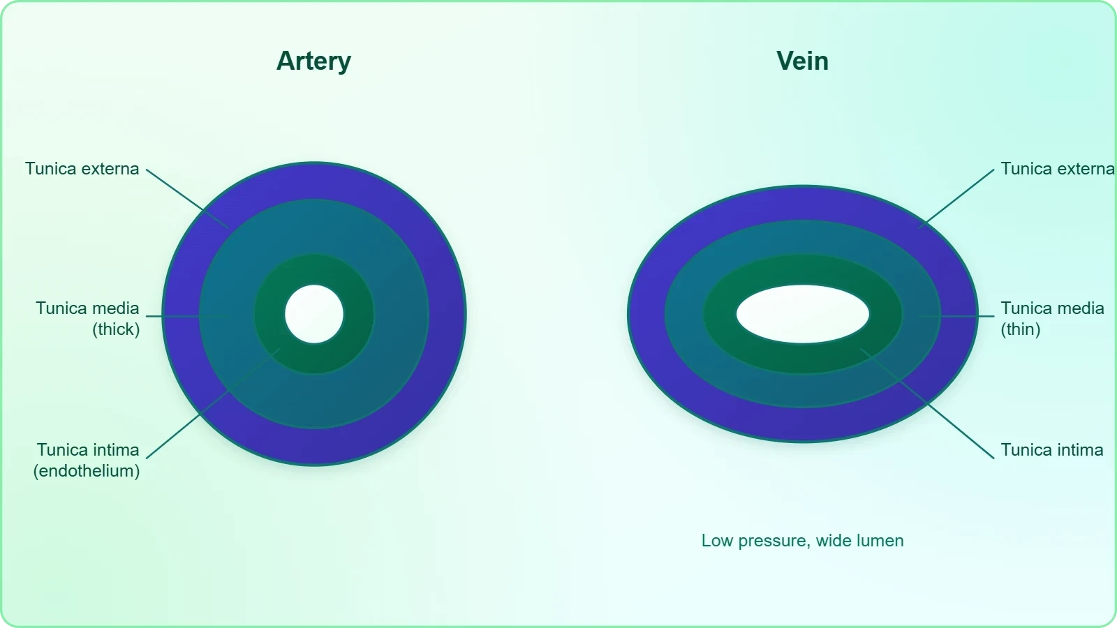
Blood flows strictly by a fixed route through blood vessels — the arteries and veins — and this fixed route is not merely a matter of anatomical convention but of structural design. Every artery and every vein, regardless of size or location, is constructed from the same three concentric layers, each contributing a distinct mechanical or physiological function. The inner lining, the middle muscular sheath and the outer fibrous coat work together to contain pressure, regulate flow and anchor the vessel within surrounding tissue. Recognising this common plan is the first step towards understanding why arteries and veins behave so differently despite sharing the same basic blueprint.
The Tunica Intima and Tunica Externa
The innermost layer is the tunica intima, an inner lining of squamous endothelium. This single layer of flattened cells presents an exceptionally smooth surface to the passing blood, minimising friction and preventing the turbulent flow that would otherwise damage formed elements and promote clot formation. The endothelium is not merely passive lining; it participates in regulating vessel tone and permeability. At the opposite extreme lies the tunica externa, an external layer of fibrous connective tissue with collagen fibres. This collagen-rich sheath gives the vessel tensile strength, prevents overstretching under pressure, and tethers the vessel to the surrounding connective tissue so that it does not collapse or shift as organs move.
The Tunica Media and Its Variation
Between these two lies the tunica media, a middle layer of smooth muscle and elastic fibres. This is the functional heart of the vessel wall: the smooth muscle can contract to narrow the lumen, and the elastic fibres recoil to smooth out the pressure pulse generated by each heartbeat. The tunica media is comparatively thin in the veins, a structural detail with clear physiological consequences. Because veins conduct blood at low pressure, they require far less muscular and elastic reinforcement than arteries, which must withstand the full systolic surge. This single difference in wall thickness explains much of the visible and functional distinction between the two vessel types.
Pulmonary Circulation: Right Ventricle to Left Atrium
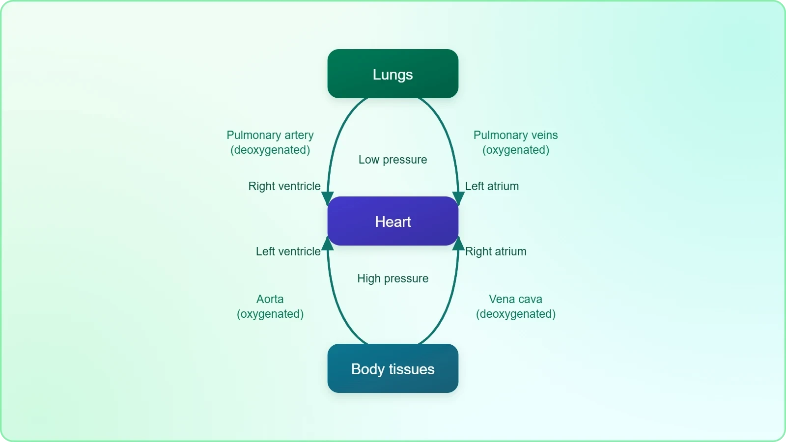
The blood pumped by the right ventricle enters the pulmonary artery, and this single fact defines the pulmonary circuit's starting point. From the right ventricle, deoxygenated blood is carried through the pulmonary artery to the lungs, where gas exchange takes place across the thin respiratory membrane. The oxygenated blood is then carried by the pulmonary veins into the left atrium. This pathway — right ventricle, pulmonary artery, lungs, pulmonary veins, left atrium — constitutes the pulmonary circulation. It is a comparatively short circuit serving a single organ, yet it is indispensable, because it is the only route by which deoxygenated blood can be reloaded with oxygen before returning to the systemic side of the heart.
Why the Pulmonary Circuit Operates at Low Pressure
The pulmonary circuit is a low-pressure system by design, and this design protects the delicate alveolar capillaries from the mechanical stress that high systemic pressures would impose. Blood arrives in the pulmonary artery at pressures far below those in the aorta, which is appropriate for a circuit whose vessels are short and whose capillary beds are thin-walled and optimised for diffusion rather than filtration. The consequence is that the right ventricle performs less muscular work than the left, and its wall is correspondingly thinner. This asymmetry between the two ventricular walls is a direct structural reflection of the two circuits' differing pressure demands.
The Significance of Complete Separation
Because the pulmonary and systemic circuits are arranged in series rather than in parallel, the output of one becomes the input of the other, and the two ventricles must therefore pump equal volumes over time. This arrangement enforces a complete separation of oxygenated and deoxygenated blood in the healthy heart, a separation that is the central functional advantage of double circulation. Blood returning from the lungs is fully oxygenated before it enters the left atrium, and blood returning from the tissues is fully deoxygenated before it enters the right atrium. The result is that systemic tissues receive blood at the highest oxygen concentration the lungs can achieve.
Systemic Circulation: Left Ventricle to Right Atrium
The left ventricle pumps blood into the aorta, and from this great vessel the oxygenated blood is carried by a network of arteries, arterioles and capillaries to the tissues. At the capillary level, the essential exchange occurs: oxygen and nutrients leave the blood, and carbon dioxide and metabolic waste enter it. The now-deoxygenated blood is collected by a system of venules, veins and vena cava and emptied into the right atrium. This is the systemic circulation, and it is the circuit that serves every organ of the body except the lungs themselves. Its scale — from the aorta's elastic trunk down to microscopic capillaries and back through the great veins — is what makes it the body's principal distributive network.
What the Systemic Circulation Delivers and Removes
The systemic circulation provides nutrients, oxygen and other essential substances to the tissues, and it takes carbon dioxide and other harmful substances away for elimination. This dual function is easy to state but profound in its implications: the circulatory system is simultaneously a delivery service and a waste-removal service, and both functions depend on the same continuous flow. If perfusion fails, tissues are starved of oxygen and simultaneously poisoned by their own accumulating waste. The systemic circuit's reach into every tissue, through progressively finer branching, is what allows this exchange to occur at the cellular scale where it is actually needed.
Arteries, Arterioles, Capillaries and Veins
The route from aorta to vena cava passes through a graded series of vessel types, each suited to its position in the pressure gradient. Arteries carry blood away from the heart at high pressure; arterioles are the principal resistance vessels, regulating distribution to capillary beds; capillaries are the thin-walled exchange vessels; venules collect blood from capillaries; and veins return it to the heart at low pressure. This progression from high-pressure conduit to low-pressure collector is the physical expression of the pressure gradient that drives flow, and it explains why the same three-layered wall plan is modified so differently along the route.
The Hepatic Portal System: A Detour Through the Liver
A unique vascular connection exists between the digestive tract and the liver, called the hepatic portal system. The hepatic portal vein carries blood from the intestine to the liver before it is delivered to the systemic circulation. This is a genuine departure from the simple artery-to-capillary-to-vein pattern, because blood passes through two successive capillary beds — first in the intestinal wall, then in the liver — before it ever reaches the general circulation. The arrangement is not an anatomical curiosity but a functional necessity: it ensures that absorbed nutrients and any absorbed foreign substances are processed by the liver before they can reach the rest of the body.
Why the Portal Detour Matters
The portal arrangement gives the liver first access to everything absorbed from the gut. Nutrients can be stored, converted or released under hepatic control; potentially harmful substances can be detoxified; and the composition of the blood entering the systemic circulation can be regulated before it reaches sensitive organs. This is why the hepatic portal vein is described as carrying blood to the liver before it is delivered to the systemic circulation — the word "before" carries the entire physiological rationale. The liver thus functions as a biochemical gatekeeper positioned precisely where the products of digestion first enter the bloodstream.
Portal Veins as a General Principle
A portal system, in the general physiological sense, is any arrangement in which blood passes through two capillary networks in series before returning to the heart. The hepatic portal system is the largest and most familiar example, but the principle illustrates something important about circulatory design: the body is willing to insert an extra capillary bed wherever intermediate processing is required. Recognising this pattern helps you interpret other specialised circulations and prevents the mistake of assuming that all blood follows the simplest possible route from artery to vein.
The Coronary System: Supply to the Cardiac Musculature
A special coronary system of blood vessels is present in our body exclusively for the circulation of blood to and from the cardiac musculature. The heart, despite being filled with blood, cannot nourish its own muscle from the blood passing through its chambers; the thick ventricular walls are far too dense for diffusion from the lumen to suffice. Instead, the coronary arteries branch from the aorta and deliver oxygenated blood directly to the myocardium, and the coronary veins return deoxygenated blood to the right atrium. This dedicated supply is essential because cardiac muscle works continuously and has one of the highest oxygen demands of any tissue.
The Functional Logic of a Dedicated Supply
The coronary circulation illustrates a general principle: any tissue with a high and uninterrupted metabolic demand requires its own dedicated blood supply rather than relying on diffusion from a neighbouring region. Cardiac muscle contracts roughly once per second throughout life, and it cannot pause to recover. Its oxygen extraction from coronary blood is already near maximal at rest, meaning that increased demand must be met by increased flow rather than by extracting more oxygen from each unit of blood. This is why the coronary arteries are functionally among the most critical vessels in the body.
Coronary Flow and the Cardiac Cycle
Coronary flow has a distinctive rhythm tied to the cardiac cycle. During ventricular contraction, the contracting muscle compresses the coronary vessels and impedes flow; during relaxation, the vessels open and perfusion rises sharply. This means that the heart's own supply is greatest during diastole, the resting phase. The dependence of coronary perfusion on diastolic relaxation explains why very rapid heart rates, which shorten diastole disproportionately, can compromise the heart's own oxygen supply even when arterial pressure is adequate.
Reading the Pathways Together: A Unified View
When the pulmonary and systemic circuits are viewed together, the elegance of double circulation becomes apparent. Blood follows a single figure-of-eight route: right ventricle to lungs to left atrium, then left ventricle to tissues to right atrium, and back to the right ventricle. Each circuit has one pump, one arterial outflow, one capillary exchange region and one venous return. The two circuits are linked in series, so that the output of the right heart is the input of the left heart and vice versa. This unified view is the mental model you should carry forward into the study of cardiac regulation, because every adjustment of heart rate or contractility acts on this entire loop.
Pressure, Resistance and Flow in the Two Circuits
Flow through any circuit depends on the pressure difference driving it and the resistance opposing it, a relationship expressed compactly as:
Here ##Q## is flow, ##\Delta P## is the pressure difference between the circuit's arterial and venous ends, and ##R## is the total resistance of the circuit. The systemic circuit has a far larger ##\Delta P## than the pulmonary circuit, which is why the left ventricle must generate much greater pressure to drive the same volume of blood. The arterioles, as the principal resistance vessels, are the main site where ##R## is adjusted to redistribute flow between organs without changing total cardiac output.
Connecting Structure to the Previous and Next Lessons
The electrical events recorded in the previous lesson on the electrocardiogram are the trigger for the mechanical events that drive these pathways. The QRS complex initiates ventricular contraction, which is what propels blood into the pulmonary artery and aorta; the T-wave marks the end of systole, when the ventricles relax and coronary perfusion rises. In the next lesson, regulation of cardiac activity, we will see how the heart's intrinsic myogenic rhythm is moderated by the autonomic nervous system and by adrenal medullary hormones, adjusting the rate and strength of contraction that move blood through the circuits described here.
Going Further
The observation that the tunica media is comparatively thin in veins rewards a moment of reflection. Because venous pressure is low, a thick muscular coat would be mechanically unnecessary, and the thinner wall allows veins to be highly distensible. This distensibility turns the venous system into a capacious reservoir holding a large fraction of the total blood volume at any moment. When the body needs to redistribute blood — during exercise or haemorrhage — sympathetic stimulation can constrict these thin-walled vessels and shift their contents towards the heart, effectively using the venous reservoir as a rapid-response volume buffer.
The hepatic portal vein's role in delivering blood to the liver before it reaches the systemic circulation also merits a further thought. Because the liver receives this blood first, it can regulate the concentration of glucose and other metabolites entering the general circulation, smoothing out the surges that follow a meal. This buffering function is a direct consequence of the portal arrangement's position in the flow path, and it illustrates how a modest anatomical detour can produce a major physiological capability. The coronary system, meanwhile, reminds us that no tissue can rely on diffusion alone when its metabolic demand is continuous and high.
Also Read
- No links available.
From our network :
- Analyzing Trump Deportation Numbers Insights Into The 2026 Immigration Crackdown
- 98% of Global MBA Programs Now Prefer GRE Over GMAT Focus EditionExplore why 98% of global MBA programs now prefer the GRE over GMAT Focus. Learn about the shorter GRE format, verbal logic importance, and strategic versatility for students.
- 10 Physics Numerical Problems with Solutions for IIT JEEPrepare for IIT JEE with 10 physics numerical problems, complete with detailed solutions and conceptual explanations. Ideal for mastering kinematics, thermodynamics, electromagnetism, and optics.
- Trump Political Strategy How Geopolitical Stunts Serve As Media Diversions
- AI-Powered 'Precision Diagnostic' Replaces Standard GRE Score ReportsDiscover the new GRE Precision Diagnostic. Learn how AI-powered behavioral analytics and time-to-solve tracking are replacing standard GRE score reports to empower students.
- Vite 6/7 'Cold Start' Regression in Massive Module GraphsIn-depth analysis of Vite 6/7 cold start regressions in massive module graphs. Learn about barrel file issues, pre-bundling optimizations, and how to improve Vite dev server performance.
- Mastering DB2 12.1 Instance Design: A Technical Deep Dive into Modern Database ArchitectureComprehensive guide to DB2 12.1 Instance Design. Explore architectural components, HADR setup, buffer pool tuning, and security policies for IBM DB2 12.1. Includes 10 functional SQL and Shell samples.
- EV 2.0: The Solid-State Battery Breakthrough and Global Factory ExpansionDiscover the impact of the Solid-State Battery Breakthrough on the EV market. Learn about CATL and BYD stock growth, global factory expansion in Europe, and the future of EV 2.0 technology.
- Mastering DB2 LUW v12 Tables: A Comprehensive Technical GuideComprehensive guide on DB2 LUW v12 tables. Explore CREATE TABLE syntax, BLU Acceleration, range partitioning, data types, and security features for IBM DB2 LUW v12.
RESOURCES
- Circulatory Pathways - SEER Training Modulestraining.seer.cancer.govThe blood vessels of the body are functionally divided into two distinctive circuits: pulmonary circuit and systemic circuit. The pump for the pulmonary circuit ...
- Pulmonary & Systemic Circulation | Circulatory Anatomy - Visible Bodyvisiblebody.comSystemic circulation moves blood between the heart and the rest of the body. It sends oxygenated blood out to cells and returns deoxygenated blood…
- Double Circulation | Definition, Related Videos & FAQs | JoVE Topicsjove.comResearchers can consider whether problems affect the separation of oxygenated and deoxygenated blood, the pulmonary pathway, the systemic pathway, or the ...
- How Your Circulatory System Works - Cleveland Clinicmy.clevelandclinic.orgSep 4, 2024 ... Your circulatory system (cardiovascular system) includes your heart and blood vessels. Your heart pumps oxygen-rich blood after your lungs ...
- In brief: How does the blood circulatory system work? - NCBI - NIHncbi.nlm.nih.govNov 21, 2023 ... This is where pulmonary circulation begins: The right ventricle pumps low-oxygen blood into the pulmonary artery, which branches off into ...
- Circulatory system - Wikipediaen.wikipedia.orgDouble circulatory systems permit blood to be repressurized after returning from the lungs, speeding up delivery of oxygen to tissues. [citation needed]. No ...
- How the Heart Works - How Blood Flows through the Heart - nhlbi, nihnhlbi.nih.govJun 25, 2025 ... Arteries and veins link your heart to the rest of the circulatory system. ... The pulmonary valve separates the right…
- 17.2D: Systemic and Pulmonary Circulation - Medicine LibreTextsmed.libretexts.orgOct 14, 2025 ... The cardiovascular system is composed of two circulatory paths: pulmonary circulation, the circuit through the lungs where blood is oxygenated; ...
- The gut-lung axis: effects and mechanisms of gut microbiota on ...pmc.ncbi.nlm.nih.gov... lungs. First, via the portal vein-systemic circulation pathway, microbial metabolites absorbed by the intestinal epithelium (e.g., SCFAs, tryptophan ...
- body fluids and circulation chapter 15 - NCERTncert.nic.inHuman circulatory system, also called the blood vascular system ... We have a complete double circulation, i.e., two circulatory pathways, namely, pulmonary and ...
- Systemic and Pulmonary Microcirculation, Double ... - Sage Journalsjournals.sagepub.comSystemic and Pulmonary Microcirculation, Double Microcirculation: From ... Oxygen-sensing pathways and the pulmonary circulation. J Physiol. 2024;602 ...
- Treatment algorithm for pulmonary arterial hypertensionpublications.ersnet.orgPulmonary arterial hypertension (PAH) therapies work through four major pathways ... routes, and potentially fewer systemic effects in general [84]. Switching ...
- The circulatory pathway is a highly organized, pressure ... - Instagraminstagram.comFeb 28, 2026 ... Understanding the Dual System of Human Blood Circulation ... Pulmonary circulation carries the same cardiac output as systemic circulation ...
- Double-Drainage Levoatrial Cardinal Vein: Cardiac CT Evaluation ...jacc.orgJan 23, 2026 ... ... pulmonary venous flow, which under normal circumstances enters the systemic arterial circulation. Although the term anomalous pulmonary ...
- Signaling pathways and targeted therapy for pulmonary hypertensionnature.comJul 1, 2025 ... ... pathway specifically in the pulmonary circulation. Targeting PPAR ... Genetic deletion of CMG2 exacerbates systemic-to-pulmonary shunt-induced ...