Blood and lymph are the body's two great transport media, and the closing lesson of this course gathers them into a single, coherent picture. Vertebrates circulate blood, a fluid connective tissue, to deliver essential substances to the cells and to carry waste substances away from them; a second fluid, lymph or tissue fluid, is also used for the transport of certain substances. Around this simple division of labour the chapter arranges an entire architecture: plasma and formed elements, the ABO and Rh groupings, coagulation, open and closed pathways, the auto-excitable heart, the cardiac cycle, the electrocardiogram, complete double circulation, and the neural and hormonal moderation of cardiac function.
This final lesson is deliberately a revisiting rather than a fresh survey. Each idea returns in the order the chapter itself follows, so that the definitions, the numbers and the relationships settle into a stable structure you can carry into an examination hall or a clinical conversation. The heart's pacemaker fires at 70 to 75 action potentials per minute, a healthy person completes 72 cardiac cycles per minute, each ventricle ejects about 70 mL per beat, and the cardiac output works out to roughly 5 litres per minute. These figures are not trivia; they are the arithmetic skeleton of the whole subject.
Because this is the last lesson of the course, it also serves as a bridge. The previous lesson examined disorders of the circulatory system, and the summary you meet here quietly explains why those disorders matter: hypertension strains the brain and kidney, coronary artery disease narrows the vessels feeding the heart muscle, angina announces oxygen shortage, and heart failure describes a pump that cannot meet the body's needs. Read the summary as a diagnostic map, not a list to be memorised.
On This Page
- Blood as a Fluid Connective Tissue
- Lymph, Tissue Fluid and the Pathways of Circulation
- The Auto-Excitable Heart and Its Pacemaker
- Recording the Heartbeat: The Electrocardiogram
- Double Circulation and Its Significance
- Moderation of Cardiac Activity and Disorders Revisited
- Consolidating the Chapter: A Revision Framework
Blood as a Fluid Connective Tissue
The chapter opens its summary with a definition that repays close attention: blood is a fluid connective tissue. The phrase sounds paradoxical until you notice that blood contains cells suspended in a matrix, exactly as bone and cartilage do, except that the matrix here is liquid. That liquid matrix is plasma, and the suspended cells are the formed elements. Because blood connects every organ to every other organ through the vessels, it earns the connective label in the functional sense as well as the histological one. This dual identity explains why questions about blood so often begin with the word "why" rather than "what".
Plasma and the Formed Elements
Blood comprises a fluid matrix, plasma, and formed elements. Red blood cells, also called erythrocytes, white blood cells, also called leucocytes, and platelets, also called thrombocytes, constitute the formed elements. Each carries a distinct responsibility that the exercises of the chapter ask you to name: erythrocytes handle gas transport, leucocytes resist infections, and thrombocytes participate in coagulation. Plasma itself is not mere water; its proteins matter for osmotic balance, clotting and defence, which is precisely why the chapter asks about the importance of plasma proteins. Keep the three formed elements and their functions paired in memory.
The white blood cells deserve a finer grain of attention because the chapter's matching exercise singles out eosinophils as resisters of infection, alongside the broader leucocyte category. Platelets, though not whole cells in the conventional sense, are indispensable to coagulation, the process that converts soluble plasma protein into an insoluble mesh when a vessel is breached. When you revise, resist the temptation to treat the formed elements as a flat list. Picture instead a division of labour: oxygen and carbon dioxide move on erythrocytes, immune defence rides on leucocytes, and haemostasis depends on thrombocytes.
ABO and Rh Grouping
Human blood is grouped into the A, B, AB and O systems based on the presence or absence of two surface antigens, A and B, on the red blood cells. A second grouping rests on the presence or absence of another antigen called the Rhesus factor, or Rh, on the red cell surface. The chapter's matching exercise links the AB group to the universal recipient, a fact worth anchoring because it follows directly from the antigen logic: a recipient with no anti-A or anti-B antibodies can accept cells bearing either antigen. Grouping is therefore not an arbitrary label but a chemical description of the cell surface.
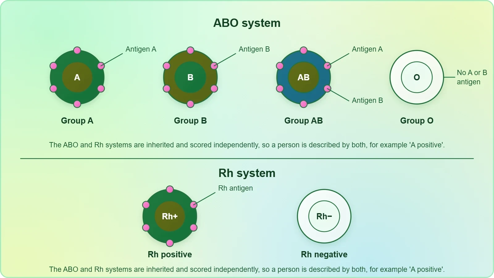
The Rh factor adds a second layer of compatibility that becomes clinically urgent during pregnancy and transfusion. Because the antigens sit on the erythrocyte membrane, the immune system reads them as identity markers, and mismatched transfusion provokes agglutination. The summary does not elaborate on this, but the logic is implicit in the antigen-antibody framing. When you answer a question on blood groups, state the antigen first, then the antibody consequence, then the practical rule. That sequence keeps the reasoning transparent and prevents the common error of memorising compatibility tables without understanding them.
Lymph, Tissue Fluid and the Pathways of Circulation
The spaces between cells in the tissues contain a fluid derived from blood, called tissue fluid. This fluid, called lymph, is almost similar to blood except for the protein content and the formed elements. That single sentence carries the whole comparison the chapter's exercises demand. Lymph is not a separate invention of the body but a filtrate of blood that has escaped the capillaries to bathe the cells, and it returns to the circulation by its own routes. Understanding this derivation makes the differences between blood and lymph logical rather than arbitrary, and it explains why lymph is described as a transport medium for certain substances only.
Open and Closed Systems
All vertebrates and a few invertebrates have a closed circulatory system. In a closed system, blood remains within vessels and the heart throughout its circuit, which permits precise direction, pressure and distribution. An open system, by contrast, lets haemolymph percolate through tissue spaces before returning. The chapter's exercise asks you to write the differences between the open and closed systems, and the cleanest way to answer is to contrast containment, pressure and efficiency of delivery. Our own circulatory system consists of a muscular pumping organ, the heart, a network of vessels, and a fluid, blood, which is the textbook definition of the closed arrangement.
Lymphatic return complements this closed design. Because tissue fluid accumulates continuously at the capillary beds, the body needs a drainage route that does not compete with the arterial circuit. Lymph provides it, carrying proteins and other substances that cannot re-enter the blood capillaries directly. When you compare blood and lymph in an answer, mention the protein content and the absence of formed elements first, then the direction of flow, then the function. This ordering mirrors the chapter's own emphasis and keeps the comparison crisp rather than sprawling.
The Auto-Excitable Heart and Its Pacemaker
The heart has two atria and two ventricles, and its musculature is auto-excitable, meaning it generates its own rhythm without waiting for a nerve impulse. The sino-atrial node, or SAN, generates the maximum number of action potentials per minute, 70 to 75, and therefore sets the pace of the heart's activities; hence it is called the pacemaker. This is why the chapter's exercise asks why we call our heart myogenic and why the SAN is called the pacemaker. Both answers converge on the same principle: the origin of the beat lies within the cardiac muscle itself, not in the nervous system.
From Action Potential to Contraction
The action potential causes the atria and then the ventricles to undergo contraction, called systole, followed by their relaxation, called diastole. The systole forces blood to move from the atria to the ventricles and onward to the pulmonary artery and the aorta. The atrio-ventricular node and the atrio-ventricular bundle matter here because they carry the impulse from the atria to the ventricles, ensuring the chambers contract in the correct sequence rather than simultaneously. Without that conduction bridge, the ventricles would receive the signal haphazardly and the pumping action would lose its coordination.
Notice how the summary compresses a sequence into a single sentence. The atria contract, the ventricles contract, both relax, and the cycle repeats. Each phase has a purpose: atrial systole tops up the ventricles, ventricular systole drives blood into the great arteries, and diastole allows the chambers to refill. The chapter's exercise on systole and diastole expects you to contrast these phases clearly. A useful discipline is to describe each phase in terms of pressure and valve position, because that framing explains the heart sounds the chapter also asks you to explain.
Cardiac Cycle, Stroke Volume and Cardiac Output
The cardiac cycle is formed by sequential events in the heart which are cyclically repeated. A healthy person shows 72 such cycles per minute. About 70 mL of blood is pumped out by each ventricle during a cardiac cycle, and this is called the stroke volume or beat volume. The volume of blood pumped out by each ventricle per minute is called the cardiac output, and it equals the product of stroke volume and heart rate, approximately 5 litres. These definitions are the most examinable content in the chapter, and they are also the most useful in clinical reasoning.
The arithmetic is worth writing out because it makes the relationship unmistakable. With a stroke volume of 70 mL and a heart rate of 72 beats per minute, the product is 5040 mL, which rounds to the stated 5 litres. The formula is therefore not a separate fact to memorise but a direct consequence of the two measurements. When heart rate rises during exercise, cardiac output can increase either through a faster rate or a larger stroke volume, and the body uses both levers. The chapter's exercise asking you to define the cardiac cycle and the cardiac output is best answered with this equation in view.
Recording the Heartbeat: The Electrocardiogram
The electrical activity of the heart can be recorded from the body surface using an electrocardiograph, and the recording is called an electrocardiogram, or ECG, which is of clinical importance. The summary states this plainly, but the exercises push further, asking you to draw a standard ECG and explain its different segments, and to distinguish the P-wave from the T-wave. The ECG is therefore not a decorative add-on to the chapter but a diagnostic instrument that translates the heart's electrical sequence into a visible trace. Each deflection corresponds to a phase of depolarisation or repolarisation in the cardiac musculature.
Reading the Waves
The P-wave reflects the electrical activity associated with atrial depolarisation, the signal that precedes atrial contraction. The QRS complex marks ventricular depolarisation, the event that triggers ventricular systole. The T-wave represents ventricular repolarisation, the recovery phase before the next cycle begins. When the chapter asks you to differentiate the P-wave and the T-wave, the essential contrast is atrial depolarisation against ventricular repolarisation. A well-drawn ECG with these three components labelled, and the intervals between them marked, answers the exercise completely and demonstrates that you understand the sequence rather than merely the shapes.
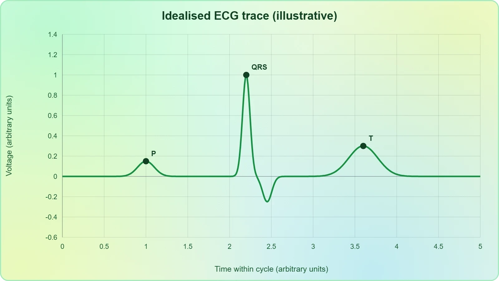
Clinical importance follows directly from this mapping. Because each wave corresponds to a specific electrical event, a disturbance in the trace points to a disturbance in conduction. The chapter does not catalogue arrhythmias in the summary, but the logic is clear: if the SAN sets the pace and the atrio-ventricular node and bundle conduct the impulse, then an abnormal ECG can localise where that chain has faltered. This is why the electrocardiograph is described as clinically important rather than merely interesting. For revision, practise sketching the trace from memory and naming each segment aloud before you check the textbook.
Double Circulation and Its Significance
We have a complete double circulation, meaning two circulatory pathways, pulmonary and systemic, are present. The pulmonary circulation starts with the pumping of deoxygenated blood by the right ventricle, which is carried to the lungs where it is oxygenated and returned to the left atrium. The systemic circulation starts with the pumping of oxygenated blood by the left ventricle to the aorta, which carries it to all the body tissues, and the deoxygenated blood from there is collected by the veins and returned to the right atrium. The two circuits are therefore serial and complete, each beginning and ending at the heart.
Why Two Circuits Matter
The significance of double circulation lies in the separation of oxygenated and deoxygenated blood and in the pressure requirements of the two circuits. The pulmonary circuit serves the lungs and needs only modest pressure, while the systemic circuit must drive blood to every tissue, including the brain and the extremities, and therefore demands higher pressure. By keeping the circuits distinct, the heart can meet both demands without compromising either. The chapter's exercise asking what is meant by double circulation and what its significance is expects exactly this reasoning: definition first, then functional consequence, then the advantage of separation.
The evolutionary dimension is also examinable. The chapter asks you to describe the evolutionary change in the pattern of heart among the vertebrates, and the summary's insistence on complete double circulation in our own species is the endpoint of that progression. Across vertebrates the heart shows increasing separation of the chambers and of the two circuits, moving from simpler arrangements toward the four-chambered organ described here. When you answer, trace the trend rather than listing species: the direction of change is toward fuller separation of oxygenated and deoxygenated blood, and our complete double circulation is the culmination of that trend.
Moderation of Cardiac Activity and Disorders Revisited
Though the heart is auto-excitable, its functions can be moderated by neural and hormonal mechanisms. This closing statement of the summary is easy to overlook, yet it reconciles two apparently competing ideas: the heart generates its own rhythm, and the body can still adjust that rhythm to circumstance. Neural input can speed or slow the pace set by the SAN, and hormonal signals can alter the force and rate of contraction. The result is a pump that is autonomous in origin but responsive in operation, which is precisely what an organism needs when demand changes from rest to exertion.
Neural and Hormonal Control
The distinction between myogenic origin and external moderation is a favourite examination target. The heart is myogenic because the impulse arises within the cardiac musculature, specifically at the SAN. It is nonetheless modulated because nerves and hormones can modify the rate and strength of contraction without initiating the beat. When you answer, separate the two claims explicitly: origin is internal, adjustment is external. This framing also explains why a transplanted or denervated heart can continue to beat, a point that follows logically from the chapter's statement even though the summary does not spell it out.
Disorders as Consequences of the Summary
The previous lesson examined disorders of the circulatory system, and the summary supplies the physiological context that makes them intelligible. Hypertension, or high blood pressure, leads to heart diseases and affects vital organs like the brain and kidney. Coronary artery disease, often called atherosclerosis, affects the vessels supplying the heart muscle and is caused by deposits of calcium, fat, cholesterol and fibrous tissues that narrow the lumen of arteries. Angina, or angina pectoris, is a symptom of acute chest pain appearing when not enough oxygen reaches the heart muscle, more common among the middle-aged and elderly.
Heart failure means the state of the heart when it is not pumping blood effectively enough to meet the needs of the body. It is sometimes called congestive heart failure because congestion of the lungs is one of its main symptoms. Crucially, heart failure is not the same as cardiac arrest, when the heart stops beating, or a heart attack, when the heart muscle is suddenly damaged by an inadequate blood supply. These distinctions are frequently confused, and the summary states them precisely. Revise them as a set of three, because examiners reward the ability to separate conditions that share a vocabulary but not a mechanism.
Consolidating the Chapter: A Revision Framework
The most efficient way to close this course is to rebuild the chapter from its own summary, in its own order. Begin with the purpose of circulation, then the two fluids, then the formed elements and groupings, then the pathways, then the heart and its cycle, then the ECG, then double circulation, and finally the moderation of cardiac activity and the disorders that follow when it fails. This sequence is not arbitrary; it mirrors the logic of the material, moving from the medium to the machinery to the measurements to the malfunctions. Reconstructing it from memory is the strongest test of whether the chapter has been absorbed.
Self-Test Against the Exercises
The chapter's exercises are a ready-made revision checklist, and each maps onto a section of the summary. Naming the formed elements and their functions tests the first section. Explaining the importance of plasma proteins tests the composition of plasma. Matching eosinophils, RBC, AB group, platelets and systole tests the groupings and the cardiac cycle. Asking why blood is a connective tissue tests the opening definition. Comparing lymph and blood, open and closed systems, systole and diastole, and the P-wave and T-wave tests the comparative core of the chapter. Working through them in order is a complete rehearsal.
The remaining exercises probe the heart's autonomy and its electrical record. Why is the heart myogenic, why is the SAN the pacemaker, and what is the significance of the atrio-ventricular node and bundle, all test the conduction story. Defining the cardiac cycle and cardiac output tests the arithmetic. Explaining heart sounds and drawing a standard ECG test the mechanical and electrical correlates of the cycle. If you can answer all of these without notes, you have met the chapter's standard. If any one falters, return to the corresponding section of this lesson rather than rereading the whole chapter.
Going further
Two observations from the summary deserve a moment of wider reflection. The first is the arithmetic of the heartbeat: about 70 mL is pumped by each ventricle per beat and about 5 litres per minute, which means the entire blood volume passes through the heart roughly once a minute at rest. The second is the distribution of the closed design across the animal kingdom: all vertebrates and a few invertebrates have a closed circulatory system, so the arrangement described here is the vertebrate norm rather than a peculiarity of our species. Together these points connect the chapter's physiology to its comparative biology.
Beyond the summary, it is worth noting that the same variables used here, stroke volume and heart rate, remain the working vocabulary of clinical cardiology, and the ECG remains a first-line diagnostic recording. The chapter's insistence on separating heart failure from cardiac arrest and heart attack reflects a distinction that matters enormously in practice, where confused terminology can delay appropriate response. None of this contradicts the material; it simply shows that the summary is a foundation rather than a closed box. Carry the definitions and the numbers forward, and the rest of the subject will arrange itself around them.
Also Read
- No links available.
From our network :
- 98% of Global MBA Programs Now Prefer GRE Over GMAT Focus EditionExplore why 98% of global MBA programs now prefer the GRE over GMAT Focus. Learn about the shorter GRE format, verbal logic importance, and strategic versatility for students.
- AI-Powered 'Precision Diagnostic' Replaces Standard GRE Score ReportsDiscover the new GRE Precision Diagnostic. Learn how AI-powered behavioral analytics and time-to-solve tracking are replacing standard GRE score reports to empower students.
- 10 Physics Numerical Problems with Solutions for IIT JEEPrepare for IIT JEE with 10 physics numerical problems, complete with detailed solutions and conceptual explanations. Ideal for mastering kinematics, thermodynamics, electromagnetism, and optics.
- Mastering DB2 LUW v12 Tables: A Comprehensive Technical GuideComprehensive guide on DB2 LUW v12 tables. Explore CREATE TABLE syntax, BLU Acceleration, range partitioning, data types, and security features for IBM DB2 LUW v12.
- Vite 6/7 'Cold Start' Regression in Massive Module GraphsIn-depth analysis of Vite 6/7 cold start regressions in massive module graphs. Learn about barrel file issues, pre-bundling optimizations, and how to improve Vite dev server performance.
- EV 2.0: The Solid-State Battery Breakthrough and Global Factory ExpansionDiscover the impact of the Solid-State Battery Breakthrough on the EV market. Learn about CATL and BYD stock growth, global factory expansion in Europe, and the future of EV 2.0 technology.
- Trump Political Strategy How Geopolitical Stunts Serve As Media Diversions
- Mastering DB2 12.1 Instance Design: A Technical Deep Dive into Modern Database ArchitectureComprehensive guide to DB2 12.1 Instance Design. Explore architectural components, HADR setup, buffer pool tuning, and security policies for IBM DB2 12.1. Includes 10 functional SQL and Shell samples.
- Analyzing Trump Deportation Numbers Insights Into The 2026 Immigration Crackdown
RESOURCES
- body fluids and circulation chapter 15 - NCERTncert.nic.inBlood is a special connective tissue consisting of a fluid matrix, plasma, and formed elements. 15.1.1 Plasma. Plasma is a straw coloured, viscous fluid ...
- Body Fluids and Circulation Overview | PDF | Heart - Scribdscribd.comBody Fluids and Circulation Overview ... Circulation involves the transport of nutrients, gases, and waste through the body via blood and lymph. Simple organisms ...
- Important Notes for NEET – Body Fluids and Circulation - BYJU'Sbyjus.comPlasma constitutes 55% of blood volume. It is a viscous fluid and contains 90-92% water. Other than water, plasma contains 6-8% proteins (fibrinogens, albumins ...
- Body fluids and circulation: Quiz 1 | Blood - Khan Academykhanacademy.orgReview the following recommended lessons to help you learn: {list of lessons covered by quiz}
- body fluids and circulation chapter 18 - AWSgrab-guidance.s3.ap-south-1.amazonaws.comBlood is a special connective tissue consisting of a fluid matrix, plasma, and formed elements. 18.1.1 Plasma. Plasma is a straw coloured, viscous fluid ...
- Overview of MicroRNA Biogenesis, Mechanisms of Actions, and ...pmc.ncbi.nlm.nih.govAug 3, 2018 ... Many studies have detected extracellular/circulating miRNAs in biological fluids, such as plasma and serum (117, 118), cerebrospinal fluid (119) ...
- MDCG 2021-24 Rev.1 Guidance on classification of medical deviceshealth.ec.europa.euin contrast, a surgically created opening to allow access to the circulatory system should not be considered to be a body orifice. ... body…
- Water, Hydration and Health - PMC - NIHpmc.ncbi.nlm.nih.govIn summary, hydration status consistently affected self-reported ... Blood and Other Body Fluids. Washington DC: Federation of American Societies ...
- Buy Body Fluids & Circulation Study Notes PDF | Blood Plasma and ...etsy.comBody Fluids & Circulation topics overview – covers blood components, lymphatic system, human circulatory. zoom image 3 of 7. High ...
- Body Fluid - an overview | ScienceDirect Topicssciencedirect.comAdvanced kinetic analysis (see later “Body Fluid Dynamics” section) ... circulation. When ventricular preload is increased, cardiac output is augmented ...
- Fluid Physiology Part 1: Volume and Distribution of Water and Its ...link.springer.comNov 30, 2023 ... This chapter focuses on the compartmentalization of body ... The modern view of body fluid circulation is an actively pumped…
- Chapter 15 Fluids and Electrolytes - Nursing Fundamentals - NCBIncbi.nlm.nih.govBody fluids are found in two main areas of the body called intracellular and extracellular compartments. See Figure 15.1 for an illustration of intracellular ...
- Lymphatic System: Function, Conditions & Disordersmy.clevelandclinic.orgJul 31, 2023 ... ... fluids throughout your body. Lymphatic system organs include your ... fluid (lymph) back into your circulatory system (your bloodstream).
- 26.1 Body Fluids and Fluid Compartments - Anatomy ... - OpenStaxopenstax.orgApr 20, 2022 ... ... circulation. Because the left side of the heart is unable to pump out ... 1-body-fluids-and-fluid-compartments. © Jun 12, 2026…
- Adult Dehydration - StatPearls - NCBI Bookshelf - NIHncbi.nlm.nih.govAug 17, 2026 ... [1][20] The body depends on a complex system to maintain euvolemia, the stable, normal volume of blood and extracellular fluid.…