A protein does not behave like a stiff rod suspended in the cell. Once the amino acid sequence is joined and the local helices and sheets have formed, the entire thread folds back on itself into a compact, three-dimensional shape — the material describes this as folding "like a hollow woollen ball." That image is worth holding onto, because it captures the essential idea of tertiary structure: a single continuous chain, coiled and packed into a defined globular form whose surface and interior are arranged for a purpose. This lesson examines that folding, then extends the picture to proteins built from more than one such folded chain.
The distinction between tertiary and quaternary structure is one of the most frequently confused points in the study of biomacromolecules, and it repays careful attention. Tertiary structure concerns one polypeptide chain and the way it folds upon itself; quaternary structure concerns the architecture that emerges when two or more folded polypeptides, called subunits, are arranged with respect to one another. Adult human haemoglobin, with its four subunits, is the example the material supplies, and it will anchor the second half of this lesson. Both levels build directly on the primary and secondary structures covered previously.
Because tertiary structure is described as absolutely necessary for the many biological activities of proteins, the lesson also considers what the shape is for. A folded chain is not decoration; it is the physical basis of what a protein does. The bonds that hold the fold together, the difference between a single-chain protein and a multi-subunit assembly, and the consequences of losing the fold are all treated here in the order the material presents them, with a short "Going further" block reserved for well-established background that extends the core content.
On This Page
- From a Folded Thread to a Folded Ball: Defining Tertiary Structure
- The Bonds and Forces That Hold the Fold
- Quaternary Structure: The Architecture of Subunits
- Comparing the Four Levels Side by Side
- Why the Fold Is Necessary: Function Follows Shape
- Consolidating the Lesson: Key Points and Self-Check
- Working Through the Concepts: A Short Computational Illustration
From a Folded Thread to a Folded Ball: Defining Tertiary Structure
The previous lesson established that a protein thread is not an extended rigid rod and that only some portions of it are arranged as a helix, with other regions folded into the forms of secondary structure. Tertiary structure begins exactly where that account leaves off. The long protein chain, already carrying its helices and sheets, is folded upon itself like a hollow woollen ball. The result is a three-dimensional view of a protein, and the material is explicit that this level of organisation is absolutely necessary for the many biological activities of proteins.
Two features of this definition deserve emphasis. First, tertiary structure belongs to a single polypeptide chain; it is the shape of one thread, not an assembly of several. Second, the folding is global rather than local. Secondary structure describes recurring local arrangements such as the right-handed helix, whereas tertiary structure describes how the whole chain, with all its local arrangements, is packed into one compact form. The hollow woollen ball is a deliberately homely comparison, and it works because a ball of wool is one continuous strand wound into a stable, rounded mass.
The Hollow Woollen Ball as a Working Image
The comparison to a hollow woollen ball carries more information than it first appears to. A ball of wool is continuous — the strand never breaks — yet its overall form is compact and roughly spherical, with the strand turning back on itself repeatedly. So it is with a globular protein: the chain remains one unbroken sequence from the N-terminal amino acid to the C-terminal amino acid, but its spatial path is folded and refolded until the whole occupies a defined three-dimensional region. The word "hollow" in the material's image also hints that the interior is not solid mass but a packed arrangement of side chains.
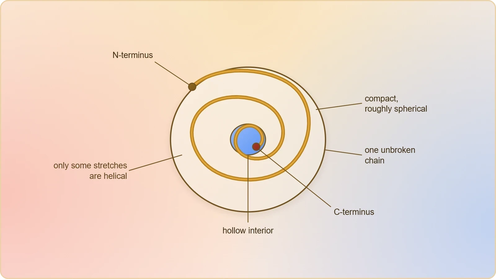
This image also clarifies why the earlier description of the thread as a helix was qualified. Only some portions of the protein thread are arranged in the form of a helix, and in proteins only right-handed helices are observed. The remaining regions are folded into other secondary forms. Tertiary structure is what happens when all of these regions, helical and otherwise, are brought together into a single folded object. The helix is a local motif; the woollen ball is the whole.
Why the Three-Dimensional Shape Matters
The material states plainly that tertiary structure is absolutely necessary for the many biological activities of proteins. The reason is structural: the activities of a protein depend on the precise arrangement of chemical groups in space, and that arrangement is created by the fold. A chain that remained an extended rod would present its groups in a line; a folded chain brings distant parts of the sequence into proximity, forming pockets, surfaces and interfaces that a linear thread could not provide. The three-dimensional view is therefore not a convenience for description but the functional reality of the molecule.
It follows that the fold and the function are linked. The material does not develop this point at length, but it states the necessity clearly enough to justify the emphasis. When later lessons turn to enzymes, the same principle will reappear: an enzyme, like any protein, has a tertiary structure, and the shape of that structure is what allows the active site to exist. Tertiary structure is thus the bridge between the sequence of amino acids and the work a protein performs in the living organism.
The Bonds and Forces That Hold the Fold
A folded chain must be held in its folded state, and the material identifies the bonds involved in its figure of the levels of protein structure. Alongside the polypeptide backbone and the hydrogen bonds of the helix and the beta-pleated sheet, the figure marks disulphide bonds as part of the architecture. These are the covalent links that can tie different parts of the chain together, and they are shown in the same diagram that distinguishes primary, secondary, tertiary and quaternary levels. The fold is therefore stabilised by a combination of bond types rather than by any single interaction.
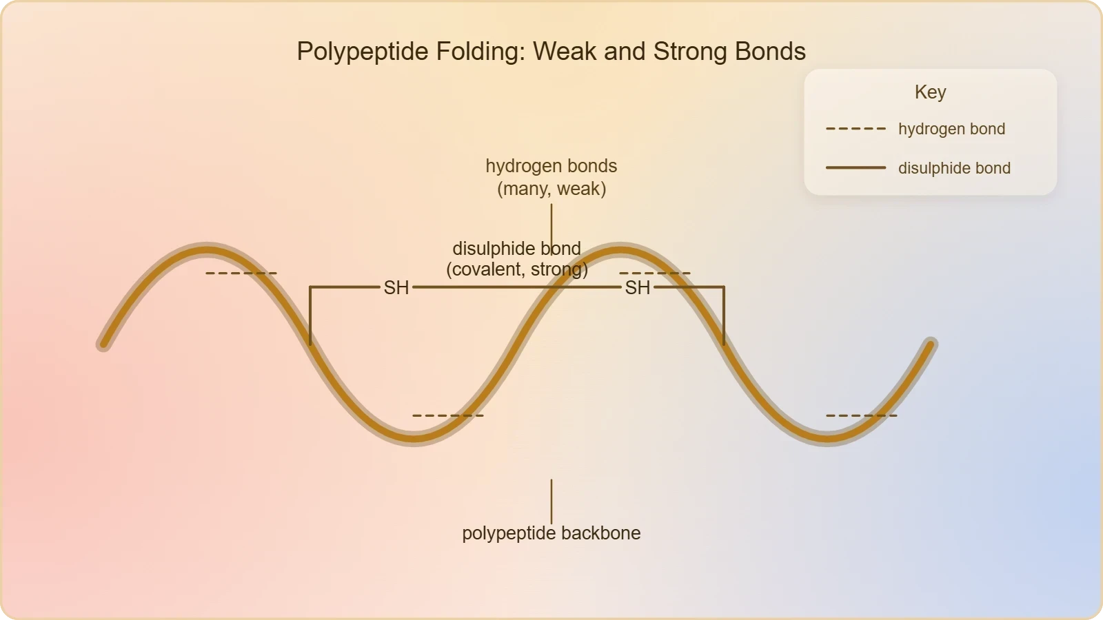
The presence of both hydrogen bonds and disulphide bonds in the figure is instructive. Hydrogen bonds are comparatively weak individually but numerous, and they are the interactions responsible for the recurring secondary motifs. Disulphide bonds are stronger covalent connections and can hold distant regions of the chain in a fixed relationship. Together they illustrate a general principle of protein architecture: stability comes from the accumulation of many interactions, some weak and some strong, distributed across the folded structure. The hollow woollen ball stays a ball because these links keep the strand in place.
Reading the Diagram of the Four Levels
The figure accompanying the material presents the levels of protein structure in sequence, labelled (a) primary, (b) secondary, (c) tertiary and (d) quaternary. The secondary panel shows the alpha-helix and the beta-pleated sheet, with hydrogen bonds indicated. The tertiary panel shows the folded chain as a whole, with disulphide bonds marked. The quaternary panel shows the arrangement of subunits. Reading the figure from (a) to (d) is a useful discipline, because it makes the hierarchy visible: each level is built upon the one before, and the labels keep the levels distinct.
For examination purposes, the figure rewards close attention to what is shown at each stage. The primary level is the sequence; the secondary level is the local folding into helix and sheet; the tertiary level is the folding of the whole chain upon itself; the quaternary level is the assembly of subunits. The bonds marked in the diagram belong to particular levels, and confusing them is a common error. A disulphide bond drawn across a folded chain belongs to the tertiary account of the molecule, not to the primary sequence, even though the cysteines it joins are specified by that sequence.
Weak Interactions and Covalent Links Together
The material's figure names hydrogen bonds and disulphide bonds, and these two categories illustrate the range of interactions at work. Hydrogen bonds form between suitable groups and are central to the helices and sheets of secondary structure; they also contribute to the packing of the tertiary fold. Disulphide bonds are covalent and can act as cross-links. Neither category alone accounts for the stability of a folded protein; the fold is a cooperative arrangement in which many such interactions reinforce one another across the whole three-dimensional object.
This cooperative character explains why the material can describe tertiary structure as a single folded entity rather than a collection of independent local folds. The helices and sheets are held by hydrogen bonds, the chain is cross-linked where disulphide bonds occur, and the whole is packed into the hollow woollen ball. The next section turns from the internal organisation of one chain to the arrangement of several chains with respect to one another, which the material calls the architecture of a protein.
Quaternary Structure: The Architecture of Subunits
Some proteins are an assembly of more than one polypeptide or subunits. The material introduces quaternary structure at precisely this point: the manner in which these individual folded polypeptides or subunits are arranged with respect to each other is the architecture of a protein, otherwise called the quaternary structure. The examples given for that arrangement are a linear string of spheres, or spheres arranged one upon each other in the form of a cube or a plate. The vocabulary of architecture is deliberate, because quaternary structure is about the relative placement of already-folded units.
The definition contains an important boundary. Each subunit has its own primary, secondary and tertiary structure; quaternary structure begins only when those folded subunits are considered together. A protein made of a single polypeptide has no quaternary structure, however elaborate its tertiary fold. The level applies to assemblies of more than one polypeptide, and the material's phrasing — "some proteins" — signals that it is not universal. This boundary is the single most useful safeguard against the confusion that surrounds the topic.
Arrangements of Subunits
The material offers three geometric images for the arrangement of subunits: a linear string of spheres, spheres arranged one upon each other in the form of a cube, and spheres arranged as a plate. These are illustrations of the principle rather than an exhaustive catalogue, and their purpose is to make the idea of relative arrangement concrete. The subunits are individual folded polypeptides, each a hollow woollen ball in its own right, and quaternary structure describes how those balls are positioned with respect to one another in the finished assembly.
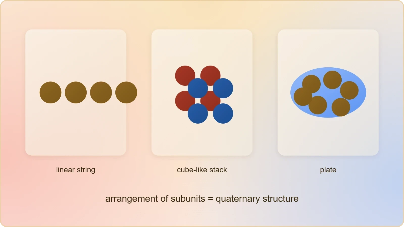
The images also make clear that quaternary structure is a matter of geometry and contact. Whether the subunits lie in a line, stack in a cube-like arrangement, or spread as a plate, the description concerns their spatial relationship. The material does not specify the bonds holding subunits together, and this lesson does not add any beyond what the figure shows; the point to retain is the definition itself. Quaternary structure is the architecture of a protein built from more than one polypeptide or subunit.
Adult Human Haemoglobin as the Model Assembly
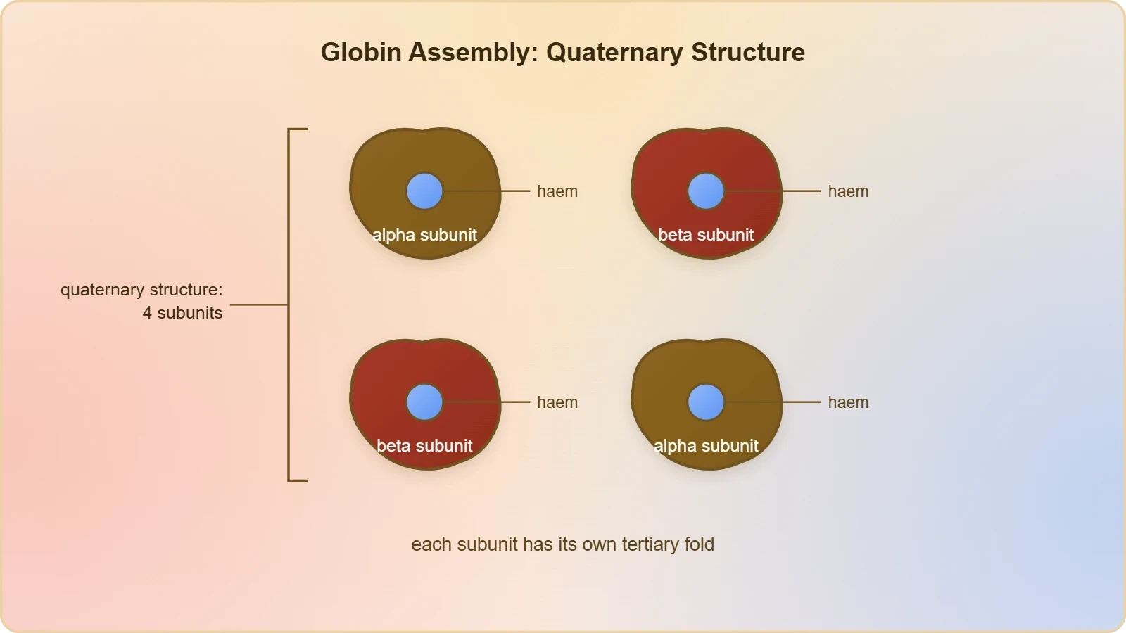
Adult human haemoglobin consists of 4 subunits. Two of these are identical to each other, and the material states that two subunits of alpha type and two subunits of beta type together constitute the human haemoglobin (Hb). The assembly is therefore described by its composition: four subunits in total, drawn from two types, with two copies of each type. This is the concrete example the material supplies for quaternary structure, and it should be learned as stated, including the numbers and the designations alpha and beta.
Haemoglobin illustrates why the level is called architecture. Each of the four subunits is a folded polypeptide with its own tertiary structure; the quaternary structure is the arrangement of the four with respect to one another. The material's figure of the levels of protein structure shows the quaternary panel as an assembly of subunits, and haemoglobin is the named instance of that panel. The example also prepares the ground for the next lesson, where enzymes and their active sites are considered, since it demonstrates that proteins can function as organised assemblies rather than isolated chains.
Comparing the Four Levels Side by Side
The four levels of protein structure are best understood as a hierarchy, and the material's figure presents them in that order. Primary structure is the amino acid sequence, running from the N-terminal amino acid to the C-terminal amino acid. Secondary structure is the local folding of portions of the thread into the right-handed helix and other forms such as the beta-pleated sheet. Tertiary structure is the folding of the long chain upon itself like a hollow woollen ball. Quaternary structure is the arrangement of subunits with respect to one another. Each level presupposes the one before it.
Keeping the levels distinct is essential for accurate description. A statement about sequence belongs to the primary level; a statement about a helix belongs to the secondary level; a statement about the overall three-dimensional shape of one chain belongs to the tertiary level; a statement about how several chains are arranged belongs to the quaternary level. The table below sets out the distinctions in a form suitable for revision, drawing only on the terms and examples the material provides.
Where Tertiary Ends and Quaternary Begins
The boundary between the two levels is a matter of counting chains. Tertiary structure describes the folding of one long protein chain upon itself; quaternary structure describes the arrangement of more than one polypeptide or subunit. A protein consisting of a single folded chain has reached its tertiary structure and stops there. A protein such as adult human haemoglobin, with four subunits, has both: each subunit is folded, and the four are arranged with respect to one another. The presence of subunits is the signal that quaternary structure is in play.
This boundary also clarifies the language of the material. When it says that some proteins are an assembly of more than one polypeptide or subunits, the word "some" is doing real work. The quaternary level is not a stage that every protein passes through; it is a description that applies to assemblies. Recognising this prevents the common mistake of attributing quaternary structure to a single-chain protein simply because its tertiary fold is complex. Complexity of fold and number of chains are separate questions.
Common Misreadings to Avoid
Several misreadings recur in the study of this topic. The first is to treat tertiary structure as a synonym for three-dimensional shape in general, when the material uses it specifically for the folding of one chain upon itself. The second is to describe quaternary structure as the folding of a chain, when it is the arrangement of subunits. The third is to assume that every protein has four levels; the material's "some proteins" makes clear that quaternary structure is not universal. Each misreading can be corrected by returning to the definitions as stated.
A fourth misreading concerns the helix. The material notes that only some portions of the protein thread are arranged in the form of a helix, and that in proteins only right-handed helices are observed. Treating the whole chain as a helix, or imagining left-handed helices in proteins, contradicts the material. The helix is a secondary motif found in portions of the thread; the tertiary structure is the folded whole. Keeping the levels and their vocabulary straight is the surest way to avoid these errors in description and in examination answers.
Why the Fold Is Necessary: Function Follows Shape
The material's strongest statement about tertiary structure is that it is absolutely necessary for the many biological activities of proteins. This is a claim about function, and it deserves to be treated as the centre of the lesson rather than an afterthought. The biological activities of proteins are varied, and the material does not enumerate them here; what it asserts is the necessity of the folded three-dimensional form for those activities. The fold is the condition under which a protein can act, and the three-dimensional view is therefore a functional as well as a descriptive matter.
The claim also connects forward to the next lesson. The material observes that an enzyme, like any protein, has a primary structure, a secondary structure and a tertiary structure, and that when one looks at a tertiary structure one is looking at the form on which the enzyme's activity depends. The active site and the chemical reactions catalysed there belong to the next lesson, but the foundation is laid here: without the folded tertiary form, the activities of proteins, enzymes among them, would not be possible.
Shape, Activity and the Lesson Ahead
The sequence of the course reflects the logic of the material. Primary and secondary levels were treated previously; tertiary and quaternary levels are treated here; enzymes follow. The progression is not arbitrary. Enzymes are proteins, and the material says so directly: almost all enzymes are proteins, with ribozymes as the nucleic acid exception. An enzyme has a tertiary structure, and the shape of that structure is what the next lesson will examine when it considers the active site and the chemical reactions that occur there.
Understanding tertiary structure therefore prepares the learner for the enzyme lesson in a specific way. The hollow woollen ball is not merely a shape; it is the arrangement that makes biological activity possible. When the next lesson describes an enzyme's active site, it will be describing a feature of a folded tertiary structure. The quaternary example of haemoglobin adds a second preparation: it shows that proteins can be assemblies of subunits, which is relevant to enzymes that consist of more than one polypeptide chain.
Going further
Two well-established points extend the material without altering it. First, haemoglobin carries oxygen in the blood, and its four-subunit structure allows it to bind oxygen cooperatively: the binding of oxygen to one subunit makes it easier for the remaining subunits to bind oxygen as well. This is a functional consequence of the quaternary arrangement the material describes, and it explains why the assembly of four subunits matters beyond mere architecture. The alpha and beta subunits work together rather than independently.
Second, loss of a protein's tertiary shape usually means loss of its function. Because the material states that tertiary structure is absolutely necessary for the many biological activities of proteins, the converse follows naturally: a protein that has lost its folded three-dimensional form has generally lost the ability to perform its biological role. This principle is the reason the study of protein folding is central to biology, and it reinforces the lesson's main point that shape and function are inseparable in proteins.
Consolidating the Lesson: Key Points and Self-Check
The lesson can be consolidated around a small number of statements taken directly from the material. Tertiary structure is the long protein chain folded upon itself like a hollow woollen ball, giving a three-dimensional view of a protein, and it is absolutely necessary for the many biological activities of proteins. Quaternary structure is the architecture of a protein: the manner in which individual folded polypeptides or subunits are arranged with respect to each other, whether as a linear string of spheres, as spheres arranged in the form of a cube, or as a plate. Adult human haemoglobin, with four subunits, two of alpha type and two of beta type, is the named example.
Around these statements cluster the supporting details: the thread is not an extended rigid rod; only some portions are arranged as a helix; only right-handed helices are observed in proteins; other regions are folded into other secondary forms; the figure marks hydrogen bonds and disulphide bonds; and enzymes, like any protein, have primary, secondary and tertiary structure. The table below gathers the quantitative and named facts that are most likely to be tested, so that they can be checked at a glance before moving to the next lesson on enzymes.
Self-Check Questions
A short self-check helps fix the material before the next lesson. Can you define tertiary structure in the material's own image, and explain why it is described as absolutely necessary for the biological activities of proteins? Can you state what quaternary structure is, and give the material's example of a protein that has it, including the number and types of subunits? Can you explain why a single-chain protein has no quaternary structure? Can you name the two bond types marked in the figure of the levels of protein structure? Answering these without reference to the text is a reliable test of understanding.
A further set of questions probes the finer points. Why does the material say that only some portions of the protein thread are arranged in the form of a helix, and what does it say about the handedness of helices in proteins? What are the three geometric images the material gives for the arrangement of subunits? What does the material say about enzymes and their structures, and what is the exception to the rule that enzymes are proteins? These questions draw only on the material and prepare directly for the lesson on enzymes, the active site and chemical reactions.
Preparing for Enzymes
The next lesson turns to enzymes, and the material has already set the stage. Almost all enzymes are proteins, and there are some nucleic acids that behave like enzymes, called ribozymes. An enzyme, like any protein, has a primary structure, a secondary structure and a tertiary structure. When the next lesson examines the tertiary structure of an enzyme, it will be examining the folded form whose shape makes catalysis possible. The hollow woollen ball of this lesson is therefore the direct precursor of the active site of the next.
The quaternary example also carries forward. Adult human haemoglobin, with its four subunits, shows that proteins can be assemblies, and some enzymes are likewise assemblies of more than one polypeptide. The distinction between tertiary and quaternary structure will remain useful when describing such enzymes. With the four levels now complete — primary, secondary, tertiary and quaternary — the learner is equipped to approach the enzyme lesson with the structural vocabulary the material requires, and with the central principle that shape and biological activity are bound together.
Working Through the Concepts: A Short Computational Illustration
Although the material is descriptive rather than computational, a small calculation can make the hierarchy concrete. Consider a hypothetical single-chain protein of known length and ask how many peptide bonds it contains; the answer follows from the sequence, which is the primary level. Then consider an assembly of four subunits and ask how many polypeptide chains it contains; the answer belongs to the quaternary level. The two questions look similar but test different levels, and distinguishing them is exactly the skill the material's definitions demand.
The illustration below uses a simple script to count chains and subunits for the examples discussed in this lesson. It is not part of the material and introduces no new biology; it merely restates the counts in a form that makes the tertiary-versus-quaternary distinction explicit. The haemoglobin entry uses the composition stated in the material: four subunits in total, two of alpha type and two of beta type. The single-chain entry represents a protein that has tertiary structure but no quaternary structure.
# Counting polypeptide chains: tertiary vs quaternary level
proteins = {
    "single_chain_protein": {"subunits": 1, "types": ["alpha"]},
    "adult_human_haemoglobin": {"subunits": 4, "types": ["alpha", "alpha", "beta", "beta"]},
}

for name, info in proteins.items():
    n = info["subunits"]
    level = "tertiary only" if n == 1 else "tertiary + quaternary"
    print(f"{name}: {n} subunit(s) -> {level}")
    print(f"  subunit types: {info['types']}")
The output confirms the boundary drawn in the lesson. The single-chain protein has one subunit and therefore reaches only the tertiary level; the haemoglobin assembly has four subunits and therefore has quaternary structure in addition to the tertiary structure of each subunit. The script does not model folding, bonds or geometry, and it should not be read as doing so; it simply counts chains, which is the operational test for whether the quaternary level applies. The biological content remains exactly as the material states it.
Reading the Counts Correctly
The counts also guard against a subtle error. A protein with four subunits does not have four tertiary structures in the sense of four separate molecules; it has one quaternary structure composed of four folded polypeptides, each with its own tertiary fold. The material's phrase "individual folded polypeptides or subunits" captures this precisely: the subunits are already folded before their arrangement is described. The quaternary level is therefore an additional layer of organisation, not a replacement for the tertiary level of each chain.
For haemoglobin specifically, the material states that two of the four subunits are identical to each other, and that two subunits of alpha type and two of beta type together constitute the molecule. The count of four is thus a count of chains, and the count of two types is a count of distinct kinds of chain. Both numbers should be retained. The distinction between the number of chains and the number of chain types is a useful habit when describing any multi-subunit protein, and it follows directly from the material's wording.
From Counts to Architecture
Counting chains establishes whether quaternary structure applies, but it does not describe the architecture. The material's geometric images — a linear string of spheres, spheres arranged one upon each other in the form of a cube, or a plate — are what turn a count into a description of arrangement. For haemoglobin, the material supplies the composition but not a geometric image, and this lesson does not invent one. The learner should be able to say that haemoglobin has four subunits, two of alpha type and two of beta type, and that their arrangement constitutes its quaternary structure.
This is the appropriate level of detail for the lesson. The material's purpose is to establish the definition and the example, not to catalogue the geometry of every assembly. The same restraint applies to the bonds: the figure marks hydrogen bonds and disulphide bonds, and the lesson has noted them without adding further categories. The next lesson will build on this foundation when it considers enzymes, their active sites and the chemical reactions they catalyse, completing the structural picture with the functional one.
Also Read
- No links available.
From our network :
- EV 2.0: The Solid-State Battery Breakthrough and Global Factory ExpansionDiscover the impact of the Solid-State Battery Breakthrough on the EV market. Learn about CATL and BYD stock growth, global factory expansion in Europe, and the future of EV 2.0 technology.
- Trump Political Strategy How Geopolitical Stunts Serve As Media Diversions
- Mastering DB2 12.1 Instance Design: A Technical Deep Dive into Modern Database ArchitectureComprehensive guide to DB2 12.1 Instance Design. Explore architectural components, HADR setup, buffer pool tuning, and security policies for IBM DB2 12.1. Includes 10 functional SQL and Shell samples.
- AI-Powered 'Precision Diagnostic' Replaces Standard GRE Score ReportsDiscover the new GRE Precision Diagnostic. Learn how AI-powered behavioral analytics and time-to-solve tracking are replacing standard GRE score reports to empower students.
- 98% of Global MBA Programs Now Prefer GRE Over GMAT Focus EditionExplore why 98% of global MBA programs now prefer the GRE over GMAT Focus. Learn about the shorter GRE format, verbal logic importance, and strategic versatility for students.
- Analyzing Trump Deportation Numbers Insights Into The 2026 Immigration Crackdown
- Mastering DB2 LUW v12 Tables: A Comprehensive Technical GuideComprehensive guide on DB2 LUW v12 tables. Explore CREATE TABLE syntax, BLU Acceleration, range partitioning, data types, and security features for IBM DB2 LUW v12.
- Vite 6/7 'Cold Start' Regression in Massive Module GraphsIn-depth analysis of Vite 6/7 cold start regressions in massive module graphs. Learn about barrel file issues, pre-bundling optimizations, and how to improve Vite dev server performance.
- 10 Physics Numerical Problems with Solutions for IIT JEEPrepare for IIT JEE with 10 physics numerical problems, complete with detailed solutions and conceptual explanations. Ideal for mastering kinematics, thermodynamics, electromagnetism, and optics.
RESOURCES
- Protein structure: Primary, secondary, tertiary & quatrenary (article)khanacademy.orgTertiary structure is the 3-D folding of a polypeptide chain, and quaternary structure is simply several chains folded into tertiary structure all tangled up ...
- modelling protein tertiary and quaternary structure using ... - PubMedpubmed.ncbi.nlm.nih.govApr 29, 2014 ... Protein structure homology modelling has become a routine technique to generate 3D models for proteins when experimental structures are not ...
- I'm about to get my PHD and I still don't understand the difference ...reddit.comJul 27, 2022 ... The tertiary structure is the folding of the protein into a structure defined by charge, hydrophobicity, and non-local interactions. The ...
- Single nucleotide variation catalog from clinical isolates mapped on ...pubmed.ncbi.nlm.nih.govAug 6, 2024 ... Single nucleotide variation catalog from clinical isolates mapped on tertiary and quaternary structures of ESX-1-related proteins reveals ...
- 13.4: Secondary, tertiary, and quaternary structure of proteinschem.libretexts.orgMay 11, 2023 ... Primarily, the interactions among R groups create the protein's complex three-dimensional tertiary structure. The nature of the R groups in…
- Biochemistry, Tertiary Protein Structure - StatPearls - NCBI Bookshelfncbi.nlm.nih.govDec 1, 2025 ... Tertiary structure denotes the unique 3-dimensional conformation adopted by a single polypeptide chain. · Tertiary structures frequently contain ...
- Levels of Protein Organizationcomis.med.uvm.eduQuaternary structure in proteins is the most intricate degree of organization still considered a single molecule. To be considered to have quaternary structure, ...
- Protein Quaternary Structure - an overview | ScienceDirect Topicssciencedirect.comQuaternary structure refers to a structure achieved by proteins composed of more than one polypeptide chain. Each polypeptide chain, called a subunit, has its ...
- Allosteric Communication Occurs via Networks of Tertiary and ...journals.plos.orgCrystal structures have revealed that most allosteric proteins are complex systems with both tertiary and quaternary structural changes [11]. Previously, we ...
- 3.9: Proteins - Protein Structure - Biology LibreTextsbio.libretexts.orgThe tertiary structure of a polypeptide chain is its overall three-dimensional shape, once all the secondary structure elements have folded together among each ...
- Insights into the quaternary association of proteins through structure ...pmc.ncbi.nlm.nih.gov... proteins with very similar tertiary structures but varied quaternary structures. ... protein structures have also been used to understand protein structure ...
- Protein tertiary structure - Wikipediaen.wikipedia.orgProtein tertiary structure is the three-dimensional shape of a protein. The tertiary structure will have a single polypeptide chain "backbone" with one or ...
- Uncovering protein structure - PMC - NIHpmc.ncbi.nlm.nih.gov... protein structure and how proteins fold into ... Currently, there are approximately 165000 protein structures, and their tertiary and quaternary structures ...
- Protein structures (primary, secondary, tertiary, quaternary) L3 - Quizletquizlet.comStudy with Quizlet and memorise flashcards containing terms like Protein structures - Primary - Secondary - Tertiary - Quaternary, Secondary structure ...
- Tertiary and quaternary structural basis of oxygen affinity in human ...nature.comSep 7, 2017 ... Human hemoglobin (Hb) is a benchmark protein of structural biology that shaped our view of allosterism over 60 years ago, ...