Protein structure is one of those ideas that appears simple until you try to define it precisely. In everyday speech, "structure" suggests a fixed scaffold, something rigid and unchanging. In biochemistry, the word carries a layered meaning: a protein has a structure at the level of its amino acid sequence, another at the level of local folding patterns, another at the level of its overall three-dimensional shape, and sometimes yet another at the level of how separate polypeptide subunits assemble together. Each of these levels answers a different question about the same molecule, and confusing them is one of the most common mistakes learners make when they first meet the topic.
This lesson concentrates on the first two of those levels. It examines how a protein thread is written, so to speak, from its N-terminal amino acid to its C-terminal amino acid, and then how that thread behaves when it is not stretched out as an extended rigid rod. The material shows that portions of the chain coil into a helix resembling a revolving staircase, that other regions fold into different arrangements, and that only right-handed helices are observed in proteins. These two levels form the foundation on which the later discussion of tertiary and quaternary structure rests, so the terminology introduced here deserves careful attention.
The lesson follows the order in which the chapter develops these ideas: the definition of primary structure, the description of the helix, the broader category of secondary structure, and the significance of handedness. Along the way, the distinction between sequence and shape is kept firmly in view, because that distinction governs everything that follows in the study of proteins and enzymes. By the end, you should be able to describe a polypeptide in the language of terminals, helices and folds, and to explain why a protein thread cannot be imagined as a straight, stiff rod.
On This Page
- What Structure Means at the Molecular Level
- The Protein Thread and Its Departure from a Rigid Rod
- Secondary Structure as the Local Folding of the Chain
- Terminology, Bonds and the Order of Ideas
- From Sequence to Shape: Building the Conceptual Bridge
- Consolidating the Lesson Through Structured Comparison
- Worked Examples and Self-Check Questions
- Summary and Forward Look
What Structure Means at the Molecular Level
The word structure, when applied to a protein, refers to the spatial arrangement of its constituent parts at a defined level of organisation. At the simplest level, structure means the order in which amino acids are joined together. At a slightly higher level, it means the local shapes that segments of the chain adopt. At a higher level still, it means the overall three-dimensional form of the entire chain, and finally the arrangement of separate chains with respect to one another. Each level is described by its own vocabulary, and each depends on the level beneath it.
This layered scheme is not an arbitrary convenience invented for examinations. It reflects the physical reality that a long chain of amino acids can be described in several equally valid ways, depending on the scale at which you observe it. A sequence of letters describes the molecule at one scale; a coiled segment describes it at another; a compact globular shape describes it at a third. The chapter presents these levels in sequence, and the present lesson is concerned with the first two of them, leaving the compact folding and subunit assembly for the lessons that follow.
Sequence as the First Level of Description
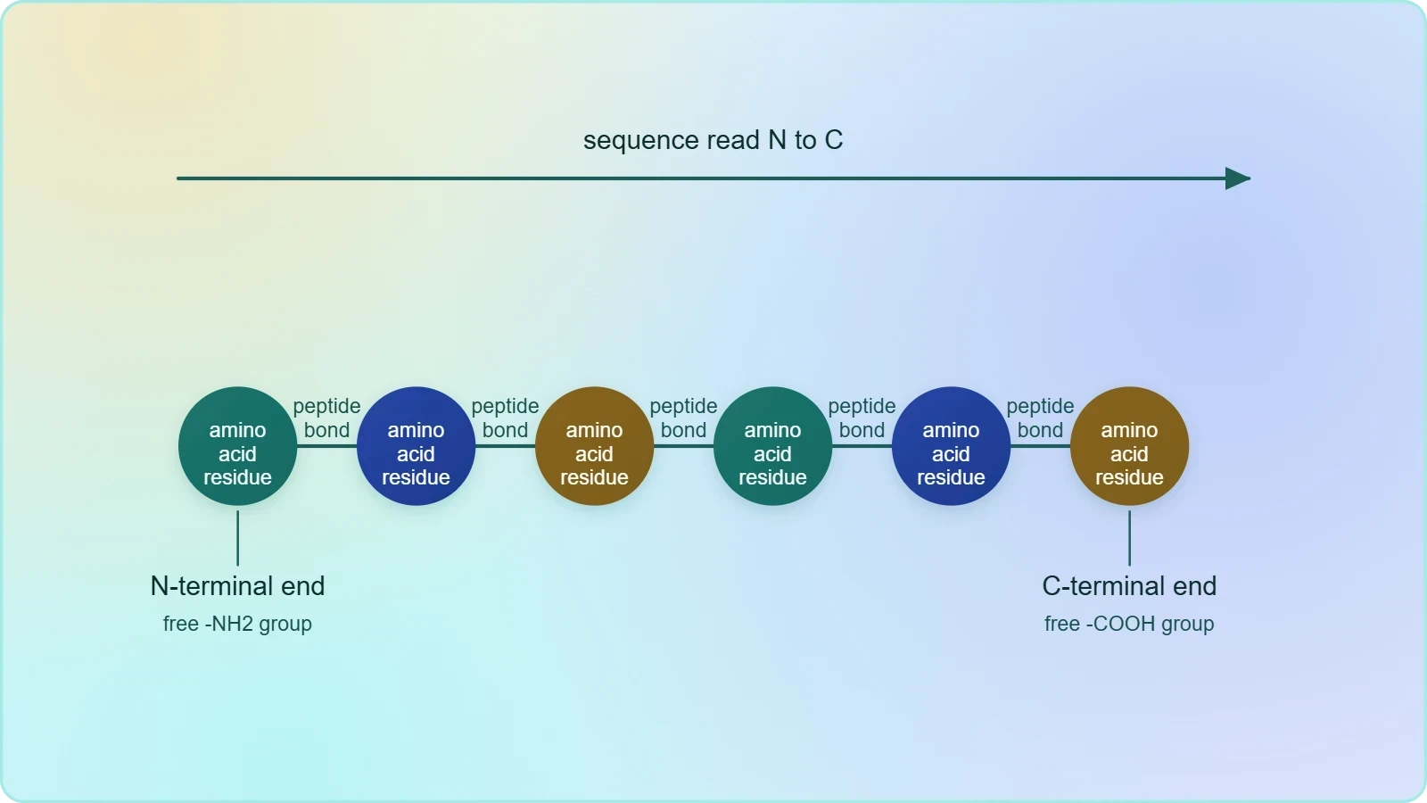
The primary structure of a protein is its amino acid sequence. The chapter states this plainly: an enzyme, like any protein, has a primary structure, meaning the amino acid sequence of the protein. The sequence is read directionally. The first amino acid is called the N-terminal amino acid, and the last amino acid is called the C-terminal amino acid. This directional convention matters because a sequence written backwards describes a different molecule, just as a sentence read in reverse loses its meaning.
The terms N-terminal and C-terminal refer to the chemical ends of the polypeptide chain. The N-terminal end carries the free amino group, and the C-terminal end carries the free carboxyl group. When the chapter says that the first amino acid is the N-terminal amino acid and the last is the C-terminal amino acid, it is establishing a reading direction that runs from the amino end to the carboxyl end. Every subsequent description of protein structure in the chapter assumes this direction, so it is worth fixing firmly in mind before moving on.
Why the Sequence Is Called Primary
The adjective primary signals that this level is fundamental and comes first, both in the order of description and in the logic of the molecule. The sequence is the information content of the protein; the folded forms are consequences of that information. The chapter does not develop this point at length, but it treats the primary structure as the baseline description against which the secondary and tertiary structures are understood. An enzyme, the chapter notes, has a primary structure just as any other protein does, which reinforces that this level is universal among proteins.
It is useful to recognise that the primary structure is a one-dimensional description. It specifies order along the chain but says nothing directly about shape in space. A list of amino acids in sequence could, in principle, belong to a molecule that is stretched out or tightly coiled; the sequence alone does not tell you which. The transition from this one-dimensional description to the three-dimensional reality of the folded protein is precisely what the higher levels of structure are designed to capture.
The Protein Thread and Its Departure from a Rigid Rod
A common mental image of a protein is that of a long, straight thread, rather like a piece of string pulled taut. The chapter explicitly corrects this image. A protein thread, it states, does not exist throughout as an extended rigid rod. Instead, the thread is folded. This correction is important because it prepares the reader for the idea that the shape of a protein is not a trivial consequence of its length but a specific, organised arrangement of the chain in space. The folding is not random; it follows patterns that can be described and named.
The phrase "throughout" in the chapter's statement is significant. It does not claim that no part of a protein is ever extended; it claims that the thread is not extended as a rigid rod throughout its entire length. Some segments may be relatively extended, while others are coiled or folded. The protein is therefore a mosaic of local arrangements rather than a uniform object. This mosaic character is exactly what the concept of secondary structure is designed to describe, and it explains why the chapter moves directly from the correction of the rod image to the description of the helix.
The Helix as a Revolving Staircase
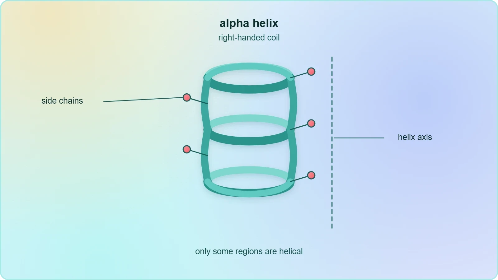
The chapter describes the folded thread as taking the form of a helix, and it offers a memorable comparison: the helix is similar to a revolving staircase. The image is apt because a spiral staircase rises while turning, so that each step is displaced both vertically and rotationally from the one below it. A polypeptide helix behaves in an analogous way, with the chain turning as it advances along the axis of the helix. The comparison gives a beginner a concrete picture of a shape that is otherwise difficult to visualise from a flat diagram.
Crucially, the chapter adds a qualification: only some portions of the protein thread are arranged in the form of a helix. The helix is therefore a local feature, not a description of the whole molecule. A protein may contain helical segments separated by regions that adopt other arrangements. This partial, segmental character of the helix is essential to the concept of secondary structure, which the chapter defines as the folding of other regions of the protein thread into other forms. The helix is one pattern among several, not the sole pattern.
Handedness: Only Right-Handed Helices Are Observed
The chapter makes a precise and memorable observation: in proteins, only right-handed helices are observed. A helix can, in principle, twist in either of two directions, just as a screw thread can be cut to turn clockwise or anticlockwise as it advances. The observation that proteins use only one of these two possibilities is a striking fact about biological molecules, and the chapter states it without elaboration. For the learner, the key point is that the statement is categorical: among the helices found in proteins, the right-handed form is the one that occurs.
This single sentence carries more weight than its brevity suggests. It tells us that the helical arrangement in proteins is not a matter of indifference but is constrained to one handedness. The chapter does not explain the physical reason for this constraint, and the lesson need not supply one beyond noting the observation itself. What matters for examination purposes is the clear, unambiguous statement that only right-handed helices are observed in proteins, a point that is easy to remember and easy to test.
Secondary Structure as the Local Folding of the Chain
Secondary structure is the term the chapter uses for the local folded arrangements that regions of the protein thread adopt. The chapter introduces it by saying that other regions of the protein thread are folded into other forms, and this is what is called the secondary structure. The definition is deliberately broad: it covers the helix and it covers the other forms into which non-helical regions fold. Secondary structure is therefore a category rather than a single shape, and the helix is its most prominently described member in the chapter's account.
The relationship between primary and secondary structure is one of dependence. The secondary structure describes how the thread, whose sequence is given by the primary structure, arranges itself locally in space. The chapter presents the two levels in immediate succession, moving from the sequence to the folding without an intervening concept, which reflects their close connection. The sequence provides the thread; the secondary structure describes the local shapes that thread adopts. Understanding this pairing is the central aim of the present lesson.
The Alpha Helix and the Beta-Pleated Sheet
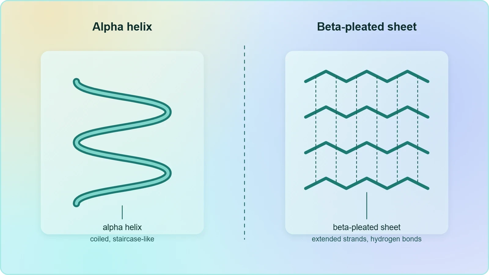
The chapter's figure labels the secondary level with two named patterns: the alpha helix and the beta-pleated sheet. The alpha helix corresponds to the coiled, staircase-like arrangement described in the text. The beta-pleated sheet is the other principal pattern, in which segments of the chain lie alongside one another in an extended, sheet-like arrangement. The figure also indicates the hydrogen bond as the interaction associated with these arrangements, and it shows the disulphide bond in the context of the higher levels of structure.
For the purposes of this lesson, the essential point is that secondary structure encompasses more than one pattern. The helix is described in the running text, and the beta-pleated sheet appears in the accompanying figure as the companion pattern. Together they illustrate the chapter's statement that the thread is folded into various forms. The learner should be able to name both patterns and to associate the helix with the revolving-staircase image and the sheet with an extended, pleated arrangement of neighbouring chain segments.
Reading the Figure: Panels and Labels
The chapter's figure presents the levels of protein structure in four labelled panels. Panel (a) shows the primary level, panel (b) the secondary level, panel (c) the tertiary level, and panel (d) the quaternary level. The secondary panel carries the labels alpha helix and beta-pleated sheet, while the figure as a whole marks the hydrogen bond and the disulphide bond. Reading the figure in this order reinforces the sequence of ideas in the text and helps the learner connect each named level with a visual representation.
The figure is best read alongside the text rather than in isolation. The text supplies the definitions and the qualifying statements, such as the observation that only some portions of the thread are helical and that only right-handed helices occur. The figure supplies the visual vocabulary, including the two named secondary patterns and the bond types. Used together, text and figure give a complete picture of the first two levels of structure and a preview of the two that follow in later lessons.
Terminology, Bonds and the Order of Ideas
The chapter's account is built on a small set of terms that recur throughout the study of proteins. The N-terminal and C-terminal amino acids define the direction of the sequence. The helix, described as a revolving staircase, names the coiled arrangement. The right-handed qualification restricts the handedness of that arrangement. Secondary structure names the category of local folded forms, of which the alpha helix and the beta-pleated sheet are the named examples. The hydrogen bond and the disulphide bond appear in the figure as the interactions associated with the folded levels. Keeping these terms distinct prevents the confusion that arises when sequence and shape are conflated.
The order of ideas in the chapter is itself instructive. It begins with the sequence and its terminals, corrects the rigid-rod image, introduces the helix and its staircase comparison, qualifies the helix as partial and right-handed, and then names the broader category of secondary structure. Only after this does it move to the tertiary and quaternary levels. Following this order ensures that each concept is in place before the next is introduced, and it mirrors the logical dependence of the higher levels on the lower ones.
A Compact Reference Table of the Key Terms
A short reference table helps consolidate the vocabulary before the lesson turns to the broader context of enzymes and to the forward look at the next levels of structure. The table below gathers the terms introduced in this lesson, pairs each with the meaning given in the chapter, and notes the level of structure to which it belongs. It is intended as a revision aid rather than a substitute for the explanatory paragraphs, and it deliberately keeps the chapter's own wording wherever the wording is definitional.
Enzymes Share These Levels of Structure
The chapter's treatment of enzymes reinforces the universality of the structural levels described here. Almost all enzymes are proteins, and the chapter notes that some nucleic acids behave like enzymes; these are called ribozymes. An enzyme, like any protein, has a primary structure, that is, the amino acid sequence of the protein. It also has secondary and tertiary structure. The mention of enzymes in this context is a reminder that the structural vocabulary is not confined to a special class of molecules but applies across the protein world, including the catalysts that drive cellular chemistry.
The reference to ribozymes is a useful qualification. It prevents the learner from concluding that catalysis is exclusively a protein function, while still affirming that almost all enzymes are proteins. For the present lesson, the key takeaway is that the primary and secondary levels are general features of proteins, and that enzymes are proteins for the purposes of structural description. The chapter's line diagram of an enzyme, mentioned in the text, is simply a schematic way of depicting such a molecule, and it presupposes the structural levels already described.
From Sequence to Shape: Building the Conceptual Bridge
The movement from primary to secondary structure is the first step in a larger progression from a one-dimensional description to a three-dimensional one. The sequence tells us the order of amino acids; the secondary structure tells us how local segments of that sequence arrange themselves in space. The chapter's next steps, the tertiary structure in which the long chain folds upon itself like a hollow woollen ball, and the quaternary structure in which separate subunits assemble, complete the progression. The present lesson stops at the second step, but it deliberately leaves the reader prepared for the third and fourth.
This preparation takes the form of a clear distinction between what has been established and what remains to be explained. We have established that the thread is not a rigid rod, that portions of it are helical, that only right-handed helices occur, and that other regions fold into other forms collectively called secondary structure. We have not yet explained how the entire chain folds into a compact three-dimensional shape, nor how multiple subunits assemble. Those questions belong to the lessons that follow, and the terminology introduced here will be used there without redefinition.
What the Next Lesson Will Address
The next lesson takes up the tertiary and quaternary levels. The tertiary structure is described in the chapter as the folding of the long protein chain upon itself like a hollow woollen ball, giving a three-dimensional view of a protein, and the chapter stresses that tertiary structure is absolutely necessary for the many biological activities of proteins. The quaternary structure is the architecture of a protein made of more than one polypeptide or subunit, and the chapter's example is adult human haemoglobin, which consists of four subunits, two of alpha type and two of beta type.
Knowing what lies ahead helps the learner place the present lesson correctly. The primary and secondary levels are not ends in themselves; they are the groundwork for understanding the compact, functional forms of proteins. The helix and the sheet are local patterns that, together with the connecting regions, fold into the overall shape described at the tertiary level. The sequence, meanwhile, is the information that ultimately determines that shape. The bridge from sequence to shape is therefore built in stages, and this lesson has laid the first two spans.
Going further
Beyond the chapter's own statements, two pieces of well-established background help the learner appreciate why the primary structure is called primary. The sequence of amino acids is encoded by DNA and determines how the protein folds; the order of amino acids carries the information that specifies the final three-dimensional shape. This is why the sequence is regarded as the fundamental level of description, and why changes in sequence can alter the folded form and, with it, biological function. The point is background rather than part of the chapter's explicit argument, but it explains the logic of the layered scheme.
A second piece of background concerns handedness. A right-handed helix twists in the same direction as a standard screw thread, so that it advances as it turns in the clockwise sense when viewed along its axis. This comparison makes the chapter's observation that only right-handed helices are observed in proteins easier to visualise. The chapter states the observation without explaining it, and the lesson has followed that restraint; the screw-thread comparison is offered only as an aid to the imagination, not as an addition to the chapter's claims.
Consolidating the Lesson Through Structured Comparison
The final section draws the lesson together by comparing the two levels of structure side by side and by revisiting the chapter's order of ideas. Comparison is a powerful learning device because it forces attention onto the differences that matter. The primary structure is a sequence; the secondary structure is a set of local shapes. The primary structure is read from the N-terminal to the C-terminal amino acid; the secondary structure is described in terms of helices and other folded forms. The primary structure is one-dimensional; the secondary structure is the first step into three-dimensional arrangement. Keeping these contrasts sharp prevents the two levels from blurring together.
The chapter's own sequence of ideas provides a ready-made revision path. It moves from the terminals, to the correction of the rigid-rod image, to the helix and its staircase comparison, to the partial and right-handed character of the helix, to the naming of secondary structure, and then onward to the tertiary and quaternary levels. A learner who can reproduce this order, with the definitions attached to each step, has understood the lesson. The tables in this section are designed to support that reproduction by presenting the contrasts and the order in compact form.
Primary Versus Secondary Structure
The comparison table below sets the two levels against each other across the dimensions that the chapter treats. It is deliberately simple, because the distinctions themselves are simple once stated clearly. The aim is to make the contrasts memorable and to provide a quick check before the learner moves on to the tertiary and quaternary levels in the next lesson.
The Chapter's Order of Ideas
The final table records the order in which the chapter develops the material, so that the learner can check their understanding against the original sequence. Each step is paired with the key statement it establishes. Reproducing this order from memory is a reliable test of whether the lesson has been absorbed, and it also prepares the ground for the next lesson, which continues the same progression at the tertiary and quaternary levels.
Worked Examples and Self-Check Questions
Applying the terminology to concrete questions is the surest way to consolidate it. The examples below are framed in the style of short examination questions, and each is answered using only the statements and definitions established in this lesson. They are not intended to introduce new material; they are intended to make the existing material operational. Working through them before reading the answers is more valuable than reading the answers directly, because the effort of retrieval strengthens memory far more than passive review does.
The questions also serve as a check on the distinctions that learners most often blur. Can you state the direction of the sequence without hesitation? Can you correct the rigid-rod image in a single sentence? Can you name the two secondary patterns shown in the figure? Can you state the handedness observation exactly as the chapter does? If any of these produces uncertainty, the relevant section above should be revisited before proceeding to the next lesson, where the tertiary and quaternary levels build directly on this foundation.
Short Answer Practice
Consider the question of what the primary structure of a protein is. The answer is the amino acid sequence of the protein, read from the N-terminal amino acid to the C-terminal amino acid. A second question asks why the protein thread cannot be described as an extended rigid rod throughout. The answer is that the thread is folded, with some portions arranged as a helix and other regions folded into other forms. A third question asks which handedness of helix is observed in proteins, and the answer is that only right-handed helices are observed.
A fourth question asks what secondary structure means. The answer is the folding of regions of the protein thread into forms such as the helix, with the alpha helix and beta-pleated sheet named as the patterns shown in the figure. A fifth question asks whether enzymes have these levels of structure. The answer is that they do: an enzyme, like any protein, has a primary structure, that is, its amino acid sequence, and it also has secondary and tertiary structure. Each answer should be given in the chapter's own terms.
Checking the Distinctions
The most common error is to treat the sequence and the shape as interchangeable. They are not. The sequence is the order of amino acids; the shape is the arrangement of the thread in space. A second common error is to assume that the whole thread is helical. It is not; only some portions are arranged as a helix, and other regions fold into other forms. A third error is to overlook the handedness observation, which is stated categorically and is easy to test. Attending to these three points covers most of the ground in this lesson.
A further check concerns the relationship between this lesson and the next. The tertiary structure is the folding of the long chain upon itself like a hollow woollen ball, and the quaternary structure is the arrangement of separate subunits with respect to one another. Neither is described in detail here, and the learner should be able to say so without conflating them with the secondary level. Knowing the boundary of the present lesson is itself a form of understanding, and it keeps the progression from sequence to shape orderly and clear.
Summary and Forward Look
This lesson has established the first two levels of protein structure as the chapter presents them. The primary structure is the amino acid sequence, read from the N-terminal amino acid to the C-terminal amino acid. The protein thread does not exist throughout as an extended rigid rod; it is folded, with some portions arranged as a helix similar to a revolving staircase, and only right-handed helices are observed in proteins. Other regions fold into other forms, and this is called the secondary structure, with the alpha helix and beta-pleated sheet named in the figure. Enzymes, being almost all proteins, share these levels.
The lesson has also prepared the ground for what follows. The tertiary structure, in which the long chain folds upon itself like a hollow woollen ball, and the quaternary structure, in which separate subunits assemble, will be treated next. The chapter's example of adult human haemoglobin, with four subunits, two of alpha type and two of beta type, will illustrate the quaternary level. The terminology established here, from terminals to helices to secondary structure, will be used there without redefinition, so a firm grasp of the present lesson is the best preparation for the next.
Key Points to Carry Forward
Carry forward the directional convention: first amino acid, N-terminal; last amino acid, C-terminal. Carry forward the correction of the rigid-rod image: the thread is folded, not extended throughout. Carry forward the staircase comparison for the helix, the qualification that only some portions are helical, and the categorical observation that only right-handed helices are observed. Carry forward the definition of secondary structure as the folding of regions of the thread into forms such as the helix, and the names of the two patterns shown in the figure. These points form a compact set that can be revised quickly.
Finally, carry forward the sense of progression. The sequence is the information; the local folds are the first expression of that information in space; the compact three-dimensional shape and the assembly of subunits complete the picture. Each level depends on the one beneath it, and each is described with its own vocabulary. The next lesson continues this progression, and the learner who has mastered the first two levels will find the third and fourth considerably easier to absorb.
Preparing for Tertiary and Quaternary Structure
Before moving on, it is worth noting how the chapter's figure anticipates the next lesson. Panel (c) shows the tertiary level, and panel (d) the quaternary level, and the figure marks the disulphide bond alongside the hydrogen bond. The learner who has read the figure in order will already have a visual preview of what is to come. The present lesson has deliberately confined itself to panels (a) and (b), so that the first two levels are secure before the more complex three-dimensional descriptions are attempted.
The transition is therefore smooth rather than abrupt. The helix and the sheet are local patterns; the tertiary structure is the overall fold of the whole chain; the quaternary structure is the assembly of separate chains. Each step adds a layer of organisation without discarding the one before it. With the sequence and the local folds firmly in place, the learner is ready to see how the entire protein acquires the three-dimensional form that the chapter says is absolutely necessary for its many biological activities.
Also Read
- No links available.
From our network :
- Mastering DB2 12.1 Instance Design: A Technical Deep Dive into Modern Database ArchitectureComprehensive guide to DB2 12.1 Instance Design. Explore architectural components, HADR setup, buffer pool tuning, and security policies for IBM DB2 12.1. Includes 10 functional SQL and Shell samples.
- Analyzing Trump Deportation Numbers Insights Into The 2026 Immigration Crackdown
- Vite 6/7 'Cold Start' Regression in Massive Module GraphsIn-depth analysis of Vite 6/7 cold start regressions in massive module graphs. Learn about barrel file issues, pre-bundling optimizations, and how to improve Vite dev server performance.
- EV 2.0: The Solid-State Battery Breakthrough and Global Factory ExpansionDiscover the impact of the Solid-State Battery Breakthrough on the EV market. Learn about CATL and BYD stock growth, global factory expansion in Europe, and the future of EV 2.0 technology.
- Trump Political Strategy How Geopolitical Stunts Serve As Media Diversions
- AI-Powered 'Precision Diagnostic' Replaces Standard GRE Score ReportsDiscover the new GRE Precision Diagnostic. Learn how AI-powered behavioral analytics and time-to-solve tracking are replacing standard GRE score reports to empower students.
- 98% of Global MBA Programs Now Prefer GRE Over GMAT Focus EditionExplore why 98% of global MBA programs now prefer the GRE over GMAT Focus. Learn about the shorter GRE format, verbal logic importance, and strategic versatility for students.
- Mastering DB2 LUW v12 Tables: A Comprehensive Technical GuideComprehensive guide on DB2 LUW v12 tables. Explore CREATE TABLE syntax, BLU Acceleration, range partitioning, data types, and security features for IBM DB2 LUW v12.
- 10 Physics Numerical Problems with Solutions for IIT JEEPrepare for IIT JEE with 10 physics numerical problems, complete with detailed solutions and conceptual explanations. Ideal for mastering kinematics, thermodynamics, electromagnetism, and optics.
RESOURCES
- Protein structure: Primary, secondary, tertiary & quatrenary (article)khanacademy.orgThe simplest level of protein structure, primary structure, is simply the sequence of amino acids in a polypeptide chain. For example, the hormone insulin…
- Four levels of protein structure (video) - Khan Academykhanacademy.orgThe four levels of protein structure are primary, secondary, tertiary, and quaternary. It is helpful to understand the nature and function of each level…
- Levels of Protein Organizationcomis.med.uvm.eduWhile primary structure describes the sequence of amino acids forming a peptide chain, secondary structure refers to the local arrangement of the chain in…
- Levels of Protein Structure - Creative Biostructurecreative-biostructure.comTo understand how a protein molecule forms its final conformation, we need to understand the four structural levels of proteins: primary structure, secondary ...
- Uncovering protein structure - PMC - NIHpmc.ncbi.nlm.nih.govProtein secondary structure refers to the way the primary structure of a protein ... level depending on both the overall shape and connectivity of…
- Protein structure prediction: recognition of primary, secondary, and ...pubmed.ncbi.nlm.nih.gov... structural features of proteins from their amino acid sequences. At the primary structure level, methods are considered for detection of remotely related ...
- What are the 4 levels of protein structure? | AAT Bioquestaatbio.comJun 22, 2020 ... There are two main types of secondary structures, the ?-helix and the ?-strand or ?-sheets, who are defined by pattern…
- Levels of protein structure – primary - EMBL-EBIebi.ac.ukLevels of protein structure – primary ... The primary protein structure refers to the sequence of amino acids and the location of disulfide bonds…
- Physiology, Proteins - StatPearls - NCBI Bookshelf - NIHncbi.nlm.nih.gov... structural levels: primary, secondary, tertiary, and quaternary. The first level is the primary structure because it is the most basic protein structure.
- 3.9: Proteins - Protein Structure - Biology LibreTextsbio.libretexts.org... levels of protein structure: primary, secondary, tertiary, and quaternary. Primary Structure. A protein's primary structure is the unique sequence of amino ...
- Guide to Understanding PDB Data: Protein Hierarchical Structurepdb101.rcsb.orgThe levels of the hierarchy may be explored using several tools at the RCSB PDB. The primary and secondary structures can be explored in…
- Everything you need to know about: Proteins (Add anything!!) : r/Mcatreddit.comDec 3, 2020 ... 42 votes, 26 comments. - primary structure: amino acid sequence (break using acid-base catalysis/enzymes) -secondary structure: alpha helix, ...
- Protein structure - Student Academic Success - Monash Universitymonash.eduThese levels are labelled primary, secondary, tertiary and quaternary. The carboxyl group on one amino acid bonds readily to the amino group on another…
- Protein structure - Wikipediaen.wikipedia.orgSecondary structure. edit. An α-helix with hydrogen bonds (yellow dots). Main article: Protein secondary structure ... At the 1D level, secondary structure and ...
- 4 Levels of Protein Structure Organizationproteinstructures.comPrimary & Secondary Structure Levels. The first of the 4 levels of protein structure organization is the amino acid sequence, often called the primary…