The skeletal system is the body's living architecture: a framework of bones and a few cartilages that gives shape, permits movement, and shields the delicate organs within. Bone and cartilage are specialised connective tissues, and their difference lies in the matrix. Bone possesses a very hard matrix owing to calcium salts deposited within it, while cartilage carries a slightly pliable matrix because of chondroitin salts. In human beings this system is made up of 206 bones and a few cartilages, grouped into two principal divisions — the axial skeleton and the appendicular skeleton.
This lesson, the eighth in the sequence on locomotion and movement, concentrates on the axial skeleton. It comprises 80 bones distributed along the main axis of the body, and it is constituted by the skull, the vertebral column, the sternum and the ribs. Having established in the previous lesson how red and white muscle fibres differ in myoglobin content, mitochondrial density and fatigue behaviour, we now turn to the rigid elements those fibres pull against. The appendicular skeleton and the joints that articulate it belong to the lesson that follows, so they are mentioned here only where the axial framework demands it.
Numbers matter in this material, and they are retained exactly as given: 80 bones in the axial skeleton, 22 in the skull, 26 vertebrae, 12 pairs of ribs. Each figure carries anatomical meaning, and each regional division explains a function — protection of the brain, passage of the spinal cord, attachment for ribs and back musculature. Read the counts as a map of the body's central column, and the structure will hold together far more easily than a list of isolated facts.
On This Page
- The Skeletal System as Framework and the Place of the Axial Division
- The Skull: Cranial and Facial Bones, Hyoid and Ear Ossicles
- The Dicondylic Skull and Its Articulation with the Vertebral Column
- The Vertebral Column: Twenty-Six Units in Five Regions
- The Sternum and the Ribs: True, False and Floating Pairs
- Consolidating the Axial Skeleton: Structure, Function and What Comes Next
The Skeletal System as Framework and the Place of the Axial Division
The skeletal system consists of a framework of bones and a few cartilages, and this system has a significant role in the movement shown by the body. The point is easy to overlook until one imagines the alternatives: chewing food without jaw bones, or walking around without the limb bones. Muscles supply the force, but bones supply the lever, the anchor and the shape. The 206 bones of the human body are therefore not a static scaffold but the mechanical partner of every contraction described in earlier lessons.
Bone and Cartilage as Specialised Connective Tissues
Bone and cartilage are both specialised connective tissues, yet their matrices differ in a way that dictates their roles. Bone has a very hard matrix due to calcium salts in it, which is why it resists compression and bears load. Cartilage has a slightly pliable matrix due to chondroitin salts, which allows it to flex and cushion. This distinction reappears throughout the axial skeleton: the skull and vertebrae are bone, while hyaline cartilage bridges the ribs to the sternum and pads the vertebrae.
The presence of cartilage within a bony framework is not incidental. Where two rigid elements must meet without grinding, or where slight give is required, cartilage is placed. The ribs attach ventrally to the sternum with the help of hyaline cartilage, and intervertebral discs separate the vertebrae. Recognising this pattern prepares the learner for the joints of the next lesson, where cartilage and ligaments take on an even larger role in permitting and restricting motion.
Two Principal Divisions and the Meaning of the Main Axis
The skeleton is grouped into two principal divisions — the axial and the appendicular skeleton. The axial skeleton comprises 80 bones distributed along the main axis of the body, and the skull, vertebral column, sternum and ribs constitute it. The appendicular skeleton, by contrast, consists of the bones of the limbs along with their girdles. The organising principle is positional: what lies on the central axis is axial, and what hangs from it is appendicular.
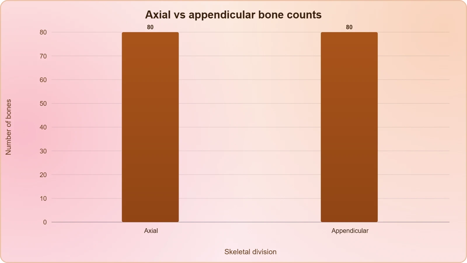
This division is more than a convenience for memorisation. The axial elements are overwhelmingly protective and supportive, enclosing the brain and spinal cord and forming the trunk's central pillar. The appendicular elements are overwhelmingly locomotive, built for reach, grasp and stride. Keeping the two divisions distinct in the mind makes the bone counts intelligible rather than arbitrary, and it explains why the axial skeleton is treated first in any systematic account of the framework.
The Skull: Cranial and Facial Bones, Hyoid and Ear Ossicles
The skull is composed of two sets of bones — cranial and facial — that total 22 bones. The cranial bones are 8 in number, and they form the hard protective outer covering, the cranium, for the brain. The facial region is made up of 14 skeletal elements, which form the front part of the skull. Beyond these two sets, the skull region also accommodates three further bony structures that are easily missed in a simple count: the hyoid bone and the ear ossicles.
Cranium and Facial Skeleton
The eight cranial bones enclose the brain in a hard case, and their names appear in the diagrammatic view of the human skull: frontal, parietal, temporal, occipital, sphenoid and ethmoid, with the parietal and temporal bones occurring as pairs. The fourteen facial elements form the front of the skull and include the nasal, lacrimal, zygomatic and maxilla bones, together with the mandible. The mandible is the lower jaw, and it is the bone whose absence would make chewing impossible.
The division between cranial and facial sets is functional as well as positional. Cranial bones are built for enclosure and protection, meeting at sutures to form a rigid vault around the brain. Facial bones are built for the openings of the senses and for the mechanics of eating: they frame the orbits, shape the nasal cavity, and carry the upper and lower jaws. Together the 22 bones give the head its form while preserving the passages on which sight, smell and ingestion depend.
The Hyoid Bone and the Ear Ossicles
A single U-shaped bone called the hyoid is present at the base of the buccal cavity. It is unusual in standing apart from the main articulations of the skull, yet it is counted within the skull region. Each middle ear contains three tiny bones — the malleus, incus and stapes — collectively called the ear ossicles. These are the smallest bones of the body, and they transmit sound vibrations across the middle ear, a mechanical function quite unlike the protective role of the cranium.
Counting carefully is worthwhile here. The skull's 22 bones comprise 8 cranial and 14 facial elements; the hyoid and the six ear ossicles are additional structures of the skull region. Learners who conflate these numbers lose the thread of the axial total. The discipline of separating what belongs to the cranium, what belongs to the face, and what merely lies within the skull region is exactly the discipline the material asks for, and it pays off when the vertebral column is approached.
The Dicondylic Skull and Its Articulation with the Vertebral Column
The skull region articulates with the superior region of the vertebral column with the help of two occipital condyles, and a skull of this kind is described as dicondylic. The occipital condyle is visible in the diagrammatic view of the human skull, sitting at the base of the occipital bone. This two-point articulation is the mechanical junction between the head and the trunk, and it is the reason the head can be balanced and moved upon the top of the vertebral column.
Why Two Condyles Matter
A single point of contact would permit rotation but offer little stability; two condyles provide a stable base while still allowing the nodding and turning movements of the head. The first vertebra, the atlas, receives these condyles, and the pairing of structures is exact. The term dicondylic therefore records a structural fact with functional consequences, and it links the skull directly to the vertebral column that is considered next.
This articulation also explains why the skull and vertebral column are treated as one continuous axial system rather than as separate topics. The skull protects the brain; the vertebral column protects the spinal cord; and the junction between them is where the central nervous system passes from cranium to canal. The occipital condyles are the physical bridge in that sequence, and the neural canal of each vertebra continues the protected passage downward.
Reading the Skull Diagram
The diagrammatic view of the human skull labels the frontal, parietal, temporal, occipital, sphenoid and ethmoid bones of the cranium, and the lacrimal, nasal, zygomatic and maxilla bones of the face, together with the mandible and the hyoid bone. The occipital condyle is marked at the base. Working through such a diagram systematically — cranium first, face second, then the separate structures — is the most reliable way to fix the 8, 14 and 22 counts in memory.
Learners should also notice what the diagram does not show: the ear ossicles lie within the middle ear and are not surface features of the skull. Their inclusion in the lesson is a reminder that the skull region is a three-dimensional assembly, not a flat outline. The same caution applies to the vertebral column, where the neural canal is internal and invisible from the lateral view except as a structural implication of each vertebra's form.
The Vertebral Column: Twenty-Six Units in Five Regions
The vertebral column is formed by 26 serially arranged units called vertebrae and is dorsally placed. It extends from the base of the skull and constitutes the main framework of the trunk. Each vertebra has a central hollow portion, the neural canal, through which the spinal cord passes. The column is differentiated into cervical, thoracic, lumbar, sacral and coccygeal regions starting from the skull, and the regional counts are among the most examinable facts in the entire chapter.
Regional Differentiation and Vertebral Counts
Starting from the skull, the regions are cervical with 7 vertebrae, thoracic with 12, lumbar with 5, sacral with 1 fused vertebra, and coccygeal with 1 fused vertebra. The number of cervical vertebrae is seven in almost all mammals, including human beings, a constancy worth noting. The sacrum and coccyx appear in the lateral view as fused terminal elements, which is why 26 units rather than 33 separate vertebrae are counted in the adult column.
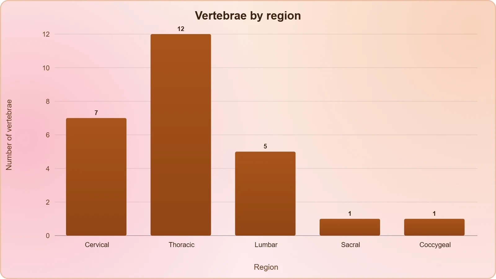
The regional counts are not arbitrary numbers to be memorised in isolation. Each region corresponds to a section of the trunk and to a set of attached structures: the cervical region supports the head, the thoracic region carries the ribs, and the lumbar region bears the weight of the upper body. The fused sacral and coccygeal elements anchor the column at its base. Reading the counts alongside the regions, rather than as a bare list, makes them far easier to retain.
Functions of the Column and the First Vertebra
The first vertebra is the atlas, and it articulates with the occipital condyles of the skull. The vertebral column protects the spinal cord, supports the head, and serves as the point of attachment for the ribs and musculature of the back. These three functions — protection, support and attachment — summarise the column's role in the axial skeleton and connect it directly to the muscular tissue discussed in the preceding lesson, whose fibres act upon it.
The neural canal deserves particular attention because it explains the column's protective function in concrete terms. Each vertebra contributes a hollow centre, and stacked in series these hollows form a continuous bony tunnel around the spinal cord. Intervertebral discs separate adjacent vertebrae, visible in the lateral view, and provide the slight pliability that a column of rigid bone would otherwise lack. The design is a compromise between strength and controlled flexibility.
The Sternum and the Ribs: True, False and Floating Pairs
The sternum is a flat bone on the ventral midline of the thorax. There are 12 pairs of ribs, and each rib is a thin flat bone connected dorsally to the vertebral column and ventrally to the sternum. Each rib has two articulation surfaces on its dorsal end and is hence called bicephalic. Thoracic vertebrae, ribs and sternum together form the rib cage. The classification of the twelve pairs into true, false and floating ribs follows directly from how each pair connects ventrally.
Bicephalic Ribs and the Three Classes
The first seven pairs of ribs are called true ribs. Dorsally they are attached to the thoracic vertebrae, and ventrally they are connected to the sternum with the help of hyaline cartilage. The 8th, 9th and 10th pairs do not articulate directly with the sternum but join the seventh rib with the help of hyaline cartilage; these are called vertebrochondral or false ribs. The last two pairs, the 11th and 12th, are not connected ventrally and are therefore called floating ribs.

The logic of the classification is purely one of ventral attachment. True ribs reach the sternum directly; false ribs reach it only indirectly, through the cartilage of the seventh rib; floating ribs do not reach it at all. Once this single criterion is grasped, the three names cease to be arbitrary labels and become descriptions. The bicephalic dorsal end, with its two articulation surfaces, is a separate feature belonging to every rib regardless of class.
The Rib Cage as a Unit
Thoracic vertebrae, ribs and sternum together form the rib cage, and the sternum's position on the ventral midline of the thorax makes it the anterior anchor of the whole assembly. The hyaline cartilage that bridges rib to sternum is the same pliable tissue noted at the outset, and its presence here allows the cage a measure of give during breathing. The cage is thus a composite structure, bony in its ribs and sternum, cartilaginous at its ventral junctions.
It is worth pausing on the arithmetic of the rib cage, because it ties the axial skeleton together. Twelve pairs of ribs articulate dorsally with the twelve thoracic vertebrae, and the sternum receives the first seven pairs directly. The count of thoracic vertebrae and the count of rib pairs therefore match, which is precisely what the regional division of the vertebral column would lead one to expect. The axial skeleton is internally consistent, and its numbers reinforce one another.
Consolidating the Axial Skeleton: Structure, Function and What Comes Next
The axial skeleton comprises 80 bones distributed along the main axis of the body, and its four components — skull, vertebral column, sternum and ribs — have now been examined in turn. The skull contributes 22 bones in cranial and facial sets, together with the hyoid and the ear ossicles, and articulates with the column through two occipital condyles. The column contributes 26 serially arranged units in five regions. The sternum and twelve pairs of ribs complete the framework of the trunk.
Protection, Support and Attachment
Three functions recur across the axial components. Protection is served by the cranium around the brain and by the neural canal around the spinal cord. Support is served by the vertebral column, which holds up the head and forms the main framework of the trunk. Attachment is served by the column as a point of attachment for the ribs and musculature of the back, and by the rib cage as the anchor for the muscles of respiration. The axial skeleton is thus both a shield and a scaffold.
These functions connect directly to the muscle physiology of the previous lesson. Red fibres, rich in myoglobin and mitochondria, and white fibres, dependent on anaerobic processes, both exert force on the bony levers described here. The fatigue that follows repeated activation of muscle is the physiological counterpart of a skeleton that must be moved repeatedly throughout life. Structure and function are two descriptions of the same organism, and the axial skeleton supplies the structural half of the account.
Preparing for the Appendicular Skeleton and Joints
The appendicular skeleton consists of the bones of the limbs along with their girdles, and each limb is made of 30 bones. That division, and the three structural forms of joints, belong to the next lesson. What should be carried forward is the organising principle established here: the skeleton divides by position relative to the main axis, and each division has its own characteristic functions. The axial skeleton protects and supports; the appendicular skeleton will be found to reach and to move.
A final check of the numbers consolidates the lesson. The axial skeleton holds 80 bones; the skull holds 22, of which 8 are cranial and 14 facial; the vertebral column holds 26 units, divided 7, 12, 5, 1 and 1; the ribs number 12 pairs, divided into 7 true, 3 false and 2 floating. The sternum is a single flat bone on the ventral midline of the thorax. Mastery of these figures is the foundation on which the appendicular counts of the next lesson will rest.
Going further
The vertebral column belongs to the axial skeleton precisely because it lies along the main axis of the body, running from the base of the skull down the dorsal midline. Everything that hangs from this central pillar — the limbs and their girdles — is appendicular by contrast. The rib cage, formed by the thoracic vertebrae, ribs and sternum, encloses and protects vital organs such as the heart and lungs, adding an internal protective function to the external framework already described.
Also Read
- No links available.
From our network :
- Mastering DB2 LUW v12 Tables: A Comprehensive Technical GuideComprehensive guide on DB2 LUW v12 tables. Explore CREATE TABLE syntax, BLU Acceleration, range partitioning, data types, and security features for IBM DB2 LUW v12.
- AI-Powered 'Precision Diagnostic' Replaces Standard GRE Score ReportsDiscover the new GRE Precision Diagnostic. Learn how AI-powered behavioral analytics and time-to-solve tracking are replacing standard GRE score reports to empower students.
- EV 2.0: The Solid-State Battery Breakthrough and Global Factory ExpansionDiscover the impact of the Solid-State Battery Breakthrough on the EV market. Learn about CATL and BYD stock growth, global factory expansion in Europe, and the future of EV 2.0 technology.
- 98% of Global MBA Programs Now Prefer GRE Over GMAT Focus EditionExplore why 98% of global MBA programs now prefer the GRE over GMAT Focus. Learn about the shorter GRE format, verbal logic importance, and strategic versatility for students.
- Vite 6/7 'Cold Start' Regression in Massive Module GraphsIn-depth analysis of Vite 6/7 cold start regressions in massive module graphs. Learn about barrel file issues, pre-bundling optimizations, and how to improve Vite dev server performance.
- Mastering DB2 12.1 Instance Design: A Technical Deep Dive into Modern Database ArchitectureComprehensive guide to DB2 12.1 Instance Design. Explore architectural components, HADR setup, buffer pool tuning, and security policies for IBM DB2 12.1. Includes 10 functional SQL and Shell samples.
- 10 Physics Numerical Problems with Solutions for IIT JEEPrepare for IIT JEE with 10 physics numerical problems, complete with detailed solutions and conceptual explanations. Ideal for mastering kinematics, thermodynamics, electromagnetism, and optics.
- Analyzing Trump Deportation Numbers Insights Into The 2026 Immigration Crackdown
- Trump Political Strategy How Geopolitical Stunts Serve As Media Diversions
RESOURCES
- Axial Skeleton: What Bones it Makes Up - Cleveland Clinicmy.clevelandclinic.orgFeb 8, 2022 ... ... (vertebrae, sacrum and tailbone) and ribcage (sternum and ribs). ... The vertebral column extends from the base of your…
- Axial Skeleton (80 bones) - SEER Training Modulestraining.seer.cancer.govSkull (28). Illustration mapping cranial bones. Cranial Bones. Parietal ... Hyoid (1). Illustration mapping the bones of the vetebral column. Vertebral Column.
- Axial Skeleton Anatomy & Physiology Study Guide | Notes - Pearsonpearson.comDefinition: The axial skeleton is composed of the skull, vertebral column, and thoracic cage (ribs and sternum), as well as associated bones such as…
- The Architecture of Enormity | The Linnean Societylinnean.orgJun 5, 2026 ... ... axial skeleton (the skull, vertebral column, ribs and associated structures) of the early sauropodomorph Macrocollum itaquii. Their findings ...
- Axial Skeleton Anatomy & Physiology Flashcards - Pearsonpearson.comIncludes the skull bones, vertebrae of the vertebral column, ribs, and sternum. Cranial bones. Bones that form the protective case around the brain. Facial ...
- axial skeleton: The skull, Vertebral column, rib cage, and fetal skullquizlet.comThe horizontal plate of the ethmoid bone separating the cranial cavity from the nasal cavity. maxilla.
- Axial skeleton | Description, Characteristics, Anatomy, & Functionbritannica.comSep 25, 2026 ... Axial skeleton, the part of the bony structure of the body that consists of the bones of the skull, the…
- The Axial Skeleton: Anatomy and Physiology Review - Albert.ioalbert.ioThe axial skeleton forms the long central axis of the body: the skull, vertebral column, ribs, sternum, and hyoid bone. The appendicular skeleton includes ...
- Human skeleton | Parts, Functions, Diagram, & Facts - Britannicabritannica.comSep 25, 2026 ... These are (1) the axial, comprising the vertebral column—the spine—and much of the skull, and (2) the appendicular, to which…
- Indore, India Dog skeleton System ♨️ As with any vertebrate ...facebook.comApr 30, 2021 ... We can divide the canine skeleton into three main sections: ✓Axial skeleton: skull, spine, ribs and sternum bones. ✓Appendicular skeleton: ...
- Anatomy, Back, Vertebral Column - StatPearls - NCBI Bookshelfncbi.nlm.nih.gov... skull, ribs, and sternum, these vertebrae make up the axial skeletal system ... Osteoporosis: Demineralization of the bones. Current U.S. Preventive ...
- 38.3: Types of Skeletal Systems - Human Axial Skeletonbio.libretexts.org1 : Axial skeleton: The axial skeleton consists of the bones of the skull, ossicles of the middle ear, hyoid bone, vertebral column, and…
- The Skeletal System Anatomical Chart Textbookwww2.american-usa.comthe skull, vertebral column, rib cage, pelvis, and limb bones, along with ... muscles, bone joints, axial skeleton, appendicular skeleton, bone types, skeletal ...
- 7.1 Divisions of the Skeletal System - Anatomy and Physiology 2eopenstax.orgApr 20, 2022 ... The axial skeleton of the adult consists of 80 bones, including the skull, the vertebral column, and the thoracic cage.…
- Exploring fetal skeletal alterations induced by gold nanoparticles in ...pmc.ncbi.nlm.nih.govThe BALB/c mouse skeleton is conventionally divided into two main structures: the axial skeleton, comprising the skull, vertebral column, ribs, and sternum, and ...