Muscle contraction is one of the most elegant mechanical events in all of biology, and the sliding filament theory remains the clearest account we have of how it happens. At its heart lies a deceptively simple proposition: a muscle fibre shortens not because its proteins compress or coil, but because thin actin filaments glide over thick myosin filaments, drawing the ends of each sarcomere toward one another. Everything else in this lesson — the nerve impulse, the chemical messenger, the calcium surge, the pivoting myosin head — exists to set that sliding in motion and, just as importantly, to bring it to a controlled stop.
This sixth lesson in the Locomotion and Movement course builds directly on the structural groundwork laid in the previous session, where actin, tropomyosin, troponin and the meromyosin monomer were introduced. Here those components are set in motion. We follow the complete sequence from the neural signal arriving at the neuromuscular junction, through acetylcholine release and the sarcolemma action potential, to calcium release into the sarcoplasm, cross bridge formation, filament sliding, and finally relaxation as calcium is pumped back. The order of ideas follows the standard treatment of the topic, and the terminology is preserved precisely so that later lessons on fibre types and fatigue rest on firm foundations.
By the end of this lesson, you should be able to narrate the contraction cycle as a continuous causal chain rather than a list of isolated facts, explain why the I band narrows while the A band holds its length, and describe the two roles ATP plays — one in powering the power stroke, the other in releasing the myosin head so the cycle can repeat. These distinctions matter, because they explain why muscles stiffen after death and why fatigue is not simply a matter of running out of energy.
On This Page
The Sliding Filament Theory: Core Statement and Evidence
The sliding filament theory states, in its own compact form, that contraction of a muscle fibre takes place by the sliding of the thin filaments over the thick filaments. This single sentence carries the entire explanatory burden of the lesson. It tells us that the filaments themselves are not elastic bands that snap shorter, nor are they consumed or broken down during a contraction. Instead, they retain their individual lengths and simply change their degree of overlap, telescoping past one another within the sarcomere. The shortening we observe at the level of the whole muscle is therefore a sum of millions of tiny relative displacements.
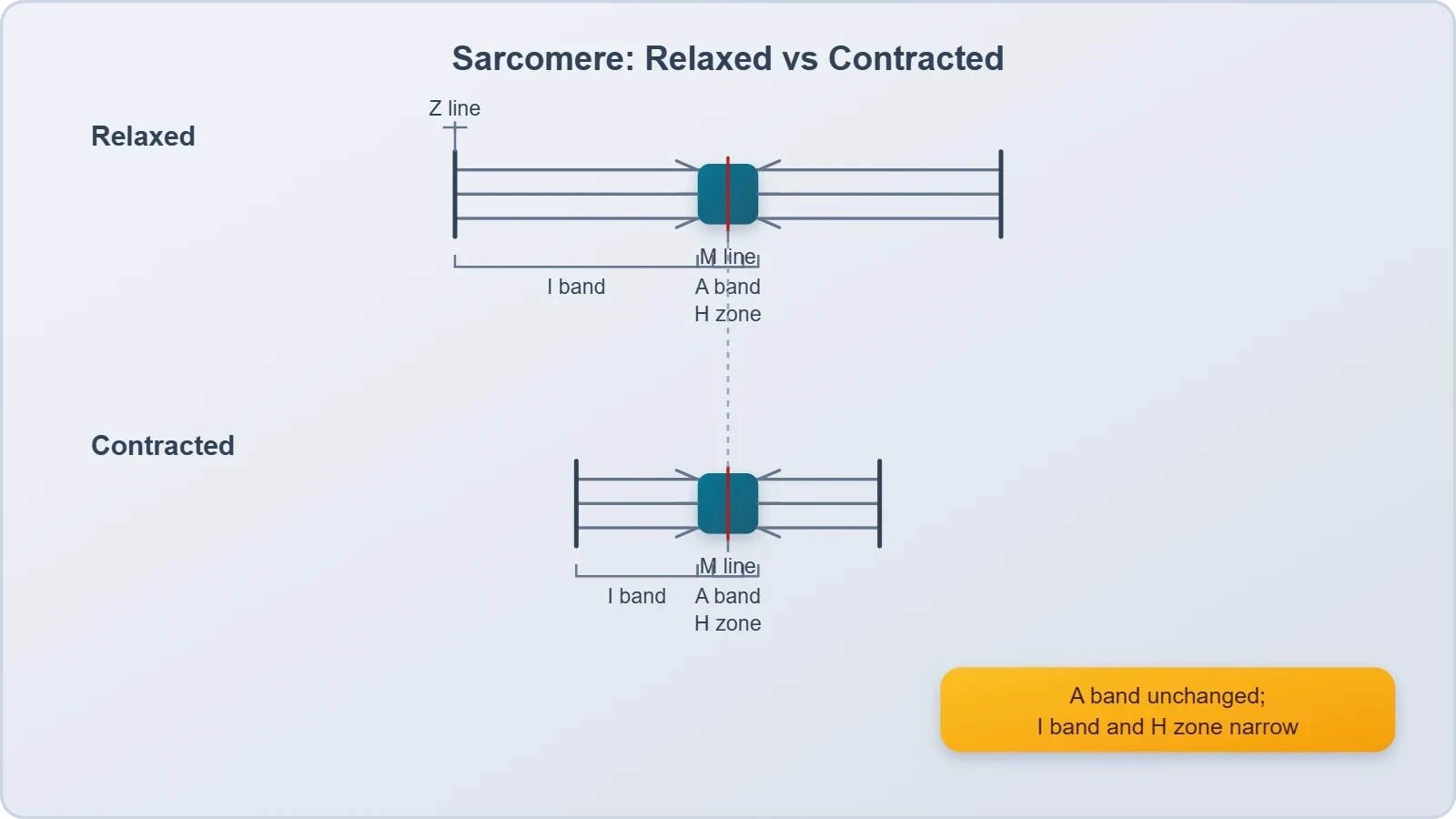
The theory earns its place because it accounts for a striking microscopic observation: during shortening, the I bands get reduced, whereas the A bands retain their length. If filaments were genuinely contracting, the A band — which is defined by the thick filaments — would shorten too. It does not. The A band is a fixed measure of thick filament length, and its constancy is direct evidence that the thick filaments are sliding rather than shrinking. Meanwhile the I band, the region containing only thin filaments, narrows as those thin filaments are drawn inward toward the centre of the sarcomere.
Why the Sarcomere Is the Functional Unit
The portion of the myofibril between two successive Z lines is considered the functional unit of contraction and is called a sarcomere. This definition is worth pausing over, because it establishes the scale at which contraction is best understood. Each sarcomere is bounded by two Z lines, and the thin filaments are firmly attached to those Z lines. When the thin filaments slide inward, they pull the Z lines with them, and the distance between successive Z lines decreases. A muscle fibre shortens because its sarcomeres shorten in series, each contributing a small reduction in length.
Within the resting sarcomere, the edges of thin filaments on either side of the thick filaments partially overlap the free ends of the thick filaments, leaving the central part of the thick filaments uncovered. This central region, not overlapped by thin filaments, is called the H zone. The H zone is a useful diagnostic feature: as sliding proceeds and the thin filaments are drawn further inward, the H zone narrows and may disappear altogether. Together with the shrinking I band, the vanishing H zone gives a precise picture of filaments gliding past one another.
Reading Band Changes as a Diagnostic
Students often memorise band behaviour without understanding why it must be so. A more reliable approach is to reason from the geometry. The A band corresponds to the full length of the thick filaments, which do not change length during contraction, so the A band is invariant. The I band corresponds to the thin filaments in the region where no thick filament is present; as thin filaments slide toward the centre, that exposed region shrinks. The H zone corresponds to the central thick filament region not yet overlapped; it too shrinks. Every observed change is consistent with sliding and inconsistent with filament shortening.
This reasoning also explains the limits of contraction. A sarcomere can shorten only until the thin filaments from opposite ends meet at the M line, at which point further sliding is geometrically impossible. The thin filaments are held in the middle of the A band by a thin fibrous membrane called the M line, and the thick filaments are gathered there. Recognising this ceiling helps explain why muscle force depends on the starting length of the sarcomere, a point that becomes important when we later consider how muscles generate tension across a range of positions.
From Nerve Signal to Calcium Release
Muscle contraction is initiated by a signal sent by the central nervous system via a motor neuron. A motor neuron along with the muscle fibres connected to it constitute a motor unit, a functional grouping that ensures a single command recruits a defined set of fibres together. The junction between a motor neuron and the sarcolemma of the muscle fibre is called the neuromuscular junction or motor-end plate. This is the physical and chemical interface where an electrical instruction travelling along a nerve becomes an electrical event in a muscle, and it is the first link in the causal chain we are tracing.
A neural signal reaching this junction releases a neurotransmitter, acetylcholine, which generates an action potential in the sarcolemma. The action potential then spreads through the muscle fibre. This propagation is the crucial relay: the signal does not travel passively but is regenerated along the surface membrane and carried inward, ensuring that the instruction reaches the interior of the fibre rather than remaining at its surface. The consequence of this spread is the release of calcium ions into the sarcoplasm, the cytoplasm of the muscle fibre, where the contractile machinery waits.
The Neuromuscular Junction as a Relay Point
The neuromuscular junction deserves attention because it is where the nervous system hands over control. The motor neuron does not physically pull the muscle; it releases a chemical messenger across a narrow gap, and that messenger depolarises the sarcolemma. Acetylcholine is the neurotransmitter responsible here, and its release is triggered by the arrival of the neural signal. The resulting action potential in the sarcolemma is the muscle's own electrical response, distinct from the nerve impulse that provoked it. Understanding this handover clarifies why drugs and toxins acting at this junction can paralyse a muscle without damaging it.
Once generated, the action potential spreads through the muscle fibre and causes the release of calcium ions into the sarcoplasm. In the resting state, calcium is sequestered away from the contractile proteins; the action potential effectively opens the gates that let it flood in. This rise in calcium concentration is the pivotal chemical switch. Everything before it is signalling; everything after it is mechanics. The elegance of the system lies in this conversion of an electrical message into a chemical one, and then into a mechanical one, with each step amplifying and specifying the next.
Calcium as the Molecular Switch
Increase in calcium level leads to the binding of calcium with a subunit of troponin on actin filaments and thereby removes the masking of active sites for myosin. Recall from the previous lesson that in the resting state a subunit of troponin masks the active binding sites for myosin on the actin filaments. Calcium binding to troponin changes its conformation, and this change is transmitted along tropomyosin, which shifts away from the sites it was blocking. The actin filament is now available for interaction. This is the moment the contractile machinery becomes competent to generate force.
The dependence on calcium is absolute, and it explains both the initiation and the termination of contraction. Without calcium, troponin keeps the active sites masked and myosin cannot bind, no matter how much ATP is available. With calcium present, the sites are exposed and the cycle can proceed. This is why the removal of calcium, rather than the exhaustion of ATP, is the normal route to relaxation. The cell must actively pump calcium back to restore the resting state, a point we return to when discussing relaxation in detail.
Cross Bridge Formation and the Power Stroke
Utilising the energy from ATP hydrolysis, the myosin head now binds to the exposed active sites on actin to form a cross bridge. This is the central mechanical event of the theory. The globular head of the myosin filament, which projects outward as a cross arm, is an active ATPase enzyme and carries binding sites for ATP and active sites for actin. When the actin sites are exposed by calcium, the head attaches, and the two filaments are physically linked. The cross bridge is not a permanent weld but a transient connection that will be made and broken repeatedly.
Once formed, the cross bridge pulls the attached actin filaments towards the centre of the A band. The Z lines attached to these actins are also pulled inwards, thereby causing a shortening of the sarcomere, that is, contraction. This pulling action is the power stroke, the working phase in which chemical energy is converted into mechanical displacement. Because the thin filaments on both sides of a thick filament are drawn inward simultaneously, the sarcomere shortens symmetrically, and the effect is multiplied across the many sarcomeres arranged in series along each myofibril.
The Myosin Head as an ATPase
The myosin head is the engine of the entire process, and its dual identity is worth emphasising. It is simultaneously an enzyme and a mechanical element. As an ATPase, it hydrolyses ATP, and the energy released by that hydrolysis is used to bind the head to actin and to drive the pull. As a structural element, it projects from the thick filament at a regular distance and angle, positioning the binding site where it can reach the actin filament. The head and short arm together form the heavy meromyosin, while the tail is the light meromyosin, and this division of labour reflects their distinct roles.
The geometry of the cross arm matters because it determines the direction of the pull. The heads project outward at regular intervals and angles, so that when they attach to actin and rotate, they sweep the thin filament in a consistent direction toward the sarcomere centre. This is why contraction is directional rather than random. The regularity of the arrangement, with many heads acting in concert, also explains how a modest displacement at each individual head sums to a substantial shortening of the whole fibre. Coordination, not brute force, produces the movement.
Breaking and Reforming the Cross Bridge
The myosin, releasing the ADP and P1, goes back to its relaxed state. A new ATP binds and the cross bridge is broken. This detail is essential and frequently misunderstood: the binding of a fresh ATP molecule, not its hydrolysis, is what detaches the myosin head from actin. The ATP is again hydrolysed by the myosin head, and the cycle of cross bridge formation repeats. Each cycle consumes one ATP and produces a small relative sliding, so sustained contraction requires a continuous supply of ATP and a continuous cycling of many heads.
This cyclical character explains why the sliding filament theory is sometimes described as a ratchet. No single cross bridge persists for the duration of a contraction; instead, heads attach, pull, detach, and reattach at new sites further along the actin filament. The cumulative effect of many such cycles is the steady drawing inward of the thin filaments. It also explains why the process can be halted at any moment: remove calcium, and the sites become masked again; withhold ATP, and the heads cannot detach, leaving the muscle locked.
Relaxation and the Return to Resting State
Relaxation is not the passive absence of contraction but an active restoration of the resting state. When the neural signal ceases, calcium is pumped back out of the sarcoplasm, the concentration of calcium falls, and troponin releases its bound calcium. The tropomyosin shifts back into its blocking position, and the active sites for myosin on the actin filaments are masked once more. With the sites hidden, cross bridges can no longer form, and the thin filaments are free to be drawn back to their resting overlap by the elastic properties of the sarcomere and its associated proteins.
The pumping of calcium back is therefore the decisive step in relaxation, and it requires energy. This is a point worth holding onto: both contraction and relaxation consume ATP. A muscle that cannot supply energy cannot relax properly, which is why the failure of calcium reuptake produces sustained stiffness rather than flaccidity. The resting state described earlier, in which the edges of thin filaments partially overlap the free ends of the thick filaments and the H zone is visible, is restored only when calcium has been cleared and the masking of active sites is complete.
Calcium Reuptake and the Resting Sarcomere
Once calcium is pumped back, the sarcomere returns to the geometry with which we began. The thin filaments sit with their edges partially overlapping the free ends of the thick filaments, leaving the central part of the thick filaments uncovered as the H zone. The I bands widen again as the thin filaments withdraw from the centre, and the distance between successive Z lines returns to its resting value. The muscle fibre is now ready for another signal. This reversibility is what allows muscle to contract repeatedly over a lifetime without permanent change to its structure.
The contrast between the contracted and relaxed sarcomere is thus a contrast in overlap, not in filament length. The thick filaments remain the same length throughout, which is why the A band is constant. The thin filaments likewise retain their length; they simply occupy different positions relative to the thick filaments. Grasping this point firmly resolves most of the confusion students encounter when first studying the theory, because it replaces a vague notion of proteins shrinking with a precise picture of filaments sliding.
Why Relaxation Requires Energy
It is a common misconception that relaxation is simply what happens when the muscle stops working. In reality, the cell must actively reclaim the calcium it released, and that reclamation depends on energy. If ATP is unavailable, calcium remains in the sarcoplasm, the active sites stay exposed, and the muscle cannot return to its resting state. This is why the cycle of contraction and relaxation is best understood as a continuous expenditure of energy in both directions, with the resting state being an actively maintained condition rather than a default.
This insight also prepares the ground for the next lesson. The rate at which a fibre can cycle cross bridges, clear calcium, and restore its resting state depends on its metabolic machinery, and fibres differ in exactly this respect. Red and white muscle fibres are distinguished partly by how they supply energy and how quickly they fatigue, topics that follow naturally once the sliding filament mechanism is secure. For now, the essential point is that relaxation is an energy-dependent return to the resting arrangement of overlapping filaments.
Energy Accounting and the Limits of the Model
Every turn of the cross bridge cycle consumes one ATP, and the cycle turns many times during a single contraction. The energy budget of a muscle is therefore dominated by the repeated hydrolysis of ATP by the myosin head, supplemented by the energy needed to pump calcium back during relaxation. This accounting explains several familiar phenomena. It explains why sustained contraction is metabolically expensive, why muscles warm up during exertion, and why the interruption of ATP supply produces rigidity rather than relaxation. The chemistry and the mechanics are inseparable.
The model also has clear limits, and acknowledging them sharpens understanding. The sliding filament theory describes what happens within the sarcomere, but it does not by itself explain how the signal is routed to the correct fibres, how force is transmitted through the tendon to the bone, or how the nervous system grades the strength of a contraction. Those questions belong to the wider study of locomotion and movement. Within its proper domain, however, the theory is remarkably complete, accounting for the band changes, the calcium dependence, and the ATP requirement in one coherent framework.
ATP Budget of a Single Cycle
Consider the cycle as a sequence of energy transactions. The myosin head, having completed a power stroke, releases ADP and P1 and returns to its relaxed state. A new ATP binds, and this binding breaks the cross bridge. The ATP is then hydrolysed by the myosin head, resetting it for the next attachment. Each of these steps is a discrete event, and the cycle as a whole consumes exactly one ATP per power stroke. Because a single contraction involves many heads cycling repeatedly, the total ATP demand scales with both the number of cross bridges and the duration of activity.
This budget has a practical consequence for how we interpret fatigue, which is the subject of the next lesson. A fibre that cycles cross bridges rapidly will deplete its ATP more quickly unless its metabolic supply keeps pace. The interplay between demand and supply, rather than any single limiting factor, determines how long a muscle can sustain a given output. For present purposes, the key point is that the sliding filament mechanism is powered by ATP at two distinct stages, and both must be satisfied for contraction and relaxation to proceed normally.
What the Theory Does Not Explain
It is worth being explicit about the boundaries of the model so that it is not asked to do more than it can. The sliding filament theory explains the relative movement of actin and myosin, the role of calcium in exposing binding sites, and the ATP dependence of the cycle. It does not, on its own, account for the recruitment of motor units, the summation of twitches, or the transmission of force through connective tissue to the skeleton. These are separate layers of explanation, and confusing them leads to muddled reasoning about how whole muscles behave.
Keeping the layers distinct is a mark of clear thinking in physiology. The sarcomere is the functional unit of contraction, and the theory describes its internal workings with precision. The muscle fibre, the motor unit, and the whole muscle are larger systems that obey additional rules. A student who can move confidently between these levels, without attributing the properties of one to another, has understood the topic properly. The next lesson extends this understanding to the different fibre types and the phenomenon of fatigue.
Going Further
Acetylcholine is a neurotransmitter that carries signals from nerve cells to muscle fibres, and its role at the neuromuscular junction is a good example of how the nervous system communicates with effector tissue. The chemical is released from the nerve ending, diffuses across the narrow junctional gap, and binds to receptors on the sarcolemma, triggering the action potential that sets the whole sequence in motion. Because the signal is chemical rather than electrical across the gap, the junction is a point of control, and substances that interfere with acetylcholine release or reception can interrupt contraction entirely.
The cross bridge cycle is repeated many times during a single muscle contraction. This is a useful corrective to the intuition that a muscle contracts by forming cross bridges once and holding them. In reality, each head attaches, pulls, detaches, and reattaches at a new site, and the cumulative effect of many such cycles is the steady sliding of the thin filaments. The repetition also explains why contraction is graded and smooth rather than jerky: at any instant, different heads are at different stages of the cycle, so the pull is continuous even though each individual interaction is brief.
Also Read
- No links available.
From our network :
- Mastering DB2 12.1 Instance Design: A Technical Deep Dive into Modern Database ArchitectureComprehensive guide to DB2 12.1 Instance Design. Explore architectural components, HADR setup, buffer pool tuning, and security policies for IBM DB2 12.1. Includes 10 functional SQL and Shell samples.
- Analyzing Trump Deportation Numbers Insights Into The 2026 Immigration Crackdown
- 10 Physics Numerical Problems with Solutions for IIT JEEPrepare for IIT JEE with 10 physics numerical problems, complete with detailed solutions and conceptual explanations. Ideal for mastering kinematics, thermodynamics, electromagnetism, and optics.
- AI-Powered 'Precision Diagnostic' Replaces Standard GRE Score ReportsDiscover the new GRE Precision Diagnostic. Learn how AI-powered behavioral analytics and time-to-solve tracking are replacing standard GRE score reports to empower students.
- EV 2.0: The Solid-State Battery Breakthrough and Global Factory ExpansionDiscover the impact of the Solid-State Battery Breakthrough on the EV market. Learn about CATL and BYD stock growth, global factory expansion in Europe, and the future of EV 2.0 technology.
- 98% of Global MBA Programs Now Prefer GRE Over GMAT Focus EditionExplore why 98% of global MBA programs now prefer the GRE over GMAT Focus. Learn about the shorter GRE format, verbal logic importance, and strategic versatility for students.
- Mastering DB2 LUW v12 Tables: A Comprehensive Technical GuideComprehensive guide on DB2 LUW v12 tables. Explore CREATE TABLE syntax, BLU Acceleration, range partitioning, data types, and security features for IBM DB2 LUW v12.
- Trump Political Strategy How Geopolitical Stunts Serve As Media Diversions
- Vite 6/7 'Cold Start' Regression in Massive Module GraphsIn-depth analysis of Vite 6/7 cold start regressions in massive module graphs. Learn about barrel file issues, pre-bundling optimizations, and how to improve Vite dev server performance.
RESOURCES
- The Sliding Filament Theory of Muscle Contraction - Naturenature.comWithin the sarcomere, myosin slides along actin to contract the muscle fiber in a process that requires ATP. Scientists have also identified many of…
- Muscle contraction: Sliding filament history, sarcomere dynamics ...pmc.ncbi.nlm.nih.govIn this 1951 paper “The structure of hair, muscle and related proteins” they argued that muscles contain continuous filaments through the whole sarcomere and ...
- 16 Muscle Contraction The sliding filament theoryanaesthetics.ukzn.ac.zaA sarcomere (Fig 2.) is the functional unit of skeletal muscle. The overlapping actin and myosin filaments form the scaffolding for the cross-bridging mechanism ...
- Re‑examining the mechanism of eccentric exercise‑induced skeletal ...pmc.ncbi.nlm.nih.gov2. Overview of the sliding filament theory ... Until the 1950s (24), researchers widely considered that the force produced during muscle contraction was directly ...
- Sliding filament theory - Wikipediaen.wikipedia.orgThe sliding filament theory explains the mechanism of muscle contraction based on muscle proteins that slide past each other to generate movement.
- 5.7: Sliding Filament Theory of Contraction - Medicine LibreTextsmed.libretexts.orgMar 15, 2026 ... The sliding filament theory explains how muscle contraction occurs when thin (actin) filaments slide past thick (myosin) filaments, ...
- Physiology, Muscle Contraction - StatPearls - NCBI Bookshelfncbi.nlm.nih.govApr 1, 2023 ... [13] During this type of contraction, a muscle is stimulated to contract according to the sliding filament theory. Concentric contractions ...
- Sliding Filament Model of Contraction | Biology for Majors IIcourses.lumenlearning.comThe sliding filament theory of muscle contraction was developed to fit the differences observed in the named bands on the sarcomere at different degrees…
- 13.1: Muscle Contraction - Biology LibreTextsbio.libretexts.orgMar 6, 2021 ... According to the sliding filament theory, a muscle fiber contracts when myosin filaments pull actin filaments closer together and thus…
- On the theory of muscle contraction: filament extensibility ... - PubMedpubmed.ncbi.nlm.nih.gov... filaments challenges the foundation of the theory of muscle mechanics. We have reformulated A. F. Huxley's sliding filament theory to explicitly take into ...
- Cardiac Myocytes and Sarcomeres - CV Physiologycvphysiology.comThe interactions between actin and myosin serve as the basis for the sliding filament theory of muscle contraction. Myosin and actin Myosin is a…
- A Role for the Giant Titin Protein - PubMedpubmed.ncbi.nlm.nih.govFeb 9, 2017 ... ... muscle strength. Shortly after the sliding filament theory of muscle contraction was introduced, there was a reluctant recognition that ...
- The Sliding Filament Theory Since Andrew Huxley - Annual Reviewsannualreviews.orgFeb 26, 2021 ... Two groundbreaking papers published in 1954 laid out the theory of the mechanism of muscle contraction based on force-generating ...
- Hand-held model of a sarcomere to illustrate the sliding filament ...journals.physiology.org... sliding filament mechanism in muscle contraction ... Their model has formed the basis for a hypothesis known as the sliding filament theory of…