The appendicular skeleton is the part of the human frame that makes movement possible. Where the axial skeleton forms the central axis of the body, the appendicular skeleton extends outward from it, carrying the limbs and the girdles that anchor them. Each limb is built from exactly 30 bones, and the girdles that attach these limbs to the axial frame are themselves paired structures, formed of two halves. Understanding this arrangement is the key to understanding how the body reaches, grasps, walks and runs.
This lesson examines the limb bones and girdles in the order the material presents them, beginning with the fore limb and pectoral girdle, then the hind limb and pelvic girdle. It then turns to joints, the points of contact between bones or between bone and cartilage, and to the three structural forms into which they are classified: fibrous, cartilaginous and synovial. Each form is illustrated with the examples the material supplies, so that the classification is anchored in concrete anatomy rather than abstract labels.
The lesson builds directly on the axial skeleton covered previously and prepares the ground for the disorders of the muscular and skeletal systems that follow. Numbers matter here: 30 bones per limb, 8 carpals, 5 metacarpals, 14 phalanges, 7 tarsals, and a patella that covers the knee ventrally. These figures are not incidental detail; they are the vocabulary of the subject, and they recur in examination questions and in clinical description alike.
On This Page
The Appendicular Skeleton: Limbs and Their Girdles
The bones of the limbs, together with their girdles, constitute the appendicular skeleton. This definition is compact but carries a great deal of anatomical consequence. The limbs themselves are the instruments of locomotion, but they cannot act on the body unless they are tethered to the axial frame, and that tethering is the work of the girdles. Each limb is made of 30 bones, a figure that holds for both the upper and the lower limb, though the bones themselves differ in shape, size and function.
The Fore Limb and the Pectoral Girdle
The bones of the hand, or fore limb, are the humerus, the radius and ulna, the carpals (wrist bones, 8 in number), the metacarpals (palm bones, 5 in number) and the phalanges (digits, 14 in number). The pectoral girdle helps in the articulation of the upper limb with the axial skeleton, and each girdle is formed of two halves. Each half of the pectoral girdle consists of a clavicle and a scapula. The scapula is a large triangular flat bone situated in the dorsal part of the thorax between the second and the seventh ribs.
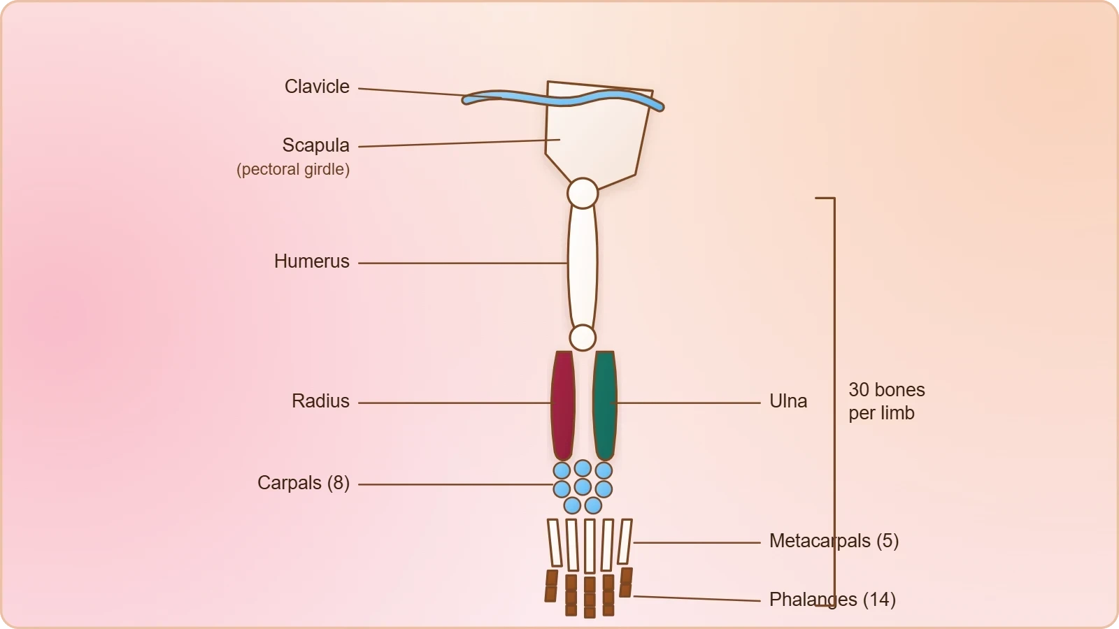
The dorsal, flat, triangular body of the scapula has a slightly elevated ridge called the spine, which projects as a flat, expanded process called the acromion. The clavicle articulates with this acromion. Below the acromion lies a depression called the glenoid cavity, which articulates with the head of the humerus to form the shoulder joint. Each clavicle is a long slender bone with two curvatures, commonly called the collar bone. The shoulder joint, therefore, is not a single articulation but a meeting of several bony landmarks, each with a name worth remembering.
The Hind Limb and the Pelvic Girdle
The bones of the legs, or hind limb, are the femur (the thigh bone, and the longest bone), the tibia and fibula, the tarsals (ankle bones, 7 in number), the metatarsals (5 in number) and the phalanges (digits, 14 in number). A cup-shaped bone called the patella covers the knee ventrally; it is the knee cap. The pelvic girdle consists of two coxal bones, and each coxal bone is formed by the fusion of three bones: the ilium, the ischium and the pubis. At the point of fusion of these bones is a cavity called the acetabulum, to which the thigh bone articulates.
The two halves of the pelvic girdle meet ventrally to form the pubic symphysis, which contains fibrous cartilage. This ventral meeting is a structural contrast with the pectoral girdle, whose halves remain separate. The pelvic girdle therefore forms a closed ring, while the pectoral girdle does not. That difference in architecture reflects a difference in function: the lower limb bears the weight of the body and requires a stable, load-bearing ring, whereas the upper limb requires mobility and reach.
Girdles as Bridges to the Axial Skeleton
The pectoral and pelvic girdle bones help in the articulation of the upper and the lower limbs respectively with the axial skeleton. This sentence is the conceptual hinge of the lesson. Without girdles, the limbs would be free appendages with nothing to push against; with them, the limbs become levers anchored to a stable frame. Each girdle is formed of two halves, and the two girdles differ markedly in how those halves relate to one another and to the rest of the body.
Landmarks of the Pectoral Girdle
The scapula is a large triangular flat bone situated in the dorsal part of the thorax between the second and the seventh ribs. Its dorsal, flat, triangular body carries a slightly elevated ridge called the spine, which projects as a flat, expanded process called the acromion. The clavicle articulates with the acromion. Below the acromion is a depression called the glenoid cavity, which articulates with the head of the humerus to form the shoulder joint. Each clavicle is a long slender bone with two curvatures, commonly called the collar bone.
These landmarks are worth learning as a sequence rather than a list. The spine of the scapula leads to the acromion; the acromion receives the clavicle; below the acromion lies the glenoid cavity; the glenoid cavity receives the head of the humerus. Following that chain from the flat body of the scapula to the shoulder joint gives a mental map of the upper limb's attachment. The two curvatures of the clavicle are equally practical: they allow the collar bone to act as a strut that holds the shoulder away from the thorax.
Landmarks of the Pelvic Girdle
The pelvic girdle consists of two coxal bones, and each coxal bone is formed by the fusion of three bones: the ilium, the ischium and the pubis. At the point of fusion of these bones is a cavity called the acetabulum, to which the thigh bone articulates. The two halves of the pelvic girdle meet ventrally to form the pubic symphysis, which contains fibrous cartilage. The acetabulum is thus a composite socket, fashioned at the meeting point of three fused bones, and it receives the femur.
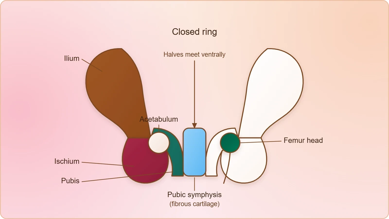
The contrast with the pectoral girdle is instructive. The pectoral girdle's halves are separate, and its socket, the glenoid cavity, is a shallow depression on a single bone. The pelvic girdle's halves meet ventrally at the pubic symphysis, and its socket, the acetabulum, is formed at a three-bone fusion. The pelvic ring is therefore closed and the shoulder arrangement open. This is a structural fact with functional consequences, and it is the kind of comparison that rewards careful reading rather than memorisation.
Joints: Definition and the Fibrous Form
Joints are essential for all types of movements involving the bony parts of the body, and locomotory movements are no exception. The material defines joints as points of contact between bones or between bone and cartilage, and classifies them into three structural forms: fibrous, cartilaginous and synovial. This threefold scheme is the organising principle of the remainder of the lesson. Each form is distinguished by the tissue that occupies the contact, and each is illustrated with examples drawn from the skeleton already described.
The Definition and Its Scope
The definition is deliberately broad. A joint is a point of contact between bones, or between bone and cartilage, and this wording admits contacts that are not freely movable as well as those that are. The classification that follows is structural rather than functional: it asks what kind of tissue lies at the contact, not how much movement the contact permits. Keeping that distinction clear prevents a common confusion, in which students assume that every joint must be a moving joint. The material's scheme allows for contacts that are almost immovable and for contacts that move freely.
Because joints are essential for all movements involving the bony parts of the body, the study of joints is not a separate topic from locomotion but its mechanical foundation. Muscles can only move the body if bones can move relative to one another, and bones can only move relative to one another at joints. The three structural forms therefore describe three different mechanical arrangements, each suited to a different demand, and the examples chosen for each form show how the skeleton distributes those demands across the body.
Fibrous Joints and Their Character
Fibrous joints are the first of the three structural forms named in the material. In this form the contact between the bones is occupied by fibrous tissue, and the arrangement permits little movement. The material places fibrous joints at the head of its classification, and the examples it supplies belong to the category of contacts where stability is the priority rather than mobility. The sutures of the skull, where flat bones of the cranium meet, are the classic illustration of this form, and they belong naturally with the axial skeleton already studied.
The logic of the fibrous form is straightforward once the tissue is understood. Fibrous tissue is tough and inextensible, so a contact filled with it holds the bones firmly together. That is precisely what the cranium requires, since the skull must protect the brain rather than articulate. The same principle appears elsewhere in the body wherever two bones must be held in a fixed relationship. Reading the classification in this way, as a correspondence between tissue and mechanical demand, makes the three forms easier to retain than a bare list of names.
Cartilaginous Joints and the Role of Cartilage
Cartilaginous joints form the second of the three structural forms. Here the contact between the bones is occupied by cartilage, and the material's own descriptions supply ready examples. The first seven pairs of ribs are attached dorsally to the thoracic vertebrae and ventrally connected to the sternum with the help of hyaline cartilage. The 8th, 9th and 10th pairs of ribs do not articulate directly with the sternum but join the seventh rib with the help of hyaline cartilage. These are the vertebrochondral, or false, ribs.
Hyaline Cartilage at the Rib Attachments
The rib attachments give the clearest illustration of cartilaginous contact in the material. The first seven pairs, the true ribs, are connected to the sternum by hyaline cartilage. The 8th, 9th and 10th pairs, the false ribs, reach the sternum only indirectly, joining the seventh rib by the same tissue. The last two pairs, the 11th and 12th, are not connected ventrally at all and are therefore called floating ribs. The rib cage as a whole is formed by the thoracic vertebrae, the ribs and the sternum together.
What the rib series shows is that cartilage can serve as a flexible bridge between bones that must move slightly relative to one another during breathing. The tissue is firm enough to hold the parts in place and pliable enough to permit a small amount of give. The distinction between true, false and floating ribs is thus not merely a labelling convention; it records three different degrees of ventral connection, and the tissue involved in the first two is hyaline cartilage. The floating ribs, lacking ventral connection, complete the series.
Fibrous Cartilage at the Pubic Symphysis
The pubic symphysis offers a second and different cartilaginous example. The two halves of the pelvic girdle meet ventrally to form the pubic symphysis, and the material specifies that this structure contains fibrous cartilage. This is a useful point of precision, because it shows that the cartilaginous category is not exhausted by hyaline cartilage. The symphysis is a midline meeting of two bones, and the fibrous cartilage within it holds the two halves of the pelvic girdle together while allowing the limited resilience that such a midline joint requires.
Placing the two examples side by side clarifies the category. At the ribs, hyaline cartilage connects rib to sternum and rib to rib; at the pelvis, fibrous cartilage joins the two coxal halves at the midline. Both are cartilaginous joints in the material's scheme, and both illustrate the same underlying principle: cartilage at a contact permits a measured amount of movement while maintaining continuity between the bones. The category is defined by the tissue, and the tissue varies within it.
Synovial Joints and the Moving Skeleton
Synovial joints are the third structural form, and they are the joints that make free movement possible. The material names them alongside the fibrous and cartilaginous forms, and the skeletal descriptions already given supply the principal examples. The glenoid cavity of the scapula articulates with the head of the humerus to form the shoulder joint. The acetabulum of the coxal bone receives the thigh bone. The patella covers the knee ventrally. These are the places where the appendicular skeleton's mobility is concentrated.
The Shoulder and Hip as Synovial Examples
The shoulder joint is formed where the glenoid cavity, a depression below the acromion of the scapula, articulates with the head of the humerus. The hip joint is formed where the acetabulum, the cavity at the fusion of the ilium, ischium and pubis, receives the thigh bone. Both are synovial joints in the material's classification, and both are the points at which a limb meets its girdle. The shoulder and the hip are therefore the two great junctions of the appendicular skeleton, and their structure repays close attention.
The two junctions differ in the depth of their sockets. The glenoid cavity is a shallow depression on the scapula, while the acetabulum is a cavity formed at the fusion of three bones. That difference in depth corresponds to a difference in the range and stability of the joint, and it echoes the contrast between the open pectoral girdle and the closed pelvic ring. The shoulder and the hip are thus not interchangeable examples; they are two synovial solutions to the same problem of attaching a limb to the axial frame.
The Knee and the Patella
The knee is the third great junction of the appendicular skeleton, and the material describes it through the patella. A cup-shaped bone called the patella covers the knee ventrally; it is the knee cap. The femur, the longest bone, meets the tibia and fibula at this region, and the patella sits over the front of the joint. The knee is therefore a synovial joint in the material's scheme, and the patella is the bony landmark that identifies it in the description of the hind limb.
The patella's position is worth noting precisely. It covers the knee ventrally, which is to say on the front of the joint, and it is described as cup-shaped. In the sequence of hind limb bones, it appears after the femur, tibia and fibula and before the tarsals, metatarsals and phalanges. That placement in the text mirrors its placement in the limb: it belongs to the knee region, between the thigh and the lower leg, and it protects the front of the joint where the bones are closest to the surface.
Reading the Skeleton as a Mechanical System
The appendicular skeleton and its joints can be read as a single mechanical system rather than as two separate topics. The limbs supply the levers, the girdles supply the anchor points, and the joints supply the pivots. Each of the three structural forms of joint corresponds to a different kind of pivot, and each is placed in the body where its particular properties are needed. Fibrous contacts hold the cranium firm; cartilaginous contacts give the ribs and the pubic symphysis their measured resilience; synovial joints give the shoulder, hip and knee their freedom.
From Bone Counts to Function
The numbers in the material are not arbitrary. Each limb is made of 30 bones, and the distribution of those bones along the limb tells us where flexibility is concentrated. The fore limb has 8 carpals, 5 metacarpals and 14 phalanges; the hind limb has 7 tarsals, 5 metatarsals and 14 phalanges. The many small bones of the wrist and hand, and of the ankle and foot, allow the fine adjustments that gripping and balancing require, while the long bones of the upper arm and thigh provide the leverage for larger movements.
The femur is singled out as the longest bone, and that fact is consistent with its role. A long bone gives a long lever arm, and the thigh is the segment that drives the stride. The patella, covering the knee ventrally, sits at the junction between the thigh and the lower leg, where the forces of the stride are transmitted. Reading the bone counts and the named landmarks together, the skeleton emerges as an engineered structure in which length, number and joint form are matched to the demands of movement.
Preparing for the Disorders Ahead
The structures described in this lesson are the ones most often affected when the muscular and skeletal systems go wrong. The joints, in particular, are the sites where the three structural forms become clinically relevant, because each form has its own vulnerabilities. The girdles and the long bones are the load-bearing elements, and the patella and the knee are the junctions where movement and weight meet. A clear grasp of the normal anatomy is therefore the necessary preparation for understanding the disorders that follow.
The lesson has kept to the material's order of ideas: the appendicular skeleton first, with its limb bones and girdles, then the definition of joints, then the three structural forms with their examples. That order is itself a teaching device, because it moves from the parts that move to the points at which they move. Carrying that sequence forward, the next lesson turns to what happens when these structures are disordered, and the vocabulary established here, from the glenoid cavity to the pubic symphysis, becomes the language in which those disorders are described.
Going further
A fulcrum is the pivot point about which a lever turns, and this idea helps explain how joints work with muscles. A bone acts as a lever, a muscle supplies the force, and the joint provides the fulcrum. The arrangement of the shoulder, hip and knee as synovial joints makes sense in these terms, because a freely moving pivot is what a lever requires. The knee cap, or patella, is a cup-shaped bone that covers the knee ventrally, and its position at the front of the joint places it where the lever of the lower leg turns.
Also Read
- No links available.
From our network :
- Analyzing Trump Deportation Numbers Insights Into The 2026 Immigration Crackdown
- Mastering DB2 12.1 Instance Design: A Technical Deep Dive into Modern Database ArchitectureComprehensive guide to DB2 12.1 Instance Design. Explore architectural components, HADR setup, buffer pool tuning, and security policies for IBM DB2 12.1. Includes 10 functional SQL and Shell samples.
- Mastering DB2 LUW v12 Tables: A Comprehensive Technical GuideComprehensive guide on DB2 LUW v12 tables. Explore CREATE TABLE syntax, BLU Acceleration, range partitioning, data types, and security features for IBM DB2 LUW v12.
- 98% of Global MBA Programs Now Prefer GRE Over GMAT Focus EditionExplore why 98% of global MBA programs now prefer the GRE over GMAT Focus. Learn about the shorter GRE format, verbal logic importance, and strategic versatility for students.
- AI-Powered 'Precision Diagnostic' Replaces Standard GRE Score ReportsDiscover the new GRE Precision Diagnostic. Learn how AI-powered behavioral analytics and time-to-solve tracking are replacing standard GRE score reports to empower students.
- 10 Physics Numerical Problems with Solutions for IIT JEEPrepare for IIT JEE with 10 physics numerical problems, complete with detailed solutions and conceptual explanations. Ideal for mastering kinematics, thermodynamics, electromagnetism, and optics.
- EV 2.0: The Solid-State Battery Breakthrough and Global Factory ExpansionDiscover the impact of the Solid-State Battery Breakthrough on the EV market. Learn about CATL and BYD stock growth, global factory expansion in Europe, and the future of EV 2.0 technology.
- Trump Political Strategy How Geopolitical Stunts Serve As Media Diversions
- Vite 6/7 'Cold Start' Regression in Massive Module GraphsIn-depth analysis of Vite 6/7 cold start regressions in massive module graphs. Learn about barrel file issues, pre-bundling optimizations, and how to improve Vite dev server performance.
RESOURCES
- Anatomy, Appendicular Skeleton - StatPearls - NCBI Bookshelfncbi.nlm.nih.govApr 21, 2024 ... The appendicular skeleton comprises the upper and lower extremities. The shoulder and hip are important limb segments connecting the ...
- Lab 5: Skeletal System II – Appendicular Skeleton and Jointsiastate.pressbooks.pubMost diarthrotic joints are found in the appendicular skeleton and thus give the limbs a wide range of motion. These joints are divided into…
- Joints in the appendicular skeleton: Developmental mechanisms ...pmc.ncbi.nlm.nih.govThese joints are composed of multiple tissues and structures. They all share a fibrous capsule that is continuous with tissues attached to the flanking…
- Joints in the appendicular skeleton: Developmental mechanisms ...pubmed.ncbi.nlm.nih.govHere we review past and recent studies on important aspects of joint development, including the roles of the interzone and morphogenesis of articular cartilage.
- Chapt. 6: Bones and Joints of the Appendicular Skeleton - Quizletquizlet.comStudy with Quizlet and memorize flashcards containing terms like appendicular skeleton, 64 upper extremity bones, pectoral (shoulder) girdle and more.
- Appendicular Arthritis - UW Radiology - University of Washingtonrad.uw.eduOsteoarthritis (a.k.a. degenerative joint disease) is the most commonly seen form of appendicular arthritis. The other two commonly seen arthropathies are ...
- Appendicular Skeleton | Learn Skeleton Anatomy - Visible Bodyvisiblebody.comThe bones of the appendicular skeleton make up the rest of the skeleton, and are so called because they are appendages of the axial…
- Appendicular Skeleton - an overview | ScienceDirect Topicssciencedirect.comThe appendicular skeleton includes the cartilages of the paired fins and the limb girdles. In general, skeletal elements are smaller and more flexible the…
- Bone Appendicular Skeleton, Trunk, Skull, and Facial Bonesstaging.seer.cancer.govC403 Short bones of lower limb and associated joints. C408 Overlapping lesion of bones, joints and articular cartilage of limbs. C409 Bone of limb,…
- Osteosarcoma of the appendicular skeleton in dogs - Frontiersfrontiersin.orgSep 22, 2025 ... OSA typically arises in the metaphyseal region of the bone. It does not cross the joint (except occasionally when infiltrating…
- Musculoskeletal system: Main bones, joints & muscles - Kenhubkenhub.comMain bones, Axial skeleton: bones of the skull, ribs, vertebral column, sternum, sacrum, coccyx, hyoid bone and auditory ossicles. Appendicular skeleton: bones ...
- Appendicular Skeleton (126 bones) - SEER Training Modulestraining.seer.cancer.govAppendicular Skeleton (126 bones) · Femur (2) · Tibia (2) · Fibula (2) · Patella (2) · Tarsals (14) · Metatarsals (10) · Phalanges…
- Skeletal System: What It Is, Function, Care & Anatomymy.clevelandclinic.orgJan 10, 2024 ... Giving your body shape: Your skeleton supports the weight of your body. · Helping you move: Your joints, connective tissue…
- 8: Appendicular Skeleton, Joints (Articulations) and Movementsbio.libretexts.orgSep 1, 2026 ... This lab explores the bones and joints of the appendicular skeleton, emphasizing their structure, movement, and role in everyday activities.
- Anatomy & Physiology Study Guide: Bones, Joints, Lab Prep | Notespearson.comComprehensive Anatomy & Physiology study guide covering appendicular skeleton, joints, bone structures, and lab assignments for effective exam prep.