Muscle tissue occupies a distinctive place in the biology of movement because it converts chemical energy into mechanical work on demand, and it does so in nearly every region of the human body. Roughly 40 to 50 per cent of the body weight of a human adult is contributed by muscles, a figure that immediately signals how central this tissue is to posture, locomotion and internal transport. Muscle is a specialised tissue of mesodermal origin, meaning it arises from the middle embryonic germ layer, and it carries four defining properties that separate it from other excitable tissues: excitability, contractility, extensibility and elasticity. Understanding these properties is the foundation for everything that follows about how limbs move, how food travels through the gut and how the heart sustains circulation.
This lesson builds directly on the earlier discussion of movement in human cells, where amoeboid movement driven by pseudopodia and ciliary movement along ciliated epithelium were examined. Those forms of movement rely on protoplasmic streaming and cytoskeletal elements such as microfilaments, and they operate at the level of single cells or thin epithelial sheets. Muscular movement is a different order of organisation altogether: it depends on the contractile property of muscle, which is effectively used for locomotion and other movements by human beings and the majority of multicellular organisms. Locomotion itself requires a perfect coordinated activity of muscular, skeletal and neural systems, so muscle cannot be studied in isolation from the framework it pulls against and the nerves that command it.
The material that follows classifies muscles by location into skeletal, visceral and cardiac types, and then describes their appearance and their voluntary or involuntary nature. Each classification criterion, whether location, appearance or nature of regulation, illuminates a different facet of the same tissue, and the three criteria frequently reinforce one another. Skeletal muscles look striped and obey conscious instruction; visceral muscles look smooth and work without it. By the end of this lesson, you should be able to place any named muscle into the correct category and justify that placement using the properties and criteria set out here, which prepares the ground for the detailed study of the skeletal muscle fibre and sarcomere in the next lesson.
On This Page
Muscle as a Specialised Tissue of Mesodermal Origin
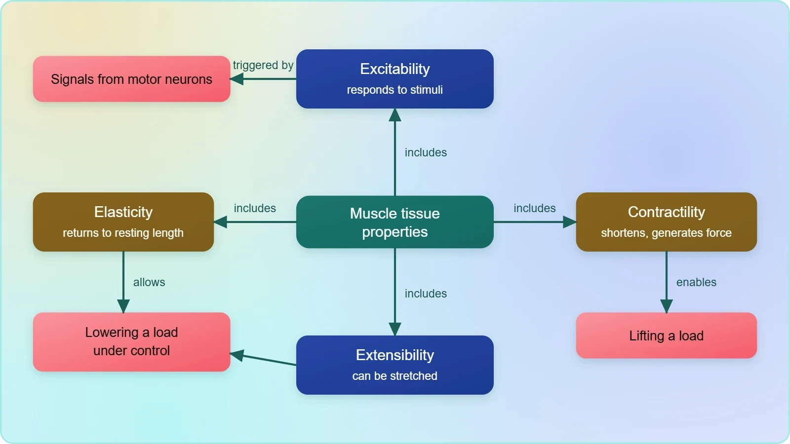
Muscle is not a generic filling material but a highly organised tissue with a defined embryonic pedigree and a measurable share of body mass. Its mesodermal origin links it developmentally to connective tissue, bone and blood, which explains why muscles and skeletal components are so closely integrated in the body. The tissue's four properties, excitability, contractility, extensibility and elasticity, are not decorative vocabulary; each describes a measurable behaviour that determines how the tissue responds to a stimulus, shortens under load, stretches without tearing and returns to its resting length. Together these properties make muscle the only tissue capable of generating the directed force required for locomotion.
Defining Properties and Their Practical Meaning
Excitability refers to the capacity of muscle tissue to receive and respond to stimuli, typically electrical signals delivered by motor neurons. Contractility is the property that allows the tissue to shorten and generate force, and it is the feature most directly responsible for movement. Extensibility permits a muscle to be stretched beyond its resting length, while elasticity ensures that it recoils to its original length once the stretching force is removed. These four properties operate in concert: a muscle that could contract but not extend would be useless for the alternating movements of walking, and one that could stretch but not recoil would offer no resistance at all.
The practical consequences of these properties appear throughout the body. When you lift a load, contractility does the work; when you lower it under control, extensibility and elasticity regulate the descent. The same quartet of properties allows the diaphragm to expand and recoil with every breath and permits the bladder wall to accommodate volume and then expel it. Because these properties are intrinsic to the tissue rather than to any particular organ, the same fundamental mechanism underlies voluntary limb movement and involuntary internal transport, even though the two are regulated in entirely different ways.
Proportion of Body Weight and Tissue Significance
The statement that about 40 to 50 per cent of the body weight of a human adult is contributed by muscles deserves emphasis because it reframes muscle as a major organ system rather than an accessory to the skeleton. This proportion reflects the sheer metabolic cost of maintaining contractile tissue, which consumes energy even at rest and substantially more during activity. It also explains why muscle mass correlates so strongly with overall metabolic rate and why prolonged immobility leads to measurable loss of both mass and function. The figure is a useful benchmark when reasoning about nutrition, exercise physiology and recovery from injury.
Because muscle constitutes such a large fraction of body mass, its classification matters for clinical and physiological reasoning alike. A disorder affecting skeletal muscle alone has a different functional signature from one affecting visceral muscle, and the two demand different investigative approaches. Recognising that the tissue is mesodermal in origin also helps explain why certain congenital conditions affect muscle alongside connective and skeletal structures. The body-weight proportion is therefore not a trivia point but a quantitative anchor for understanding why muscle health is inseparable from general health.
Classification of Muscles by Location
Muscles have been classified using different criteria, namely location, appearance and nature of regulation of their activities. Based on their location, three types of muscles are identified: skeletal, visceral and cardiac. Location is the most intuitive starting point because it immediately suggests what each muscle type is likely to do and how it is likely to be controlled. A muscle attached to bone serves posture and locomotion; a muscle embedded in the wall of a hollow organ serves internal transport; a muscle confined to the heart serves circulation. The location criterion therefore organises the entire topic into three coherent groups before any microscopic detail is introduced.
Skeletal Muscles: Location and Association with Bone
Skeletal muscles are closely associated with the skeletal components of the body. This close association is structural as well as functional: the muscles attach to bones and, when they contract, pull on those bones to produce movement at joints. Because they are primarily involved in locomotory actions and changes of body postures, skeletal muscles are the type most familiar from everyday experience of walking, running, lifting and gesturing. Their placement on the skeleton also explains why damage to this group produces immediately visible effects on movement, whereas damage to visceral muscle may produce subtler internal symptoms that are harder to localise.
The association with skeletal components carries a further implication that prepares the ground for later lessons. Skeletal muscle does not act alone; it requires the coordinated activity of the skeletal and neural systems to produce useful locomotion. A muscle that contracts without an intact bony lever produces no movement, and a muscle that receives no neural command remains silent. This is why the study of skeletal muscle naturally leads into the structure of the muscle fibre and sarcomere, and eventually into the mechanics of joints and the skeleton itself. Location, in other words, is the entry point to a whole system of interdependent parts.
Visceral Muscles: Location in Hollow Organs
Visceral muscles are located in the inner walls of hollow visceral organs of the body like the alimentary canal and the reproductive tract. Their position in the walls of hollow organs dictates their function: they assist, for example, in the transportation of food through the digestive tract and gametes through the genital tract. Because these organs must move their contents progressively rather than in a single powerful pull, visceral muscle is adapted for sustained, rhythmic activity rather than for rapid voluntary force. The location criterion thus predicts not only where the tissue is found but also the kind of work it performs.
The hollow-organ setting also explains why visceral muscle is described as involuntary. The contents of the alimentary canal must be propelled continuously, including during sleep and during periods of illness, and conscious attention to every wave of contraction would be neither possible nor desirable. Locating this muscle in organ walls rather than on the skeleton removes it from the voluntary motor pathways that govern limb movement. The distinction is functional as much as anatomical, and it is reinforced by the appearance of the tissue, which lacks the striations seen in skeletal muscle.
Appearance Under the Microscope: Striated and Smooth
Appearance is the second classification criterion, and it divides the muscle types with striking clarity. Skeletal muscles have a striped appearance under the microscope and hence are called striated muscles. Visceral muscles, by contrast, do not exhibit any striation and are smooth in appearance, which is why they are called smooth muscles or nonstriated muscle. The two names, striated and nonstriated, are simply descriptions of what the observer sees through the eyepiece, yet they correlate strongly with function and regulation. A striped pattern signals a highly ordered internal arrangement of contractile proteins, while a smooth appearance signals a less regimented arrangement suited to sustained contraction.
Striated Muscle and the Striped Pattern
The striped appearance of skeletal muscle is a direct visual consequence of its internal architecture, in which contractile units are aligned in regular repeating series along the length of the fibre. This regularity produces alternating light and dark bands when the tissue is viewed under a microscope, and the pattern is what gives the tissue its name. Because the pattern is so consistent, striation serves as a reliable diagnostic feature: a muscle showing clear striations can be assigned to the skeletal category with confidence. The next lesson examines the sarcomere, the repeating unit responsible for this banding, in detail.
It is worth noting that striation is described here as an appearance rather than a function, which keeps the classification criterion clean and observational. The striped pattern does not by itself explain why skeletal muscle is voluntary; it merely accompanies the structural organisation that makes rapid, powerful, precisely timed contraction possible. Students should resist the temptation to treat striation as a cause of voluntary control. The two features are correlated because both arise from the same underlying design, but the material presents them as separate criteria, and keeping them separate prevents confusion when the three classification schemes are compared side by side.
Smooth Muscle and the Absence of Striation
Visceral muscle does not exhibit any striation and is smooth in appearance, a characteristic that reflects a different internal arrangement of contractile elements. This smoothness is not a deficiency but an adaptation: the tissue is built for slow, sustained and rhythmic contraction rather than for the rapid, forceful pulls required of limb muscles. The absence of visible bands makes smooth muscle easy to distinguish from skeletal muscle in prepared slides, and the name nonstriated muscle records precisely this observation. The terminology is descriptive and consistent, allowing the three criteria of location, appearance and regulation to be applied independently to the same tissue.
The smooth appearance also helps explain why visceral muscle can maintain contraction over long periods without fatigue in the way skeletal muscle would. Hollow organs such as the alimentary canal must propel their contents steadily for hours, and a tissue organised for sustained rather than explosive activity suits this task. Recognising the smooth appearance therefore gives the learner a quick visual test that reinforces the functional classification. When you examine a slide and see no bands, you can reasonably predict an involuntary, hollow-organ location, and when you see clear bands, you can predict skeletal association and voluntary control.
Voluntary and Involuntary Regulation of Muscle Activity
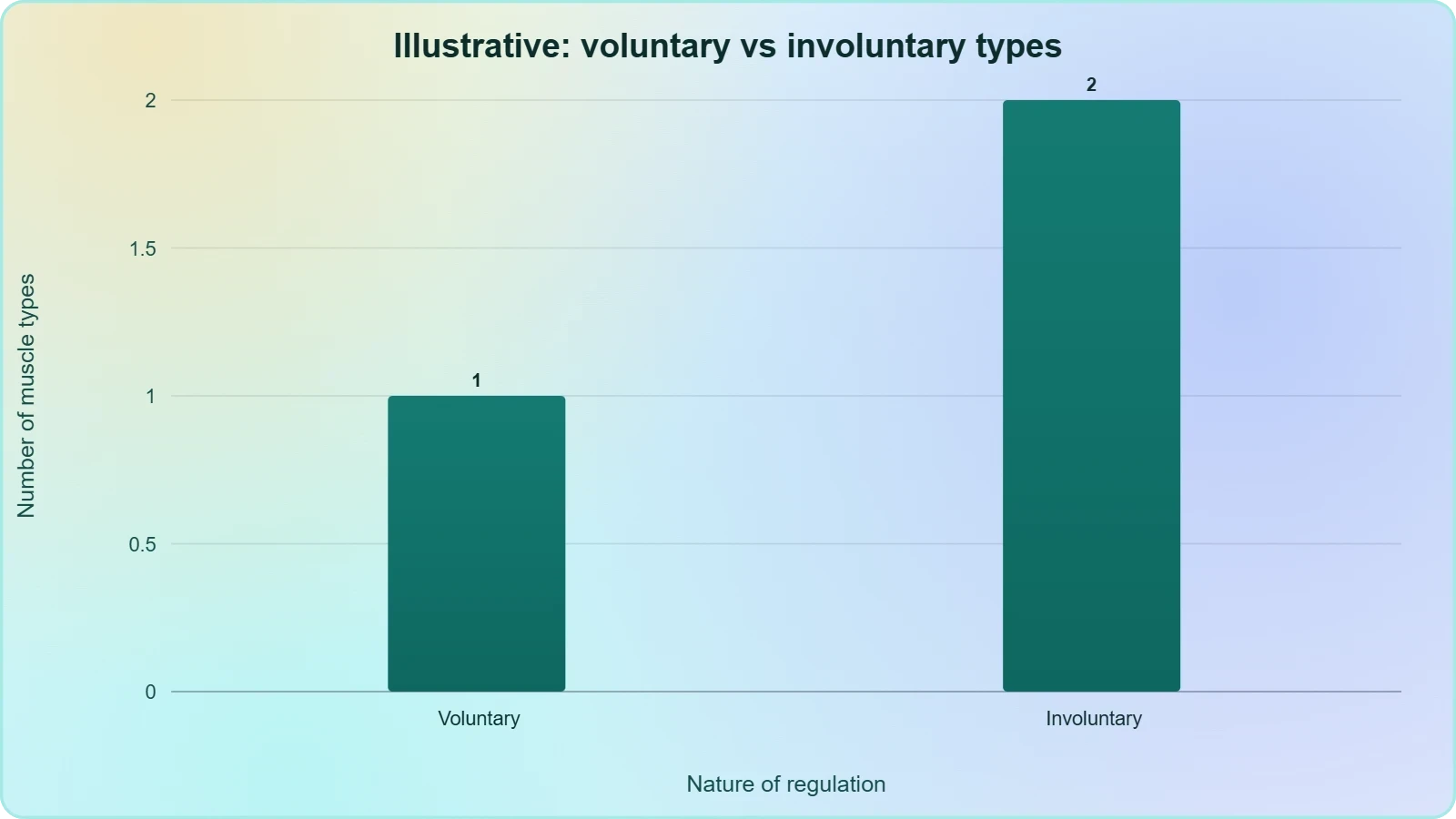
The third classification criterion is the nature of regulation of muscle activities, and it produces the familiar pairing of voluntary and involuntary muscles. Skeletal muscles are under the voluntary control of the nervous system and are therefore known as voluntary muscles. Visceral muscles are not under voluntary control and are therefore known as involuntary muscles. This criterion is arguably the most consequential for everyday life, because it determines whether a movement can be initiated at will or proceeds automatically. The distinction also explains why the two muscle types respond differently to injury, fatigue and disease, and why clinicians ask different questions when each is affected.
Voluntary Control in Skeletal Muscle
Voluntary control means that the nervous system can initiate, modulate and terminate contraction according to conscious intention. When you decide to raise your hand, the command travels through motor pathways to skeletal muscle, which responds because it is wired into the voluntary system. This arrangement gives human beings precise, rapid and adaptable control over posture and locomotion, allowing movements to be learned, refined and corrected in real time. The cost of this arrangement is that skeletal muscle depends entirely on intact neural command; when the pathway is interrupted, the muscle may remain structurally sound yet unable to produce useful movement.
The voluntary nature of skeletal muscle also explains why its activities are described as primarily involved in locomotory actions and changes of body postures. These are tasks that benefit from conscious direction and can be adjusted moment to moment as terrain, load and intention change. The same muscle can produce a delicate gesture or a powerful leap depending on the command it receives, a flexibility that involuntary muscle does not require. Recognising this helps students appreciate why the skeletal and neural systems must act in perfect coordination for locomotion, as the material emphasises.
Involuntary Control in Visceral Muscle
Involuntary control means that contraction proceeds without conscious direction, regulated by the nervous system at a level below awareness. Visceral muscle operates in this mode, which is precisely what the body requires of the alimentary canal and reproductive tract: contents must be propelled continuously, including during sleep, and conscious supervision of every contraction would be impossible. The absence of voluntary control does not mean absence of regulation; it means regulation is delegated to automatic mechanisms. This delegation frees attention for voluntary tasks while ensuring that digestion and gamete transport proceed reliably in the background.
The involuntary label also carries practical implications for how we think about health and disease. Because we cannot consciously command visceral muscle, disorders of this tissue cannot be corrected by simply deciding to contract or relax; they require medical investigation and treatment. Conversely, the automatic nature of visceral activity means that ordinary digestion proceeds without our intervention, a fact easy to overlook precisely because it is so reliable. Understanding the voluntary-involuntary divide therefore connects the microscopic classification of muscle to the lived experience of moving, eating and resting.
Muscle in the Wider Context of Locomotion
Muscle does not produce locomotion on its own, and the material is explicit that locomotion requires a perfect coordinated activity of muscular, skeletal and neural systems. The muscular system supplies the force, the skeletal system supplies the levers and the neural system supplies the commands and timing. Remove any one of the three and movement fails, even if the remaining two are perfectly healthy. This triad is the conceptual bridge between the present lesson and the lessons that follow on the skeletal muscle fibre, the sarcomere and the skeletal system. It also explains why the study of muscle is inseparable from the study of joints and bones.
From Cellular Movement to Muscular Movement
The previous lesson examined movement at the cellular scale, where macrophages and leucocytes in blood exhibit amoeboid movement effected by pseudopodia formed by the streaming of protoplasm, and where ciliary movement in the trachea removes dust particles and foreign substances inhaled with atmospheric air. Those mechanisms rely on cytoskeletal elements such as microfilaments and on the coordinated beating of cilia. Muscular movement operates at a different scale and uses a different mechanism, the contractile property of muscle, yet it serves the same fundamental purpose of displacing material, whether that material is a cell, a dust particle or the entire body.
Recognising this continuity prevents the topic from fragmenting into unrelated facts. Ciliary movement facilitates the passage of ova through the female reproductive tract, while visceral muscle assists the transport of gametes through the genital tract; both serve reproduction, but through different mechanisms. Amoeboid movement moves individual cells, while skeletal muscle moves the whole organism. The progression from cell-level to tissue-level movement is therefore a progression in scale and coordination rather than a change in basic biological purpose, and this perspective makes the classification of muscle feel like a natural extension of what came before.
Preparing for Fibre Structure and Contraction
The present lesson has established what muscle is, what properties it possesses and how it is classified. The next step is to examine how a skeletal muscle fibre is organised internally and how the sarcomere, the repeating contractile unit, produces shortening. That structural detail will make sense only if the properties of excitability and contractility are already firmly understood, because the sarcomere is the physical machinery through which those properties are realised. The classification into striated and nonstriated also prepares the learner for the observation that striations arise from the regular alignment of sarcomeres along the fibre.
Students should therefore leave this lesson with three anchors: the four properties, the three location-based types and the voluntary-involuntary distinction. These anchors will be revisited repeatedly as the course moves through muscle contraction, the axial and appendicular skeleton, the three structural forms of joints and the disorders of the muscular and skeletal systems. Mastery of the vocabulary now will pay dividends later, because the terminology introduced here, including striated, nonstriated, voluntary and involuntary, recurs throughout the remaining lessons of the course.
Going Further: Background That Aids Understanding
The word striated refers to the striped appearance of certain muscle fibres under a microscope, and the term is purely descriptive of what the observer sees rather than of any functional property. When a prepared slide of skeletal muscle is examined, alternating light and dark bands run across the fibre at regular intervals, producing the striped effect that gives the tissue its name. This pattern arises because the contractile units within the fibre are aligned in series, so their banding coincides across the width of the fibre. Recognising the origin of the term helps students remember that striated and nonstriated are observational labels, not explanations.
Why Involuntary Muscle Works Without Conscious Control
Involuntary muscles work without conscious control, which is why we do not have to think about digestion. The alimentary canal must propel its contents continuously throughout the day and night, and delegating this task to automatic regulation ensures that it proceeds reliably regardless of what occupies our attention. If digestion depended on conscious command, every meal would require sustained mental effort, and the process would falter whenever attention wandered. The involuntary arrangement is therefore an efficient division of labour between the voluntary system, which handles deliberate action, and the automatic system, which handles the continuous background tasks of internal transport.
This background note is offered separately from the core material and does not alter any of its definitions or classifications. It simply supplies the intuitive reason behind a distinction that the material states as fact. The same logic applies to the heart, whose cardiac muscle is the third type identified by location and which must beat without pause from before birth until death. Appreciating why involuntary control exists makes the voluntary-involuntary criterion feel less like an arbitrary label and more like a sensible solution to the problem of keeping a complex organism running while its owner attends to the outside world.
Also Read
- No links available.
From our network :
- Vite 6/7 'Cold Start' Regression in Massive Module GraphsIn-depth analysis of Vite 6/7 cold start regressions in massive module graphs. Learn about barrel file issues, pre-bundling optimizations, and how to improve Vite dev server performance.
- Mastering DB2 LUW v12 Tables: A Comprehensive Technical GuideComprehensive guide on DB2 LUW v12 tables. Explore CREATE TABLE syntax, BLU Acceleration, range partitioning, data types, and security features for IBM DB2 LUW v12.
- Trump Political Strategy How Geopolitical Stunts Serve As Media Diversions
- AI-Powered 'Precision Diagnostic' Replaces Standard GRE Score ReportsDiscover the new GRE Precision Diagnostic. Learn how AI-powered behavioral analytics and time-to-solve tracking are replacing standard GRE score reports to empower students.
- EV 2.0: The Solid-State Battery Breakthrough and Global Factory ExpansionDiscover the impact of the Solid-State Battery Breakthrough on the EV market. Learn about CATL and BYD stock growth, global factory expansion in Europe, and the future of EV 2.0 technology.
- Mastering DB2 12.1 Instance Design: A Technical Deep Dive into Modern Database ArchitectureComprehensive guide to DB2 12.1 Instance Design. Explore architectural components, HADR setup, buffer pool tuning, and security policies for IBM DB2 12.1. Includes 10 functional SQL and Shell samples.
- 98% of Global MBA Programs Now Prefer GRE Over GMAT Focus EditionExplore why 98% of global MBA programs now prefer the GRE over GMAT Focus. Learn about the shorter GRE format, verbal logic importance, and strategic versatility for students.
- Analyzing Trump Deportation Numbers Insights Into The 2026 Immigration Crackdown
- 10 Physics Numerical Problems with Solutions for IIT JEEPrepare for IIT JEE with 10 physics numerical problems, complete with detailed solutions and conceptual explanations. Ideal for mastering kinematics, thermodynamics, electromagnetism, and optics.
RESOURCES
- Comparison of Muscle Development in Drosophila and Vertebratesncbi.nlm.nih.gov... types defined by their structural and functional properties: skeletal, cardiac and smooth. ... As in Drosophila, vertebrate skeletal, cardiac and visceral muscle ...
- Transcriptional control of muscle development by myocyte ... - PubMedpubmed.ncbi.nlm.nih.govMetazoans contain multiple types of muscle cells that share several common properties ... skeletal, cardiac, and visceral muscle lineages. Paradoxically, MEF2 ...
- Molecular Properties of Voltage-Gated Calcium Channels - NCBI - NIHncbi.nlm.nih.govThe L-type channel is the primary route for Ca2+ entry into cardiac, skeletal, and smooth muscles. The skeletal muscle L-type channel acts as a…
- Muscle Contraction - PMC - NIHpmc.ncbi.nlm.nih.govThere are three types of muscle: skeletal, cardiac, and smooth. Myocytes are ... Cardiac and skeletal muscle cells also differ in their electrical properties.
- Muscles and muscle tissue: Types and functions | Kenhubkenhub.comskeletal muscle; visceral striated muscle; cardiac muscle. Review the structure of skeletal and smooth/cardiac muscle in these study units:.
- Adipose Tissue Distribution, Inflammation and Its Metabolic ... - PMCpmc.ncbi.nlm.nih.govFeb 25, 2020 ... ... skeletal and heart muscle, pancreas, and brain (64). ... Effects of sex on the change in visceral, subcutaneous adipose…
- Types of muscle tissue: MedlinePlus Medical Encyclopedia Imagemedlineplus.govJan 28, 2025 ... The 3 types of muscle tissue are cardiac, smooth, and skeletal ... Smooth muscle fibers are located in walls of…
- Muscle Types - SEER Training Modulestraining.seer.cancer.govIn the body, there are three types of muscle: skeletal (striated), smooth, and cardiac. Skeletal Muscle. Skeletal muscle, attached to bones, is responsible for ...
- Smooth Muscle Function & Anatomy - Cleveland Clinicmy.clevelandclinic.orgJan 30, 2025 ... Your body has three types of muscle: skeletal, cardiac and smooth. ... Visceral myopathies. These are a group of smooth…
- Associations of thoracic muscle area factor and visceral adiposity ...sciencedirect.comJul 16, 2026 ... ... skeletal muscle mass (TASM) factor and visceral fat with cardiac function. ... Cai et al. Effects of different types…
- Skeletal Muscles – Structure, Function And Types - BYJU'Sbyjus.comSkeletal Muscle Definition. Skeletal muscle is one of the three types of muscles in the human body- the others being visceral and cardiac muscles.…
- Preoperative visceral fat and muscle loss after cardiovascular ...aspenjournals.onlinelibrary.wiley.comDec 17, 2025 ... ... skeletal muscle mass following cardiac surgery compared with normal visceral fat. ... The inflammatory properties of visceral adipose ...
- BIOL 237 Class Notes - Muscle Structureunm.eduThere are three types of muscle tissue, all of which share some common properties: ... The three types of muscle: skeletal, cardiac, and visceral…
- A pig BodyMap transcriptome reveals diverse tissue physiologies ...nature.comJun 17, 2021 ... ... cardiac and head muscle ... Human masseter muscle fiber type properties, skeletal malocclusions, and muscle growth factor expression.