The male reproductive system is a study in deliberate anatomical compromise. Every structure within it — the paired testes suspended in the scrotum, the coiled duct system that carries sperm toward the exterior, the accessory glands that manufacture seminal plasma, and the erectile penis — exists to serve one biological imperative: the production, maturation, storage, and delivery of viable spermatozoa. What makes this system genuinely fascinating is that it solves a thermodynamic problem. Spermatogenesis, the process by which diploid germ cells become streamlined haploid sperm, cannot proceed efficiently at core body temperature. The architecture of the male reproductive tract is therefore organised around a single organising principle: keeping the testes roughly 2 to 2.5 °C cooler than the rest of the body.
This lesson examines that architecture in detail, moving from the external genitalia inward through the scrotum, the testes and their lobular organisation, the accessory ducts, and finally the glands whose secretions constitute semen. It builds directly on the introductory survey of human reproductive events — gametogenesis, insemination, fertilisation, implantation, gestation, and parturition — and prepares the ground for a closer histological examination of the seminiferous tubules and interstitial tissue in the next lesson. Along the way, we will consider the countercurrent heat-exchange mechanism of the pampiniform plexus, the clinical consequences of cryptorchidism, and the erectile tissue that underlies penile erection.
Because this material sits at the intersection of pure anatomy and clinical medicine, it rewards careful attention to both structure and function. A learner who can describe the path of a spermatozoon from seminiferous tubule to urethral meatus, and explain why each segment of that path exists, has understood the male reproductive system far more deeply than one who has merely memorised a list of organs. The sections that follow combine the core descriptive content with worked reasoning, comparative tables, and the kind of applied detail that distinguishes genuine comprehension from surface recall.
On This Page
- The Scrotum and the Temperature Requirement for Spermatogenesis
- Gross Anatomy of the Testis and Its Lobular Organisation
- The Accessory Duct System: From Rete Testis to Urethra
- Accessory Glands and the Composition of Seminal Plasma
- The Penis, Erectile Tissue, and External Genitalia
- Worked Reasoning and Exam-Style Practice
- Consolidating the Anatomy Before the Histological Turn
The Scrotum and the Temperature Requirement for Spermatogenesis
The testes are situated outside the abdominal cavity within a pouch called the scrotum, and this external position is not incidental but essential. The scrotum maintains the testes at a temperature approximately 2 to 2.5 °C lower than normal internal body temperature, and this thermal differential is a prerequisite for efficient spermatogenesis. The reasoning is enzymatic: the meiotic divisions and chromatin remodelling that transform spermatogonia into spermatozoa depend on a cascade of temperature-sensitive enzymes, and at core body temperature these reactions proceed too slowly or with excessive error. The scrotum therefore functions as a thermoregulatory organ, not merely a protective sac.
Thermoregulatory Anatomy of the Scrotum
The scrotal wall contains a layer of smooth muscle known as the dartos muscle, which responds to temperature by contracting in the cold and relaxing in warmth, thereby adjusting the surface area available for heat loss. Working alongside it is the cremaster muscle, a skeletal muscle extension of the internal oblique that raises and lowers the testes in response to temperature and other stimuli. Together these two muscles give the scrotum an active, dynamic capacity to modulate testicular temperature. This is why the scrotum appears taut in cold weather and relaxed in heat — a visible demonstration of physiological thermoregulation in action.

Beyond muscular adjustment, the scrotum's thin skin and lack of subcutaneous fat facilitate heat dissipation through radiation and evaporation. The rich vascular supply to the region further assists thermal exchange. When body temperature rises — during fever, for instance, or prolonged sitting in a hot environment — the efficiency of spermatogenesis can decline, and sustained thermal stress is a recognised contributor to reduced fertility. This explains the widely reported association between prolonged heat exposure, tight clothing, or occupational heat stress and impaired semen quality, a point that connects basic anatomy to practical reproductive health advice.
The Pampiniform Plexus and Countercurrent Heat Exchange
The most elegant component of testicular thermoregulation is the pampiniform plexus, a dense network of veins that surrounds the testicular artery within the spermatic cord. Warm arterial blood descending toward the testis passes in close proximity to cooler venous blood ascending from the testis, and heat flows from the warmer artery to the cooler vein. This countercurrent heat-exchange mechanism pre-cools the arterial blood before it reaches the testicular tissue, contributing substantially to the temperature gradient. The arrangement is a classic example of biological engineering, analogous to the countercurrent systems found in the limbs of arctic animals and in the gills of fish.
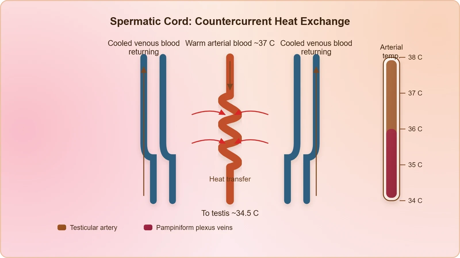
The efficiency of this exchange depends on the close anatomical apposition of artery and veins, which is why the spermatic cord is such a compact structure. When this arrangement is disrupted — for example, by a varicocele, an abnormal dilation of the pampiniform plexus — the countercurrent exchange becomes less effective, testicular temperature rises, and spermatogenesis may be impaired. Varicocele is in fact one of the more common identifiable causes of male subfertility, and its correction sometimes improves semen parameters. The clinical lesson is direct: the anatomy of the spermatic cord is not a passive conduit but an active thermal regulator.
Gross Anatomy of the Testis and Its Lobular Organisation
In adults, each testis is oval in shape, measuring approximately 4 to 5 cm in length and 2 to 3 cm in width. It is covered by a dense fibrous covering, the tunica albuginea, which gives the organ its firm consistency and sends inward septa that divide the glandular interior into compartments. Each testis contains roughly 250 such compartments, called testicular lobules. This lobular architecture is not merely descriptive detail; it reflects the testis's organisation as a collection of independent secretory units converging on a shared drainage system, a design that maximises the surface area available for sperm production within a compact organ.
Lobules, Seminiferous Tubules, and Cellular Residents
Each lobule contains one to three highly coiled seminiferous tubules, and it is within these tubules that sperms are produced. The tubule wall is lined internally by two distinct cell populations: the male germ cells, or spermatogonia, and the Sertoli cells. The germ cells undergo meiotic divisions that ultimately yield spermatozoa, while the Sertoli cells provide nutrition and structural support to the developing germ cells. This division of labour — germ cells as the productive lineage, Sertoli cells as the nurturing and organising stroma — is a recurring theme in reproductive biology and will be examined histologically in the next lesson.

The total length of seminiferous tubules within a single testis is remarkable, running to hundreds of metres when uncoiled, which explains how an organ only a few centimetres long can sustain continuous sperm output for decades. The coiled arrangement packs this enormous epithelial surface into a small volume. Each tubule is bounded by a basement membrane and a layer of contractile myoid cells that help propel sperm toward the rete testis. Understanding this geometry helps explain why testicular volume correlates so strongly with sperm-producing capacity, and why testicular atrophy — from any cause — has such direct consequences for fertility.
Interstitial Spaces and Leydig Cells
The regions outside the seminiferous tubules are called interstitial spaces, and they contain small blood vessels along with interstitial cells, also known as Leydig cells. Leydig cells synthesise and secrete the testicular hormones called androgens, of which testosterone is the principal and most potent. Other immunologically competent cells are also present in the interstitium, reflecting the testis's status as an immunologically privileged site — a necessary adaptation, since haploid germ cells arising after puberty carry antigens that the immune system would otherwise attack. This privilege is maintained by tight junctions between Sertoli cells, forming the blood–testis barrier.
The functional partnership between tubule and interstitium is worth emphasising. Spermatogenesis within the tubules requires high local concentrations of testosterone, which the Leydig cells supply through the interstitial vasculature. Luteinising hormone from the anterior pituitary stimulates Leydig cells, while follicle-stimulating hormone acts primarily on Sertoli cells. This two-cell, two-gonadotropin arrangement means that any lesion affecting either compartment — tubular or interstitial — can impair fertility, and the pattern of hormonal disturbance helps clinicians localise the problem. The next lesson explores this histology in depth.
The Accessory Duct System: From Rete Testis to Urethra
Sperm produced in the seminiferous tubules must travel a defined route to reach the exterior, and the male sex accessory ducts provide that route. The sequence runs from rete testis to vasa efferentia, then epididymis, then vas deferens, and finally the ejaculatory duct and urethra. Each segment contributes something distinct: transport, maturation, storage, or mixing with glandular secretions. The seminiferous tubules of the testis open into the vasa efferentia through the rete testis, a network of anastomosing channels within the mediastinum testis that collects sperm from all the lobules and funnels them into a small number of efferent ducts.
Epididymis and Vas Deferens
The vasa efferentia leave the testis and open into the epididymis, which lies along the posterior surface of each testis. The epididymis is a single, highly coiled tube that serves not only as a conduit but as a site of sperm maturation and temporary storage; sperm acquire progressive motility and fertilising capacity as they traverse it. The epididymis then leads to the vas deferens, a muscular tube that ascends into the abdomen and loops over the urinary bladder. The thick smooth muscle of the vas deferens generates the peristaltic force of ejaculation, and its surgical interruption — vasectomy — is a common and highly effective method of male contraception.
The vas deferens receives a duct from the seminal vesicle and opens into the urethra as the ejaculatory duct. This junction marks the point where sperm become mixed with the secretions of the accessory glands to form semen. The ducts as a whole store and transport sperms from the testis to the outside through the urethra, which originates from the urinary bladder and extends through the penis to its external opening, the urethral meatus. Because the urethra is shared between the urinary and reproductive systems in males, it is described as a urogenital passage, a detail with clinical implications for both urinary and reproductive disorders.
Cryptorchidism: When Descent Fails
The testes normally descend from the abdominal cavity into the scrotum during late fetal development, but in cryptorchidism — undescended testes — one or both testes remain within the abdomen or inguinal canal. Because the abdominal environment is too warm, spermatogenesis is impaired, and the risk of testicular cancer is substantially increased in the undescended organ. This clinical entity is a direct demonstration of the temperature principle: the same tissue that functions well at 2 to 2.5 °C below core temperature fails when held at core temperature. Early surgical correction, known as orchidopexy, is recommended to preserve fertility potential and reduce malignant risk.
Cryptorchidism also illustrates a broader developmental principle: organ position is functionally significant, not arbitrary. The scrotum's external location is an evolutionary solution to a thermal constraint, and when that solution fails, the consequences follow predictably. Learners preparing for examinations should be able to explain both the mechanism of impairment and the rationale for early intervention. The condition is one of the most common congenital anomalies of the male reproductive system, affecting a meaningful proportion of term and a higher proportion of preterm male infants, and it resolves spontaneously in many cases during the first year of life.
Accessory Glands and the Composition of Seminal Plasma
The male accessory glands include paired seminal vesicles, a single prostate, and paired bulbourethral glands. Their secretions constitute the seminal plasma, which is rich in fructose, calcium, and certain enzymes. Seminal plasma is not a passive vehicle; it nourishes and protects sperm, provides the energy substrate they need for motility, and modulates the chemical environment of the female reproductive tract. The fructose contributed largely by the seminal vesicles is the principal energy source for spermatozoa, which cannot metabolise glucose as efficiently and depend on this sugar for the ATP that powers their flagellar movement.
Seminal Vesicles, Prostate, and Bulbourethral Glands
The seminal vesicles are paired structures that contribute the bulk of the seminal fluid volume, supplying fructose and prostaglandins that may influence sperm transport and function. The prostate, a single gland situated below the urinary bladder and surrounding the prostatic urethra, adds a slightly alkaline fluid containing enzymes such as acid phosphatase and prostate-specific antigen, which helps neutralise the acidity of the vaginal environment and liquefies the coagulated ejaculate. The bulbourethral glands, also called Cowper's glands, secrete a lubricating fluid that helps lubricate the penis and neutralise residual acidity in the urethra before ejaculation.

The coordinated action of these glands produces an ejaculate with a characteristic composition and a defined sequence of emission. The alkaline components are particularly important because the vaginal environment is acidic, and sperm survival depends on buffering this acidity. When the prostate is enlarged or inflamed, both urinary and reproductive functions can be affected, which is why prostate health is a recurring concern in male medicine. The bulbourethral secretions, though small in volume, contribute meaningfully to lubrication and to preparing the urethral passage for the passage of semen.
Clinical Connections and Common Misconceptions
A frequent misconception is that semen and sperm are synonymous. In reality, spermatozoa constitute only a small fraction of the ejaculate volume; the majority is seminal plasma from the accessory glands. Another common error is to assume that the prostate is essential for sperm production, when in fact it contributes only to the seminal fluid. Learners should also distinguish the prostate from the bulbourethral glands and the seminal vesicles, since examination questions often test the specific contribution of each. A further misconception concerns the role of the scrotum, which is sometimes described as merely protective rather than thermoregulatory.
Clinical conditions reinforce these distinctions. Benign prostatic enlargement can obstruct urine flow because the prostate encircles the prostatic urethra, while prostatitis can cause pain and ejaculatory disturbance. Seminal vesicle disorders are rarer but can affect semen volume and composition. Understanding the normal contribution of each gland makes it easier to interpret abnormalities in semen analysis, such as low volume, poor liquefaction, or reduced fructose content, each of which points toward a particular glandular source. This diagnostic reasoning is a valuable skill for students moving toward clinical study.
The Penis, Erectile Tissue, and External Genitalia
The penis is the male external genitalia, and its function is to facilitate insemination by delivering semen into the female genital tract. It is made up of special tissue that helps in erection, and this phrase refers specifically to erectile tissue — the paired corpora cavernosa and the single corpus spongiosum, which surrounds the urethra. These are spongy, vascular structures containing numerous blood-filled spaces. During sexual arousal, arterial inflow increases while venous outflow is restricted, engorging the erectile tissue with blood and producing rigidity. The enlarged end of the penis, called the glans penis, is covered by a loose fold of skin called the foreskin.
Erectile Tissue and the Mechanism of Erection
Erection is fundamentally a vascular event governed by neural and local chemical signals. Parasympathetic nerve activity releases nitric oxide, which relaxes the smooth muscle of the cavernosal arteries and trabeculae, allowing blood to flood the erectile spaces. As these spaces expand, they compress the draining veins against the tunica albuginea, reducing outflow and maintaining tumescence. The corpus spongiosum remains somewhat less rigid than the corpora cavernosa, which prevents compression of the urethra and allows semen to pass during ejaculation. This functional differentiation within the erectile tissue is an elegant detail that examination candidates often overlook.
Understanding erectile physiology clarifies why disorders of erection can arise from vascular, neurological, or psychological causes, and why treatments such as phosphodiesterase inhibitors work by enhancing the nitric oxide signalling pathway. The foreskin, or prepuce, protects the glans and is richly innervated; its surgical removal, circumcision, is practised for cultural, religious, and medical reasons. The glans itself is highly sensitive and is the principal site of sexual sensation. These anatomical details connect directly to clinical practice and to public health discussions, making the external genitalia a topic of both biological and social significance.
Integrating Structure and Function
The male reproductive system rewards a holistic reading: each structure's position and composition follow from its function. The scrotum's external location serves thermoregulation; the coiled tubules maximise epithelial surface area; the epididymis provides a maturation chamber; the muscular vas deferens generates propulsive force; the accessory glands supply nourishment and buffering; and the erectile penis delivers the ejaculate. When one component fails, the consequences ripple through the system, which is why clinical assessment of male fertility examines each level in turn, from hormonal signalling to ductal patency to glandular secretion.
This integrated view also prepares the learner for the next lesson, which examines the seminiferous tubules and interstitial tissue at histological resolution. Having established the gross anatomy and the functional logic of the system, we can now ask how the tubule wall is organised, how the blood–testis barrier forms, and how Leydig cells interact with the tubular compartment. The macroscopic and microscopic perspectives together produce a coherent understanding of how the male body manufactures, matures, stores, and delivers the gametes on which sexual reproduction depends.
Worked Reasoning and Exam-Style Practice
Applying the anatomy to quantitative and clinical questions consolidates understanding far more effectively than passive reading. Consider the thermal principle as a calculation: if core body temperature is 37 °C, the testes operate optimally at approximately 34.5 to 35 °C. The required differential can be expressed as a simple relationship, and reasoning about what happens when it is lost — as in cryptorchidism or varicocele — tests whether the learner has grasped the underlying mechanism rather than memorised a number. The following worked examples illustrate the style of reasoning that examination questions reward.

Worked Example: Thermal Gradient and Sperm Function
Suppose a clinical scenario describes a patient with a persistent varicocele and asks why semen quality might be reduced. The reasoning chain runs: varicocele dilates the pampiniform plexus, impairing countercurrent heat exchange, which raises testicular temperature toward core levels, which slows the temperature-sensitive enzymatic steps of spermatogenesis, which reduces sperm count and motility. Expressing the temperature differential formally clarifies the logic:
When ##\Delta T## falls toward zero, spermatogenic efficiency declines. This is the same principle that explains cryptorchidism: an undescended testis sits at core temperature, so ##\Delta T \approx 0##, and both sperm production and malignant risk are adversely affected. Framing the problem this way converts a descriptive fact into a predictive model, which is precisely the kind of transferable understanding that examinations and clinical practice both demand from students of reproductive biology.
Common Mistakes and How to Avoid Them
Several errors recur in student answers. Confusing Sertoli cells with Leydig cells is common: Sertoli cells reside within the tubule and nourish germ cells, while Leydig cells lie in the interstitium and secrete androgens. Another frequent slip is to state that the epididymis produces sperm, when it stores and matures them. Students also sometimes claim the prostate produces fructose, when the seminal vesicles are the principal source. Finally, describing the scrotum as merely protective misses its thermoregulatory role entirely. Careful attention to location and function prevents these mistakes.
A useful revision strategy is to draw the pathway from seminiferous tubule to urethral meatus and annotate each segment with its function and its clinical correlate. This single diagram consolidates duct anatomy, glandular contributions, and the thermal principle in one visual framework. Pairing the diagram with the tables in this lesson provides both spatial and tabular representations, which supports retention. Learners should also practise explaining, in their own words, why the testes are external — a question that tests the deepest organising principle of the entire system.
Consolidating the Anatomy Before the Histological Turn
The male reproductive system, viewed as a whole, is a coordinated apparatus in which each structure's design follows from a specific functional demand. The scrotum and its muscular and vascular adaptations maintain the thermal environment that spermatogenesis requires. The testis, divided into roughly 250 lobules, houses the coiled seminiferous tubules where germ cells divide and Sertoli cells sustain them, while the interstitial Leydig cells supply the androgens that drive the process. The duct system collects, matures, stores, and propels sperm, and the accessory glands furnish the seminal plasma that nourishes and protects them. The erectile penis completes the system by delivering the ejaculate.
This lesson has also introduced several ideas that extend beyond the core descriptive content: the countercurrent heat-exchange mechanism of the pampiniform plexus, the clinical significance of cryptorchidism and varicocele, and the identification of the penis's "special tissue" as erectile tissue comprising the corpora cavernosa and corpus spongiosum. These additions connect anatomy to physiology and to clinical medicine, and they demonstrate why the temperature requirement is not a peripheral fact but the organising principle of the entire male system. A learner who can explain that principle, and trace its consequences through each organ, has achieved genuine understanding.
The next lesson turns to the microscope, examining the seminiferous tubules and interstitial tissue in histological detail. There we will consider the stages of the seminiferous epithelium, the blood–testis barrier formed by Sertoli cell junctions, and the endocrine dialogue between Leydig cells, Sertoli cells, and the pituitary. The gross anatomy established here provides the map; the histology will supply the cellular terrain. Together they form the foundation for understanding gametogenesis, fertilisation, and the reproductive events that the introductory lesson surveyed.
Also Read
- No links available.
From our network :
- Vite 6/7 'Cold Start' Regression in Massive Module GraphsIn-depth analysis of Vite 6/7 cold start regressions in massive module graphs. Learn about barrel file issues, pre-bundling optimizations, and how to improve Vite dev server performance.
- Mastering DB2 12.1 Instance Design: A Technical Deep Dive into Modern Database ArchitectureComprehensive guide to DB2 12.1 Instance Design. Explore architectural components, HADR setup, buffer pool tuning, and security policies for IBM DB2 12.1. Includes 10 functional SQL and Shell samples.
- 98% of Global MBA Programs Now Prefer GRE Over GMAT Focus EditionExplore why 98% of global MBA programs now prefer the GRE over GMAT Focus. Learn about the shorter GRE format, verbal logic importance, and strategic versatility for students.
- EV 2.0: The Solid-State Battery Breakthrough and Global Factory ExpansionDiscover the impact of the Solid-State Battery Breakthrough on the EV market. Learn about CATL and BYD stock growth, global factory expansion in Europe, and the future of EV 2.0 technology.
- AI-Powered 'Precision Diagnostic' Replaces Standard GRE Score ReportsDiscover the new GRE Precision Diagnostic. Learn how AI-powered behavioral analytics and time-to-solve tracking are replacing standard GRE score reports to empower students.
- 10 Physics Numerical Problems with Solutions for IIT JEEPrepare for IIT JEE with 10 physics numerical problems, complete with detailed solutions and conceptual explanations. Ideal for mastering kinematics, thermodynamics, electromagnetism, and optics.
- Trump Political Strategy How Geopolitical Stunts Serve As Media Diversions
- Mastering DB2 LUW v12 Tables: A Comprehensive Technical GuideComprehensive guide on DB2 LUW v12 tables. Explore CREATE TABLE syntax, BLU Acceleration, range partitioning, data types, and security features for IBM DB2 LUW v12.
- Analyzing Trump Deportation Numbers Insights Into The 2026 Immigration Crackdown
RESOURCES
- Male Reproductive System: Structure & Function - Cleveland Clinicmy.clevelandclinic.orgMay 8, 2023 ... The male reproductive system mostly exists outside of your body. The external organs include your penis, scrotum and testicles. Internal…
- Male Reproductive System - SEER Training Modulestraining.seer.cancer.govThis system consists of a pair of testes and a network of excretory ducts (epididymis, ductus deferens (vas deferens), and ejaculatory ducts), seminal vesicles, ...
- Male Reproductive System - TeachMeAnatomyteachmeanatomy.infoThe male reproductive organs within the pelvis support sperm production, storage and delivery, as well as seminal fluid secretion. This section introduces the ...
- Male Reproductive System - MedlinePlusmedlineplus.govMale Reproductive System Topics. Anatomy; ART see Assisted Reproductive Technology; Artificial Insemination see Infertility · Assisted Reproductive ...
- Anatomy of the Male Reproductive System | Springer Nature Linklink.springer.comFeb 9, 2011 ... Male mammals have two testicles, which are components of both the reproductive and the endocrine system. Therefore, the two main…
- Aging changes in the male reproductive system - MedlinePlusmedlineplus.govJul 15, 2024 ... Male reproductive system. In: Standring S, ed. Gray's Anatomy. 42nd ed. Philadelphia, PA: Elsevier Limited; 2021:chap 74. van den Beld…
- Gross and Morphometric Anatomy of the Male Reproductive System ...pmc.ncbi.nlm.nih.govThe reproductive tract of male mammals usually consists of the testes and associated epididymis, vasa deferentia, accessory sex gland complex, urethra, and ...
- Chapter 6 Male Reproductive System Terminology - NCBI - NIHncbi.nlm.nih.govOther male reproductive terms, whose definitions cannot be easily built from word components, will be described in context based on the anatomy and physiology ...
- New findings on the male reproductive system and spermatozoa of ...pmc.ncbi.nlm.nih.govJul 1, 2025 ... The anatomy of the adult male reproduction system of Aedes aegypti (Linnaeus) (Diptera, Culicidae). Ann Entomol Soc Am. 1961;54:832–44 ...
- Physiology, Male Reproductive System - StatPearls - NCBI Bookshelfncbi.nlm.nih.govMay 1, 2023 ... The male reproductive system consists of internal structures, including the testes, epididymides, vasa deferentia, and prostate, ...
- 27.1 Anatomy and Physiology of the Testicular Reproductive Systemopenstax.orgApr 20, 2022 ... The testes (singular = testis) are the male gonads—that is, the male reproductive organs. They produce both sperm and androgens,…
- Anatomy of the Male Reproductive System of Sugar Gliders ...pubmed.ncbi.nlm.nih.govSep 23, 2024 ... Sugar gliders possess two pairs of bulbous bulbourethral glands, located dorsally and laterally to the rectum, but lack vesicular glands.
- Male reproductive organs: Anatomy and function - Kenhubkenhub.comThe male sex organs comprise a complex arrangement of internal and external genital organs. Their function is concerned with reproduction and sexual ...
- The gross anatomy of the male reproductive system of a neotropical ...pubmed.ncbi.nlm.nih.govThis study focuses on one of the many unexplored areas relating to the species, the male reproductive system. The results showed that the mean…
- Male reproductive system - Mayo Clinicmayoclinic.orgThe male reproductive system makes, stores and moves sperm. Testicles produce sperm. Fluid from the seminal vesicles and prostate gland mixes with sperm to ...