Human reproduction is one of the most intricate and beautifully orchestrated sequences in all of biology, a chain of events in which a single fertilised cell becomes a fully formed infant across roughly nine months. Understanding this process requires more than memorising anatomical labels; it demands an appreciation of how structure, hormonal signalling, and timing interlock with extraordinary precision. This opening lesson establishes the conceptual scaffolding for the entire course, mapping the full arc of reproductive events and contrasting the strikingly different biological timelines that govern men and women.
Humans are sexually reproducing and viviparous organisms, meaning the embryo develops inside the maternal body and is nourished directly through a placental connection until birth. That single fact shapes almost every anatomical and physiological feature you will encounter in the lessons ahead. The reproductive sequence itself unfolds as a defined cascade: gametogenesis, insemination, fertilisation, blastocyst formation and implantation, gestation, and finally parturition. Each stage depends on the one before it, and each is regulated by hormones that rise and fall in carefully timed rhythms.
What makes the human case especially instructive is the profound asymmetry between male and female reproductive timelines. Sperm production, once initiated at puberty, continues essentially uninterrupted into advanced old age, whereas ovum formation ceases in women around the age of fifty. This lesson examines that contrast in detail, corrects a common oversimplification about when egg production actually ends, and situates viviparity alongside the alternative strategies of oviparity and ovoviviparity found elsewhere in the animal kingdom.
On This Page
The Defining Features of Human Reproduction
Human beings reproduce sexually, which means that two specialised haploid cells, the sperm and the ovum, fuse to restore a diploid chromosome complement in the resulting zygote. This fusion, termed fertilisation, generates genetic variation that is the raw material of evolution and the reason siblings differ from one another. Sexual reproduction in humans is therefore not merely a mechanism of multiplication but a strategy for producing genetically diverse offspring, and every structural feature of the reproductive systems you will study exists to serve that end efficiently and safely.
Equally defining is viviparity, the condition in which the developing embryo is retained within the maternal body and nourished through a placenta rather than relying on yolk reserves sealed inside an eggshell. Viviparity imposes substantial physiological demands on the mother, including nutrient transfer, waste removal, and immune tolerance of a genetically half-foreign fetus, yet it confers a decisive advantage: the young are protected and provisioned throughout their most vulnerable developmental period. These two features, sexuality and viviparity, frame everything that follows in this course.
Viviparity, Oviparity and Ovoviviparity Compared
Reproductive strategies across the animal kingdom fall into three broad categories that are worth distinguishing precisely. Oviparous animals, such as birds and most reptiles, lay eggs in which the embryo develops outside the mother's body, drawing nourishment from yolk within a protective shell. Ovoviviparous animals, including certain sharks and some snakes, retain eggs internally until they hatch, so the young emerge live but were nourished by yolk rather than by a placental connection. Viviparous animals, mammals like humans among them, retain the embryo internally and sustain it through a direct maternal supply.
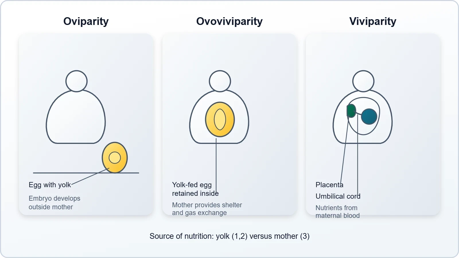
The distinction between ovoviviparity and true viviparity is subtle but biologically important. In ovoviviparity, the egg provides the nutrients and the mother merely provides shelter and gas exchange; in viviparity, the maternal bloodstream supplies nutrients and oxygen through a specialised placental interface. Humans exemplify the most derived form of viviparity, in which an elaborate chorioallantoic placenta mediates exchange and endocrine communication. Recognising these categories helps explain why human pregnancy is physiologically so demanding compared with egg-laying strategies.
Why Viviparity Demands an Integrated System
Because the human embryo develops internally for roughly nine months, the female reproductive system must accomplish far more than producing gametes. It must provide a receptive tract for sperm, a site for fertilisation, a prepared uterine lining for implantation, a placental interface for exchange, and a muscular organ capable of expelling the fetus at term. The mammary glands then extend the system's function beyond birth by supplying nutrition to the newborn. This structural and functional integration is why the female reproductive system is studied as a coordinated whole rather than as a collection of separate organs.
Viviparity also creates an immunological paradox. The fetus inherits half its genes from the father and is therefore antigenically foreign to the mother, yet it is not normally rejected. The placenta and maternal immune system achieve a state of local tolerance through mechanisms you will meet in later lessons. For now, it is enough to recognise that viviparity is not simply internal incubation; it is an active, hormonally managed partnership between two genetically distinct individuals, sustained by structures that exist only for the duration of pregnancy.
The Sequence of Reproductive Events
The reproductive events in humans follow a logical and largely invariant order, and grasping that order is the single most useful organising principle for this course. The sequence begins with gametogenesis, the formation of sperms in males and ova in females. It proceeds through insemination, the transfer of sperms into the female genital tract, and then fertilisation, the fusion of male and female gametes to form a zygote. From the zygote arise the blastocyst and its attachment to the uterine wall, a step called implantation, which marks the true beginning of pregnancy.
Following implantation comes gestation, the period of embryonic and fetal development during which organs form and mature. The sequence concludes with parturition, the delivery of the baby, and lactation, the production of milk to sustain the newborn. Each of these events occurs only after puberty, when the reproductive systems become functional under hormonal drive. The elegance of the sequence lies in its dependency: fertilisation cannot occur without gametes, implantation cannot occur without a blastocyst, and gestation cannot proceed without implantation.
From Gametes to Zygote: The Early Events
Gametogenesis is the production of haploid gametes through meiosis, and it differs markedly between the sexes in timing and output. In males, spermatogenesis begins at puberty and generates enormous numbers of small, motile sperm continuously. In females, oogenesis begins before birth, pauses for years, and produces a single large ovum per cycle from a finite pool of follicles. These differences in gamete biology foreshadow the divergent reproductive timelines examined later in this lesson and are explored in full in the dedicated gametogenesis lesson.
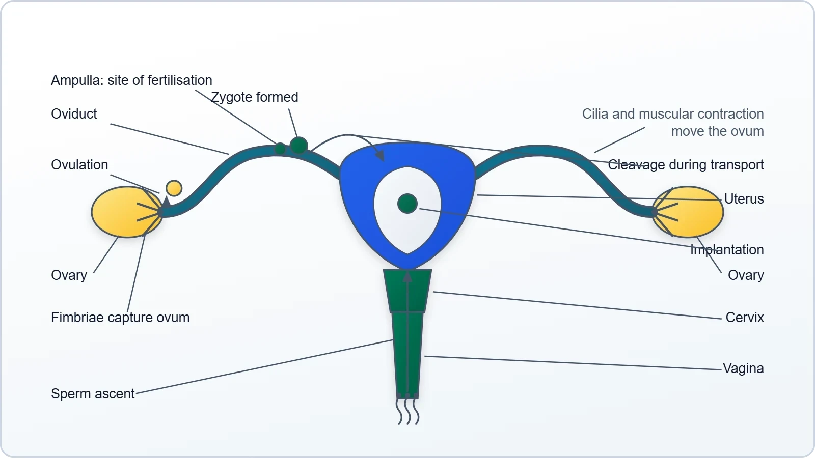
Insemination delivers sperm into the female genital tract, where they undergo capacitation and travel toward the ovum. Fertilisation typically occurs in the ampullary region of the oviduct, producing a diploid zygote that immediately begins cleaving as it travels toward the uterus. Within about five to seven days, the developing blastocyst attaches to the endometrium and implants. This transition from a free-living embryo to an implanted one is a critical checkpoint, and failure at this stage accounts for a significant proportion of early pregnancy loss.
Gestation, Parturition and Lactation
Gestation in humans lasts approximately nine months, conventionally divided into three trimesters. During this period the embryo develops into a fetus, organ systems form and mature, and the placenta mediates nutrient and gas exchange while secreting hormones that maintain pregnancy. The uterus grows dramatically, and maternal physiology adapts to support the increasing demands of the developing offspring. These adaptations are examined in detail in the later lessons on pregnancy and embryonic development.
Parturition is the process of giving birth, driven by coordinated uterine contractions regulated by hormonal signals including oxytocin. Following delivery, lactation provides the newborn with colostrum and then mature milk, delivering both nutrition and passive immunity. The mammary glands, though often described separately, are functionally part of the reproductive system because they complete the reproductive sequence by supporting the survival of the offspring. Together, gestation, parturition and lactation close the loop that began with gametogenesis.
Puberty and the Hormonal Switch
Reproductive events do not begin at birth; they are switched on at puberty by a coordinated hormonal cascade. The hypothalamus begins secreting gonadotropin-releasing hormone, or GnRH, in pulses, and this pulsatile pattern is essential because continuous GnRH actually suppresses the system. GnRH stimulates the anterior pituitary to release luteinising hormone and follicle-stimulating hormone, which in turn activate the gonads. The gonads respond by producing gametes and secreting sex steroids, and these steroids drive the visible changes of puberty and sustain reproductive function thereafter.
The timing of this switch varies between individuals but typically occurs between ages ten and fourteen, earlier in girls than in boys. The rise in GnRH secretion is thought to result from a relaxation of central inhibition rather than the sudden appearance of a new signal, and metabolic cues such as body fat and leptin influence the timing. Understanding puberty as a hormonally gated event explains why reproductive capability is absent in childhood and why disorders of GnRH secretion can delay or prevent pubertal development.
The Hypothalamic-Pituitary-Gonadal Axis
The hypothalamic-pituitary-gonadal axis is the master regulatory circuit of reproduction, and it operates through both positive and negative feedback. In the male, testosterone from Leydig cells feeds back negatively on the hypothalamus and pituitary, keeping GnRH and gonadotropin secretion within a stable range. In the female, the same axis operates cyclically, with oestrogen exerting negative feedback for most of the cycle but switching to positive feedback at mid-cycle to trigger ovulation. This difference in feedback logic is a central reason male and female reproductive patterns diverge so sharply.
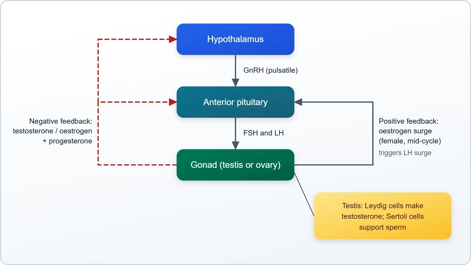
Appreciating this axis clarifies why reproductive function is vulnerable to disruption by stress, illness, and nutritional deficiency. Signals from the brain can suppress GnRH release, temporarily halting gamete production and cycling. In clinical practice, this explains amenorrhoea in athletes and in states of severe energy deficit. The axis is therefore best understood not as a fixed machine but as a responsive control system that integrates hormonal, metabolic, and neural information to decide whether conditions are suitable for reproduction.
When Reproductive Capability Ends
In females, reproductive capability ends at menopause, when the finite pool of ovarian follicles is exhausted and oestrogen production falls. Menopause typically occurs between ages forty-five and fifty-five and marks the permanent cessation of ovulation and menstruation. In males, by contrast, there is no comparable abrupt endpoint; spermatogenesis and testosterone production decline gradually with age, and fertility can persist into advanced old age. This asymmetry is one of the most striking contrasts in human reproductive biology and directly reflects the different biology of the two gamete-producing systems.
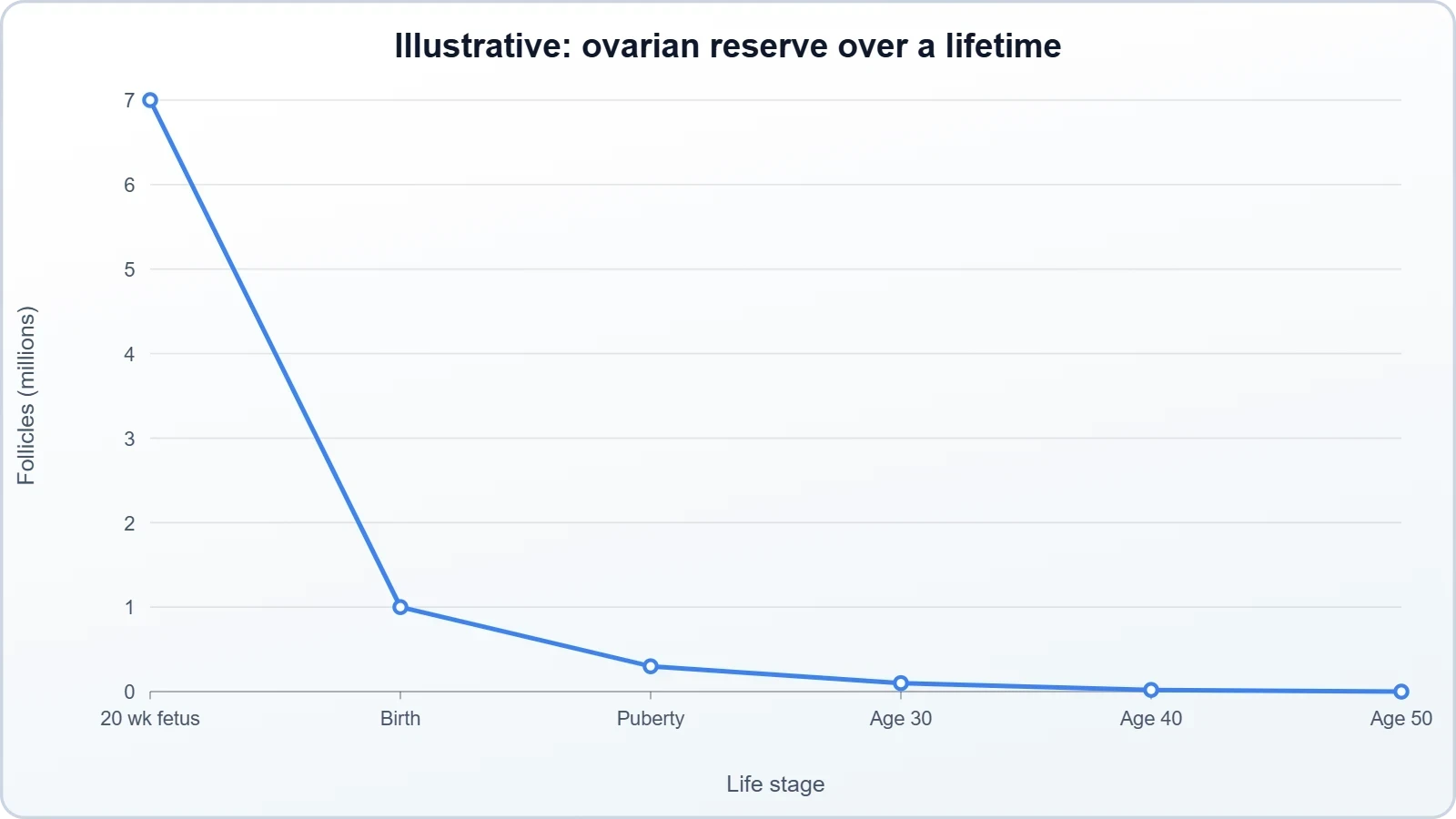
The reference material notes that ovum formation ceases in women around the age of fifty, and this is broadly correct as a description of menopause. However, it is a simplification worth correcting. Follicle depletion actually begins before birth, accelerates during childhood and adolescence, and continues throughout adult life, so the decline in ovarian reserve is a lifelong process rather than an event at fifty. Menopause is simply the point at which the remaining follicle pool becomes too small to sustain cyclic hormone production.
Anatomical Overview of the Two Systems
Although this lesson does not examine either system in full, a brief orientation is essential before the detailed lessons that follow. The male reproductive system is located in the pelvis and comprises a pair of testes along with accessory ducts, glands, and the external genitalia. The testes lie outside the abdominal cavity within the scrotum, a pouch that maintains them at a temperature two to two and a half degrees Celsius below core body temperature, a condition necessary for spermatogenesis. Each adult testis is oval, roughly four to five centimetres long and two to three centimetres wide.
The female reproductive system consists of a pair of ovaries along with a pair of oviducts, the uterus, the cervix, the vagina, and the external genitalia, all located in the pelvic region. The ovaries are the primary female sex organs, producing the ovum and several steroid hormones. Each ovary is about two to four centimetres long and is connected to the pelvic wall and uterus by ligaments. Together with the mammary glands, these structures are integrated to support ovulation, fertilisation, pregnancy, birth, and child care.
The Testes and the Scrotal Thermostat
Each testis is enclosed in a dense fibrous covering and divided internally into about 250 compartments called testicular lobules. Each lobule contains one to three highly coiled seminiferous tubules, the sites of sperm production. The tubules are lined by male germ cells, or spermatogonia, which undergo meiotic divisions to form sperm, and by Sertoli cells, which nourish the developing germ cells. The spaces between tubules, the interstitial spaces, contain blood vessels and Leydig cells, which synthesise and secrete testicular hormones called androgens.
The scrotal location of the testes is a striking anatomical solution to a physiological problem. Spermatogenesis is temperature-sensitive, and the scrotum acts as a thermostat, adjusting position to keep the testes cooler than the body core. This is why undescended testes, a condition called cryptorchidism, impair fertility if uncorrected. The arrangement illustrates a recurring theme in reproductive anatomy: structures are often positioned and organised in ways that optimise the delicate conditions required for gamete production rather than for the comfort of the organism.
The Ovaries and the Accessory Tract
The ovaries are covered by a thin epithelium enclosing the ovarian stroma, which is divided into a peripheral cortex and an inner medulla. The cortex houses the follicles at various stages of development, while the medulla contains blood vessels and connective tissue. The oviducts, uterus, cervix, and vagina form the accessory tract through which the ovum travels, where fertilisation occurs, and within which the embryo implants and develops. The uterus in particular is a remarkable organ, capable of expanding enormously during pregnancy and contracting powerfully during birth.
The functional integration of these organs is best appreciated by tracing the path of the ovum. Released from the ovary at ovulation, it is captured by the fimbriae of the oviduct and transported toward the uterus by ciliary action and muscular contraction. Fertilisation normally occurs in the ampullary region of the oviduct. The resulting zygote continues toward the uterus, where the prepared endometrium receives it. Every structure in the tract therefore has a defined role in the journey from ovulation to implantation, and the next lessons examine each in turn.
Common Misconceptions and Clinical Connections
Several persistent misconceptions surround human reproduction, and addressing them early prevents confusion later. One common error is the belief that the testes are inside the abdomen like the ovaries; in fact, they descend into the scrotum because sperm production requires a cooler temperature. Another is the assumption that male and female reproductive systems are mirror images; they are not, differing fundamentally in gamete biology, hormonal feedback, and the timing of reproductive capability. A third is the idea that menopause is a sudden event rather than the endpoint of a gradual, lifelong depletion of follicles.
Clinical connections reinforce the value of understanding the sequence of events. Failure at any stage, whether gamete production, fertilisation, implantation, or gestation, can result in infertility, and clinical investigation proceeds by identifying which stage is disrupted. Assisted reproductive technologies such as in vitro fertilisation bypass several early stages by combining gametes outside the body and transferring the resulting embryo to the uterus. Understanding the normal sequence is therefore the foundation for understanding both pathology and treatment.
Correcting the Timeline Simplification
The statement that ovum formation ceases around age fifty is a useful shorthand but conceals important biology. Female gametes are not produced continuously; instead, a fixed stock of oocytes is established before birth, peaks at several million during fetal life, and declines steadily thereafter. By puberty, only a fraction remains, and each cycle consumes a cohort of follicles. Menopause occurs when the reserve is effectively exhausted, and the average age of around fifty reflects the typical rate of depletion rather than a programmed switch.
This correction matters for several reasons. It explains why fertility declines gradually in the years before menopause, why age is a major determinant of reproductive success, and why conditions that accelerate follicle loss can cause premature ovarian insufficiency. It also highlights the contrast with the male system, where germ cells are replenished continuously by spermatogonial division. Framing the female timeline as a lifelong depletion rather than a sudden cessation gives a more accurate and clinically useful picture.
Why the Male and Female Systems Diverge
The divergence between male and female reproductive systems is rooted in the different demands of producing sperm versus ova. Sperm are small, numerous, and motile, and their continuous production is metabolically feasible because the germ line is replenished by stem cell division. Ova are large, nutrient-rich, and few, and their production is constrained by the finite pool established before birth. These gamete-level differences cascade into differences in hormonal regulation, organ structure, and reproductive lifespan, and they explain why the same hypothalamic-pituitary-gonadal axis produces such different patterns in the two sexes.
Understanding this divergence also illuminates the evolutionary logic of the two strategies. Continuous sperm production maximises the chance of fertilisation across many potential mates, while the production of a small number of well-provisioned ova reflects the heavy investment required for internal gestation. Neither strategy is superior; each is adapted to its role in a cooperative reproductive process. Recognising this helps students move beyond rote memorisation toward a genuine understanding of why the systems are built as they are.
Exam-Style Practice and Key Takeaways
Consolidating this lesson requires both recall and reasoning. Students should be able to list the reproductive events in order, define viviparity and contrast it with oviparity and ovoviviparity, explain why the testes are scrotal, and describe the hormonal trigger for puberty. Equally important is the ability to reason about the consequences of disruption at each stage. The practice questions below are designed to test understanding rather than mere memorisation, and they mirror the style of questions commonly encountered in senior secondary and early undergraduate examinations.
A useful revision technique is to draw the sequence as a flowchart and annotate each step with its hormonal regulator and its anatomical site. This forces integration of structure and function, which is precisely what examiners reward. Students should also practise explaining the male-female timeline contrast in their own words, since this is a frequent short-answer question. The worked example below illustrates how a quantitative question on gamete numbers might be approached, reinforcing the numerical reasoning that appears in later lessons.
Worked Example: Reasoning About Gamete Numbers
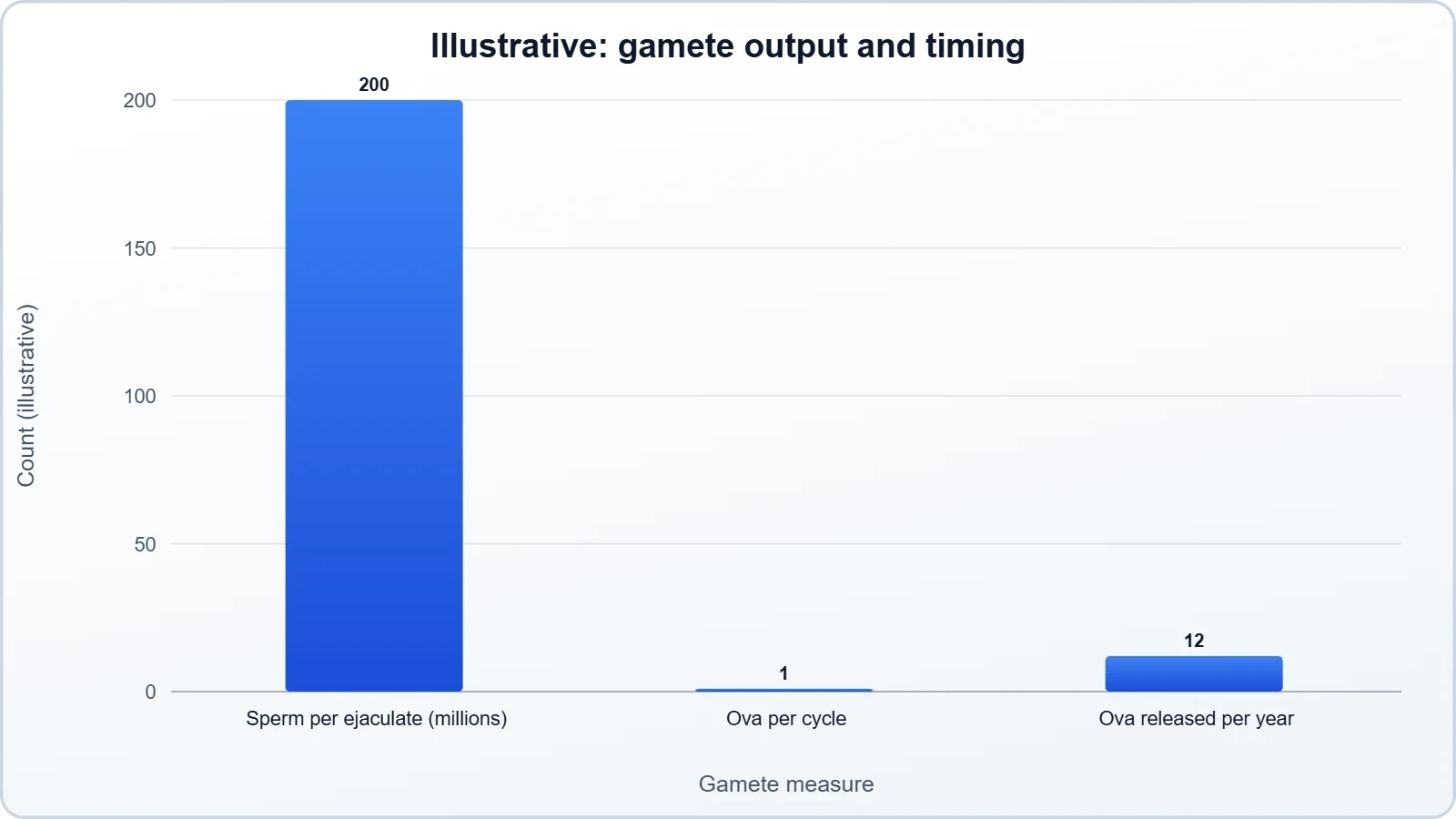
Consider a question asking why the female gamete supply is described as finite while the male supply is not. A sound answer begins with the observation that oocytes are established before birth and decline thereafter, whereas spermatogonia divide continuously after puberty. If a female is born with approximately one to two million oocytes and ovulates roughly four hundred across her lifetime, the vast majority are lost to atresia. The male, by contrast, produces millions of sperm daily. The arithmetic below illustrates the scale of the difference.
The comparison is stark and explains why the two systems are regulated so differently. A finite oocyte pool must be conserved and released sparingly, which is why ovulation is cyclic and hormonally controlled. A continuously replenished sperm supply can be produced in vast excess, which is why spermatogenesis is continuous and relatively insensitive to timing. Framing the contrast numerically makes the underlying logic memorable and provides a model for answering similar questions in examinations.
Key Takeaways for the Next Lesson
This lesson has established the sequence of reproductive events, the defining features of human reproduction, the hormonal switch at puberty, and the divergent timelines of the two sexes. It has also corrected the simplification about ovarian reserve and situated viviparity among alternative reproductive strategies. These ideas form the conceptual foundation on which all subsequent lessons build. The next lesson turns to the male reproductive system in detail, examining the anatomy of the testes, the duct system, and the accessory glands introduced briefly here.
Before moving on, students should be comfortable with the terminology of the sequence and with the distinction between the male and female timelines. They should also appreciate that reproductive anatomy is organised around function, with structures positioned and shaped to optimise gamete production, transfer, and development. With this framework in place, the detailed study of each organ system becomes far more coherent, because every structure can be understood in terms of the role it plays in the larger reproductive sequence.
Also Read
- No links available.
From our network :
- 98% of Global MBA Programs Now Prefer GRE Over GMAT Focus EditionExplore why 98% of global MBA programs now prefer the GRE over GMAT Focus. Learn about the shorter GRE format, verbal logic importance, and strategic versatility for students.
- EV 2.0: The Solid-State Battery Breakthrough and Global Factory ExpansionDiscover the impact of the Solid-State Battery Breakthrough on the EV market. Learn about CATL and BYD stock growth, global factory expansion in Europe, and the future of EV 2.0 technology.
- How to Print Cheaply: Cutting the Cost per PageA practical, measured guide to lowering what each printed page costs — duplex and draft modes, third-party ink, yield mathematics, and knowing when a new printer beats refills.
- Analyzing Trump Deportation Numbers Insights Into The 2026 Immigration Crackdown
- 10 Physics Numerical Problems with Solutions for IIT JEEPrepare for IIT JEE with 10 physics numerical problems, complete with detailed solutions and conceptual explanations. Ideal for mastering kinematics, thermodynamics, electromagnetism, and optics.
- Do You Need a New Router or Is Your Internet Plan the Problem?A practical comparison of the signs that point to router limits versus signs your broadband speed or provider is the bottleneck, so you can decide what to replace before spending money.
- AI-Powered 'Precision Diagnostic' Replaces Standard GRE Score ReportsDiscover the new GRE Precision Diagnostic. Learn how AI-powered behavioral analytics and time-to-solve tracking are replacing standard GRE score reports to empower students.
- Trump Political Strategy How Geopolitical Stunts Serve As Media Diversions
- Where to Put Your Wireless Router for Better CoverageA practical, physics-grounded guide to router placement: why the centre of the home beats the corner, how walls and metal sap signal, and how to run a simple survey that proves the improvement.
RESOURCES
- roles in gamete maturation, fertilization and embryo implantationpubmed.ncbi.nlm.nih.govConclusions: A detailed understanding of the contributions of EVs in the series of events from gametogenesis ... Human Reproduction and Embryology. All rights ...
- Gametogenesis – Human Reproduction: A Clinical Approachiastate.pressbooks.pubHuman Reproduction: A Clinical Approach. Gametogenesis, Fertilization, and Implantation. Gametogenesis. Mitosis and meiosis are the two important cell cycles ...
- The effects of smoking on fertility from gametogenesis to implantationsciencedirect.comStudies in rodents and human and nonhuman primates also suggest that cigarette smoke or nicotine alters the motility of the female reproductive tract, and…
- Implantation – Human Reproduction: A Clinical Approachiastate.pressbooks.pubHuman Reproduction: A Clinical Approach. Gametogenesis, Fertilization, and Implantation. Implantation. Implantation occurs when the conceptus adheres to the ...
- Biology and Development of Human Tissues | Adelaide Universityadelaide.edu.auThe Reproduction and Development component of the course investigates male and female reproductive systems and functions, gametogenesis, fertilisation, ...
- Embryology & Clinical Neuroanatomy (0990063) 2025-26 - Module ...york.ac.uk... human reproduction, gametogenesis, fertilisation and generation of the zygote ... Gametogenesis (spermatogenesis and oogenesis), fertilisation, implantation ...
- Human Reproduction - NCERTncert.nic.inSuch an endometrium is necessary for implantation of the fertilised ovum and other events of pregnancy. During pregnancy all events of the menstrual cycle…
- Extracellular vesicles: roles in gamete maturation, fertilization and ...pmc.ncbi.nlm.nih.govGametogenesis, fertilization, implantation and early embryo development are ... In human reproduction, a major challenge is presented by limitations in ...
- Editorial: Advances on molecular mechanisms regulating reproductionfrontiersin.orgEditorial: Advances on molecular mechanisms regulating reproduction - from gametogenesis to fertilization ... fertilization, and embryo implantation. The ...
- Mechanisms of implantation: strategies for successful pregnancypmc.ncbi.nlm.nih.govCell-specific expression of Wnt ligands, receptors and inhibitors in mouse and human endometria during the reproductive cycle and pregnancy suggests roles for ...
- OHSU researchers develop functional eggs from human skin cellsnews.ohsu.eduSep 30, 2025 ... Meiosis is strictly related to the sperm and egg cell in sexual reproduction ... In vitro gametogenesis, or IVG, has…
- Induction of male neogametogenesis in a three-dimensional ...academic.oup.comMolecular Human Reproduction, Volume 32, Issue 3, 2026, gaag036, https://doi.org/10.1093/molehr/gaag036. Published: 04 June 2026. Article history. Received:.
- Ultrastructure of Reproduction: Gametogenesis, Fertilization, and ...link.springer.com`This book is suitable for advanced students, and particularly for researchers in the fields of reproductive and developmental biology. The chapters on human ...