Human reproduction is best understood not as a list of isolated chapters but as a single, continuous biological narrative in which every structure exists to serve the next event in the chain. The course you have just completed moves deliberately from anatomy to physiology, from the paired testes and ovaries through gametogenesis, the menstrual cycle, fertilisation, implantation, gestation, parturition and lactation. Each lesson supplied a piece of the puzzle; this closing lesson assembles those pieces into one coherent picture so that the material can be recalled, applied and examined with genuine confidence rather than rote memorisation.
Integration matters because examiners increasingly reward connections rather than fragments. A question about contraception is really a question about the hypothalamic–pituitary–gonadal axis, gamete transport and endometrial receptivity all at once. A question about lactation quietly tests your understanding of neuroendocrine reflexes and mammary histology. When you can trace a single hormone from its hypothalamic origin to its target tissue and predict the physiological consequence of altering it, you demonstrate the kind of understanding that distinguishes a strong answer from a merely correct one. This lesson builds that connective tissue systematically.
The pages that follow review the whole course through six integrated lenses: the endocrine control architecture, the structural logic of the reproductive tracts, the cellular events of gametogenesis, the timing mechanisms of the cycle, the sequence from fertilisation to birth, and finally a disciplined examination workshop. Along the way you will find worked reasoning, a flowchart linking hypothalamus, pituitary, gonads and target tissues, a catalogue of the mistakes that most often cost marks, and practice questions that deliberately require you to combine topics. Treat this as a rehearsal for the real thing.
On This Page
The Endocrine Architecture That Governs Reproduction
Every reproductive event in the human body ultimately traces back to a small set of hormonal signals organised into a hierarchy. The hypothalamus sits at the apex, secreting gonadotropin releasing hormone (GnRH) in pulses. These pulses reach the anterior pituitary through the hypophyseal portal circulation and stimulate the secretion of two gonadotropins: luteinising hormone (LH) and follicle stimulating hormone (FSH). The gonads then respond in a tissue-specific manner, and their steroid products feed back upon the hypothalamus and pituitary to modulate the entire system. Understanding this three-tier arrangement is the single most powerful organising idea in the course.
The elegance of the design lies in its differential wiring. In the male, LH acts on Leydig cells to stimulate androgen synthesis, while FSH acts on Sertoli cells to support spermatogenesis. In the female, the same two hormones drive follicular development and ovulation, but the feedback logic is far more intricate, switching from negative to positive at mid-cycle. A single pair of pituitary hormones therefore produces two entirely different physiological outcomes depending on the gonadal context. This is why examiners love questions that ask you to compare male and female endocrine control.
Mapping the Hypothalamus–Pituitary–Gonadal Axis
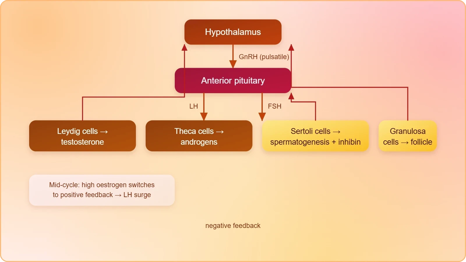
A flowchart is the most efficient revision tool for this axis because it forces you to specify direction, target and consequence. Begin with the hypothalamus releasing GnRH, which travels to the anterior pituitary. The pituitary releases LH and FSH into the systemic circulation. LH targets Leydig cells in the testis and theca cells in the ovary; FSH targets Sertoli cells in the testis and granulosa cells in the ovary. The gonads then secrete androgens, oestrogens and progesterone, which return to the hypothalamus and pituitary as feedback signals, closing the loop.
Represented as a sequence, the male axis reads: hypothalamus → GnRH → anterior pituitary → LH → Leydig cells → testosterone → negative feedback; and in parallel, anterior pituitary → FSH → Sertoli cells → spermatogenesis, with Sertoli cells secreting inhibin that selectively suppresses FSH. The female axis is more dynamic, with oestrogen exerting negative feedback at low concentrations and positive feedback at high sustained concentrations, triggering the LH surge. Inhibin from granulosa cells again selectively suppresses FSH. Drawing this once from memory will consolidate more than an hour of passive reading.
Feedback Loops and Why They Matter Clinically
Negative feedback keeps the system stable; positive feedback creates the dramatic mid-cycle event that makes ovulation possible. In the follicular phase, rising oestrogen from the developing follicle suppresses GnRH and gonadotropin release, preventing premature recruitment of additional follicles. Once oestrogen remains elevated for roughly a day and a half, the feedback polarity flips, and the pituitary discharges a surge of LH that ruptures the mature follicle. This switch is one of the most examinable concepts in reproductive endocrinology because it explains both normal cyclicity and the mechanism of combined oral contraceptives.
Clinical reasoning follows directly from the loop. A lesion in the hypothalamus reduces GnRH, lowering LH and FSH and impairing gametogenesis. Exogenous gonadotropins can bypass the block and restore follicular growth, which is the basis of ovulation induction. Conversely, continuous rather than pulsatile GnRH administration desensitises the pituitary, a principle exploited in certain therapeutic protocols. When you understand the loop, you can predict the effect of almost any intervention, and that predictive power is precisely what integrated examination questions are designed to test.
Structural Logic of the Male and Female Systems
Anatomy in this course is never decorative; every structure is placed where it is because of a functional requirement. The male system is organised around continuous, temperature-sensitive sperm production and efficient delivery. The testes lie outside the abdominal cavity in the scrotum, which maintains a temperature 2–2.5 °C below core body temperature, a condition necessary for spermatogenesis. Each adult testis is oval, roughly 4–5 cm long and 2–3 cm wide, and is divided into about 250 testicular lobules, each containing one to three highly coiled seminiferous tubules where sperm are produced.
The female system, by contrast, is organised around cyclical gamete release, internal fertilisation, gestation and lactation. A pair of ovaries, each about 2–4 cm long, produces the ovum and ovarian steroid hormones. The oviducts, uterus and vagina form the accessory duct system, while the mammary glands are integrated structurally and functionally to support ovulation, fertilisation, pregnancy, birth and child care. Recognising that the two systems share a common endocrine language but differ radically in architecture and timing is the key comparative insight the examiner expects.
From Seminiferous Tubule to Urethral Meatus
Within each seminiferous tubule, two cell populations cooperate. Male germ cells (spermatogonia) undergo meiotic divisions leading to sperm formation, while Sertoli cells provide nutrition to the germ cells. Outside the tubules, in the interstitial spaces, lie small blood vessels and Leydig cells, which synthesise and secrete testicular androgens. The duct system then collects and transports the sperm: seminiferous tubules open through the rete testis into the vasa efferentia, which open into the epididymis along the posterior surface of each testis. The epididymis leads to the vas deferens, which ascends to the abdomen and loops over the urinary bladder.
The vas deferens receives a duct from the seminal vesicle and opens into the urethra as the ejaculatory duct. The urethra originates from the urinary bladder and extends through the penis to the urethral meatus. The penis, made of special tissue that permits erection, facilitates insemination; its enlarged end, the glans penis, is covered by the foreskin. The accessory glands — paired seminal vesicles, a prostate and paired bulbourethral glands — contribute seminal plasma rich in fructose, calcium and certain enzymes, with bulbourethral secretions aiding lubrication.
Oviduct, Uterus and the Mammary Gland
Each fallopian tube is about 10–12 cm long and extends from the periphery of the ovary to the uterus. Its funnel-shaped infundibulum bears finger-like fimbriae that collect the ovum after ovulation; it leads to the wider ampulla, and the narrow isthmus joins the uterus. The single uterus, shaped like an inverted pear, opens into the vagina through the narrow cervix, whose canal together with the vagina forms the birth canal. Its wall has three layers: the thin perimetrium, the thick smooth-muscle myometrium, and the glandular endometrium that undergoes cyclical change.
The mammary gland completes the functional circuit. Each breast contains glandular tissue divided into 15–20 mammary lobes containing clusters of alveoli, whose cells secrete milk into lumens. Alveoli open into mammary tubules, which join to form a mammary duct; several ducts unite into a wider mammary ampulla connected to the lactiferous duct, opening at the nipple. The female external genitalia — mons pubis, labia majora, labia minora, hymen and clitoris — complete the picture. Note carefully that the presence or absence of the hymen is not a reliable indicator of virginity or sexual experience.
Gametogenesis as a Cellular Narrative
Gametogenesis is the bridge between anatomy and inheritance, and it rewards careful step-by-step reasoning. In the testis, immature male germ cells (spermatogonia) multiply by mitotic division. Each spermatogonium is diploid and contains 46 chromosomes. Some spermatogonia, called primary spermatocytes, periodically undergo meiosis. A primary spermatocyte completes the first meiotic division, a reduction division, producing two equal haploid secondary spermatocytes with 23 chromosomes each. These undergo the second meiotic division to produce four equal haploid spermatids, each carrying 23 chromosomes.
The spermatids are then transformed into spermatozoa by spermiogenesis. After spermiogenesis, sperm heads become embedded in the Sertoli cells and are finally released from the seminiferous tubules by spermiation. Spermatogenesis begins at puberty because of a significant increase in GnRH secretion; the elevated GnRH acts on the anterior pituitary, stimulating LH and FSH. LH acts on Leydig cells to stimulate androgen synthesis and secretion, and androgens in turn stimulate spermatogenesis, while FSH acts on Sertoli cells to support the process. This sequence is a favourite of examiners because it links every level of the axis.
Oogenesis and the Asymmetry of Gamete Production
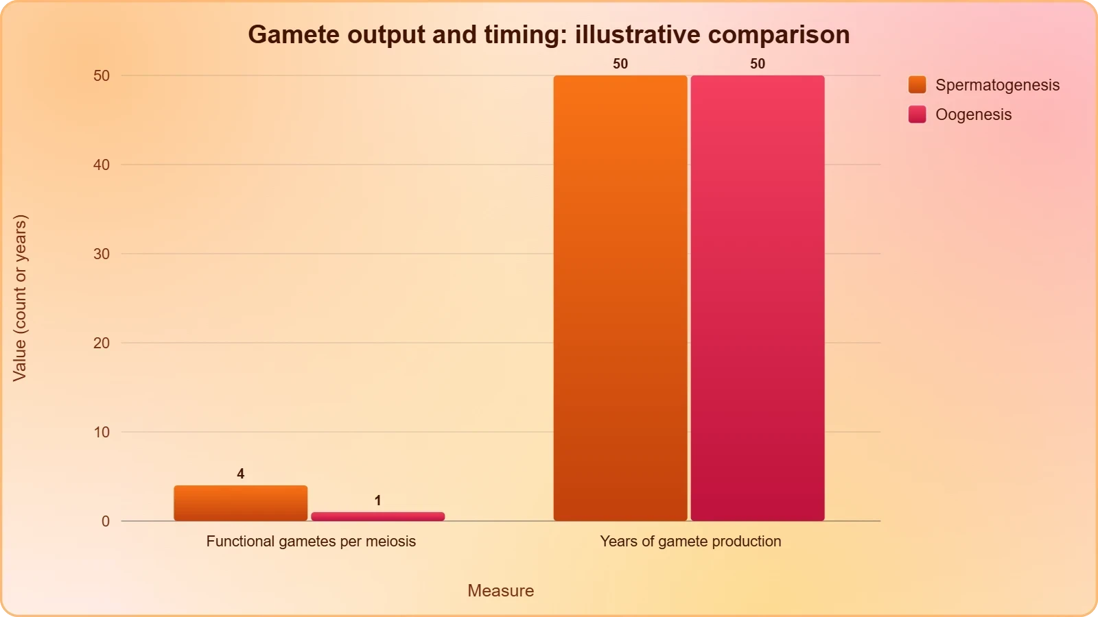
Oogenesis differs from spermatogenesis in timing, yield and cytoplasmic distribution. Whereas a primary spermatocyte yields four functional spermatids, a primary oocyte completes meiosis with unequal cytoplasmic division, producing one large functional ovum and smaller polar bodies that degenerate. This asymmetry conserves cytoplasmic reserves and organelles for the early embryo. The process begins before birth and is suspended in prophase I until puberty, after which a cohort of follicles resumes development each cycle, usually culminating in a single ovulation.
The contrast in duration is equally instructive. Sperm formation continues even in old men, but formation of the ovum ceases in women around the age of fifty years, reflecting the finite ovarian reserve established during fetal life. This difference explains why maternal age influences chromosomal risk and why the endocrine profile of the ageing ovary differs so markedly from the ageing testis. When you can articulate both the cellular and the temporal contrasts, you have genuinely integrated the two halves of the course rather than memorising them separately.
Hormonal Control of Gamete Maturation
The hormonal control of gamete maturation is best remembered as a division of labour. LH is the steroid driver: it stimulates Leydig cells in the male and theca cells in the female, providing the androgen substrate for oestrogen synthesis. FSH is the nurturing driver: it acts on Sertoli cells to sustain spermatogenesis and on granulosa cells to promote follicular growth and oestrogen production. Inhibin, secreted by Sertoli and granulosa cells, selectively suppresses FSH, providing fine control without shutting down the entire axis.
This division explains several clinical observations. Elevated FSH with low inhibin suggests failing gonadal feedback, as in diminished ovarian reserve. Suppressed FSH with low LH suggests a hypothalamic or pituitary problem rather than a gonadal one. In the male, selective FSH suppression by inhibin allows sperm production to continue under LH-driven androgen support. Reading a hormone panel as a story about which level of the axis has failed is a skill that transfers directly from this lesson to clinical and examination contexts alike.
Timing Mechanisms: The Menstrual Cycle and Its Coordination
The menstrual cycle is the temporal framework that synchronises ovulation, endometrial preparation and the possibility of implantation. Its phases are driven by the changing balance of oestrogen and progesterone, which in turn reflect follicular and luteal activity. In the follicular phase, FSH promotes follicle growth and granulosa cells secrete oestrogen, which thickens the endometrium. At mid-cycle, sustained high oestrogen triggers the LH surge and ovulation. The luteal phase follows, with the corpus luteum secreting progesterone that maintains the endometrium and suppresses further follicular development.
If fertilisation does not occur, the corpus luteum regresses, progesterone falls, and the endometrium is shed as menstruation. If implantation occurs, the developing embryo secretes signals that sustain the corpus luteum, preserving progesterone and preventing endometrial breakdown. The endometrium undergoes cyclical changes during the menstrual cycle, while the myometrium exhibits strong contraction during delivery of the baby. This dual behaviour of the uterine wall — cyclical remodelling in one layer, powerful contractility in another — is a neat illustration of how structure serves function across different timescales.
Reading the Cycle as a Hormone Graph
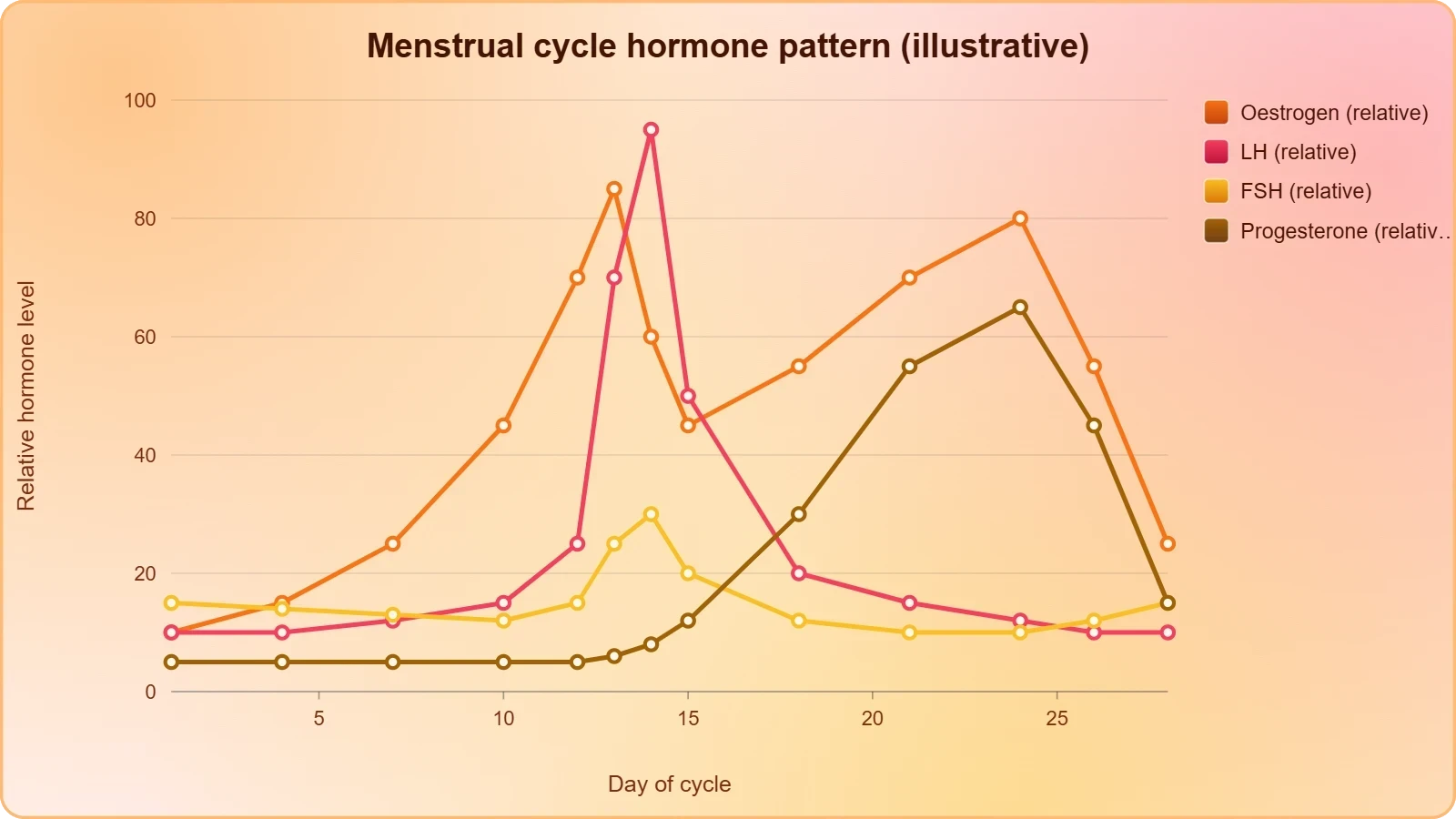
Examiners frequently present a hormone graph and ask you to identify phases and events. The reliable strategy is to anchor on the LH surge, which marks ovulation and divides the cycle into follicular and luteal halves. Before the surge, oestrogen rises steadily with follicular growth. After the surge, progesterone dominates and oestrogen shows a secondary rise. FSH rises early in the follicular phase and again briefly at mid-cycle. Menstruation coincides with the fall of both ovarian steroids. Once you can narrate the graph in this order, most cycle questions become straightforward.
It also helps to attach a physiological purpose to each rise and fall. Rising oestrogen thickens the endometrium and, at its peak, converts feedback from negative to positive. The LH surge triggers follicular rupture roughly a day and a half later. Progesterone stabilises the endometrium and raises basal body temperature slightly, which is why temperature tracking can crudely indicate ovulation. Falling progesterone withdraws support and precipitates menstruation. Every curve on the graph therefore has a downstream consequence, and describing those consequences is what earns full marks.
Linking the Cycle to Contraception
Contraceptive mechanisms become intuitive once the cycle is understood. Combined hormonal contraceptives deliver steady levels of oestrogen and progestogen, which suppress GnRH and gonadotropin release through negative feedback. Without a mid-cycle LH surge, ovulation does not occur. Progestogen additionally thickens cervical mucus, impeding sperm passage, and thins the endometrium, reducing receptivity to implantation. Barrier methods, by contrast, act mechanically before fertilisation, while intrauterine devices interfere with sperm function and endometrial environment. Each method can be mapped onto a specific point in the reproductive sequence.
This mapping is exactly the kind of integrated reasoning that higher-level questions reward. A question asking how a hormonal contraceptive prevents pregnancy is really asking you to trace the axis from hypothalamus to endometrium and identify every point of interruption. A strong answer names the feedback suppression, the absent LH surge, the altered cervical mucus and the changed endometrium, and then explains how these together prevent gamete meeting and implantation. Practise writing such answers in structured paragraphs rather than bullet fragments.
From Fertilisation to Parturition and Lactation
The reproductive events in humans include formation of gametes, transfer of sperms into the female genital tract (insemination), and fusion of male and female gametes (fertilisation) leading to formation of the zygote. This is followed by formation and development of the blastocyst and its attachment to the uterine wall (implantation), embryonic development (gestation) and delivery of the baby (parturition). These events occur after puberty, and each depends on the preceding one. Fertilisation typically occurs in the ampulla of the oviduct, and the resulting zygote travels toward the uterus while dividing.
Implantation establishes the maternal–fetal relationship and depends on a receptive endometrium primed by progesterone. Gestation then proceeds through embryonic development, with the placenta assuming endocrine and nutritive roles. Parturition is initiated by coordinated signals that increase uterine contractility, and the myometrium exhibits strong contraction during delivery. Lactation follows, with milk secreted by alveolar cells, stored in lumens, and expressed through the duct system at the nipple. Each stage is a distinct physiological event, yet all are linked by the same hormonal thread that runs through the entire course.
Sequencing the Events Correctly
Examiners often test the order of events because it reveals whether you understand causality. The correct sequence is gametogenesis, insemination, fertilisation, blastocyst formation, implantation, gestation and parturition, with lactation following birth. A common error is to place implantation before blastocyst formation, or to describe the zygote as implanting directly. In reality, cleavage produces the blastocyst, and only the blastocyst attaches to the uterine wall. Another frequent slip is to confuse the site of fertilisation with the site of implantation; the former is the oviduct, the latter the endometrium.
Precision about location and timing earns marks. Fertilisation occurs in the upper oviduct, implantation in the endometrium, and gestation within the uterus. Parturition involves cervical dilation and myometrial contraction, while lactation involves alveolar secretion and ductal transport. When you write an answer, name the structure, the event and the hormonal driver together. This habit prevents vague descriptions and demonstrates the integrated understanding that the final lesson of the course is designed to consolidate before your examination.
Lactation as an Integrated Endpoint
Lactation is a fitting endpoint because it draws on anatomy, endocrinology and neural reflexes simultaneously. The mammary gland's glandular tissue is divided into 15–20 lobes of alveoli, whose cells secrete milk into lumens; the milk then passes through mammary tubules, ducts, the ampulla and the lactiferous duct to the nipple. Suckling provides sensory input that sustains the hormonal environment required for milk production and ejection. This reflex arc is a clear example of neuroendocrine integration, linking the infant's behaviour to the mother's physiology.
Lactation also illustrates the principle of functional continuity. The same hormonal shifts that support pregnancy prepare the mammary gland for milk secretion, and the same hypothalamic–pituitary system that governed ovulation now participates in milk ejection. When you revise, treat lactation not as an isolated final topic but as the closing chord of the reproductive symphony. If you can explain how a single neuroendocrine axis coordinates gamete production, cyclical preparation, pregnancy and breastfeeding, you have achieved the integration this course set out to build.
Examination Workshop: Mistakes, Practice and Strategy
The final stage of preparation is diagnostic: identify the errors you are most likely to make and eliminate them deliberately. The most common confusion in this course is between Sertoli and Leydig cells. Sertoli cells sit inside the seminiferous tubule, nourish germ cells and respond to FSH; Leydig cells sit in the interstitial spaces, secrete androgens and respond to LH. A second frequent error is mixing the roles of LH and FSH, attributing ovulation to FSH or spermatogenesis support to LH. A third is misplacing events, such as describing the zygote as implanting directly or fertilisation as occurring in the uterus.
Other recurring mistakes include confusing the hymen with a reliable indicator of virginity, which it is not; forgetting that the scrotum maintains a temperature 2–2.5 °C below core temperature; and muddling the layers of the uterine wall, where the endometrium undergoes cyclical change while the myometrium contracts during delivery. Students also lose marks by omitting the direction of feedback, writing simply that oestrogen inhibits the pituitary without specifying the concentration-dependent switch to positive feedback at mid-cycle. Each of these errors is avoidable with a short, targeted checklist.
Worked Practice Questions
Consider this integrated question: explain how a combined oral contraceptive prevents pregnancy. A complete answer traces the axis. Steady exogenous oestrogen and progestogen suppress GnRH and gonadotropin secretion through negative feedback, so no mid-cycle LH surge occurs and ovulation is prevented. Progestogen thickens cervical mucus, hindering sperm passage, and thins the endometrium, reducing the chance of implantation. The answer should also note that these effects are reversible on discontinuation. Structuring the response as feedback suppression, then gamete transport, then endometrial receptivity demonstrates the multi-topic linkage examiners reward.
A second question might ask you to compare spermatogenesis and oogenesis. A strong response covers site, timing of initiation, duration, meiotic products, cytoplasmic division and hormonal control. Spermatogenesis begins at puberty and continues into old age, yielding four haploid spermatids per primary spermatocyte through equal divisions. Oogenesis begins before birth, is suspended until puberty, ceases around age fifty, and yields one functional ovum with polar bodies through unequal division. Both depend on the hypothalamic–pituitary axis, but the ovarian feedback pattern is cyclical while the testicular pattern is stable. Such comparative answers score highly.
Revision Strategy for the Final Week
Effective revision for this course is active rather than passive. Draw the endocrine flowchart from memory daily, then check it against the table in this lesson. Narrate the menstrual cycle as a hormone graph without looking at notes, anchoring on the LH surge. Write one integrated answer per day, such as the contraceptive question or a comparison of the two gametogenies. Finally, rehearse the sequence from gametogenesis to lactation as a single spoken narrative, because fluency in speech usually predicts fluency in writing under examination conditions.
Pay particular attention to terminology, since marks are lost for imprecise words. Distinguish insemination from fertilisation, blastocyst from zygote, and gestation from parturition. Use the correct terms for the layers of the uterine wall and the parts of the oviduct. When describing hormonal effects, always name the source, the target and the consequence. This discipline transforms a vague answer into a precise one. With the whole course now integrated into a single narrative, you are prepared not merely to recall facts but to reason across topics, which is the true measure of mastery in reproductive biology.
Also Read
- No links available.
From our network :
- 10 Physics Numerical Problems with Solutions for IIT JEEPrepare for IIT JEE with 10 physics numerical problems, complete with detailed solutions and conceptual explanations. Ideal for mastering kinematics, thermodynamics, electromagnetism, and optics.
- Analyzing Trump Deportation Numbers Insights Into The 2026 Immigration Crackdown
- Do You Need a New Router or Is Your Internet Plan the Problem?A practical comparison of the signs that point to router limits versus signs your broadband speed or provider is the bottleneck, so you can decide what to replace before spending money.
- How to Print Cheaply: Cutting the Cost per PageA practical, measured guide to lowering what each printed page costs — duplex and draft modes, third-party ink, yield mathematics, and knowing when a new printer beats refills.
- EV 2.0: The Solid-State Battery Breakthrough and Global Factory ExpansionDiscover the impact of the Solid-State Battery Breakthrough on the EV market. Learn about CATL and BYD stock growth, global factory expansion in Europe, and the future of EV 2.0 technology.
- Where to Put Your Wireless Router for Better CoverageA practical, physics-grounded guide to router placement: why the centre of the home beats the corner, how walls and metal sap signal, and how to run a simple survey that proves the improvement.
- 98% of Global MBA Programs Now Prefer GRE Over GMAT Focus EditionExplore why 98% of global MBA programs now prefer the GRE over GMAT Focus. Learn about the shorter GRE format, verbal logic importance, and strategic versatility for students.
- AI-Powered 'Precision Diagnostic' Replaces Standard GRE Score ReportsDiscover the new GRE Precision Diagnostic. Learn how AI-powered behavioral analytics and time-to-solve tracking are replacing standard GRE score reports to empower students.
- Trump Political Strategy How Geopolitical Stunts Serve As Media Diversions
RESOURCES
- The Future of IVF: The New Normal in Human Reproduction - PMCpmc.ncbi.nlm.nih.govJan 3, 2022 ... Integration of microfluidics into the IVF laboratory may give rise ... practice. Indeed, microfluidics has been used for the isolation ...
- Sperm DNA fragmentation: how to test, when to test, and what to do ...academic.oup.comApr 28, 2026 ... ... integration of SDF testing into clinical practice. Rather than ... C. Improving standard practices in studies using results from…
- ESHRE good practice recommendations on recurrent implantation ...pmc.ncbi.nlm.nih.govJun 15, 2023 ... Deciphering the effect of reproductive tract microbiota on human reproduction. ... WHO Laboratory Manual for the Examination and Processing of ...
- Guidance on qualifications for fertility counselors: a committee opinionintegration.asrm.org... reproduction undergo a psychosocial consultation with psychological testing when appropriate. ... Assisted human reproduction counselling practice guidelines, ...
- Diagnosing adenomyosis: an integrated clinical and imaging ...pubmed.ncbi.nlm.nih.govApr 15, 2020 ... ... examination and imaging, is proposed to adequately identify and characterise adenomyosis. ... Human Reproduction and Embryology. All rights ...
- MemoNeet: Neet Revision App - App Storeapps.apple.comMemoNeet: NEET Exam Prep 2026 Welcome Future Doctors! Prepare smarter ... Human Reproduction 26. Reproductive Health 27. Principles of Inheritance and ...
- Recommendations From the 2023 International Evidence-based ...pubmed.ncbi.nlm.nih.govSep 18, 2023 ... ... Reproductive Medicine, Endocrine Society, European Society for Human Reproduction and Embryology, and the European …
- NASW Standards for Integrating Genetics into Social Work Practicesocialworkers.org... testing and referral resources for such testing. In cases where ... human reproduction, medical intervention and the value of life with major disabilities.
- Integrating Sexual and Reproductive Health Services Within HIV ...frontiersin.orgIntegration of HIV testing services into family planning ... Does integration of HIV and SRH services achieve economies of scale and scope in practice?
- Clinical utility of sperm DNA fragmentation testingtau.amegroups.orgSperm DNA is an integral element in the success of human reproduction. There ... Practice Committee of the American Society for Reproductive Medicine.
- Testing and interpreting measures of ovarian reserve: a ... - ASRMasrm.orgThe process of reproductive aging has traditionally centered on the principle that human oocytes peak in number during fetal life, undergo ovulation or atresia ...
- Expanding access to sexual and reproductive health and rights ...gh.bmj.comApr 2, 2025 ... ... integration of SRHR into broader health policy dialogues. ... practice guidelines for development used in training (0=lowest adherence ...
- Sexual and reproductive health and rightswho.intThe World Health Organization defines sexual health as a state of physical, emotional, mental and social well-being related to sexuality; it is not merely ...
- Reproduction unit test key File PDFwww2.american-usa.comImplementing Reproduction Unit Test Keys in Practice ... Record the reproduction key details and automate the test as part of the continuous integration.
- Opinions on integrating couple counselling and female sexual ...journals.plos.orgSep 9, 2022 ... Integrating female Sexual and Reproductive Health (SRH) services into male-dominated service could be more effective than trying to integrate ...