Spermatogenesis is the continuous, hormonally governed production line that converts a diploid stem cell in the wall of a seminiferous tubule into a streamlined, motile, enzyme-armed spermatozoon capable of traversing the female reproductive tract and delivering a haploid genome to an oocyte. It begins at puberty, when rising gonadotropin-releasing hormone (GnRH) from the hypothalamus switches on the anterior pituitary, and it continues, in principle, for the remainder of a man's life. Understanding this process requires holding three pictures in mind at once: the cellular sequence of mitotic multiplication, meiotic reduction and morphological remodelling; the endocrine loop of GnRH, luteinising hormone (LH), follicle stimulating hormone (FSH) and androgens; and the structural logic of the mature sperm, whose every compartment exists to serve fertilisation.
The clinical and educational value of this topic is considerable. Male infertility assessment, contraceptive research, toxicology and even forensic biology all rest on accurate knowledge of sperm production, sperm morphology and sperm motility. Equally, the topic is a favourite of examiners because it rewards precise numerical reasoning: chromosome counts at each stage, the arithmetic of one primary spermatocyte yielding four spermatids, and the timing of meiotic events. Learners who confuse spermatogenesis with spermiogenesis, or who assume that meiosis in the male is completed before birth as it is in the female, lose marks and, more importantly, lose the conceptual thread that connects this lesson to the one on oogenesis that follows.
This lesson builds directly on the anatomy of the male reproductive system and prepares the ground for oogenesis. It traces the lineage from spermatogonium to spermatozoon, examines the hormonal control loop in detail, dissects the sperm's head, neck, middle piece and tail, and extends the core syllabus with the acrosome reaction, mitochondrial ATP supply, the roughly 64–74 day duration of the process, and the modern World Health Organization semen analysis criteria that refine the simplified textbook figures. Worked calculations, a programming model of the cell divisions, and comparison tables are included so that the material can be revised quickly and applied confidently.
On This Page
- The Cellular Sequence: From Spermatogonia to Spermatids
- Spermiogenesis and Spermiation: Building and Releasing the Sperm
- Hormonal Control: The Hypothalamic–Pituitary–Testicular Axis
- The Structure of a Spermatozoon
- Transport, Maturation and the Making of Semen
- Worked Examples, Comparisons and Revision
- Clinical Connections and Exam-Style Practice
The Cellular Sequence: From Spermatogonia to Spermatids
Spermatogenesis unfolds inside the seminiferous tubules, the coiled ducts that constitute the bulk of testicular tissue. The immature male germ cells, the spermatogonia, lie on the inner wall of these tubules. Each spermatogonium is diploid and contains 46 chromosomes. Beginning at puberty, these cells multiply by mitotic division, replenishing their own population while periodically committing some daughter cells to the meiotic pathway. This mitotic reserve is the reason sperm production can continue indefinitely, a point of sharp contrast with the female system, where the entire stock of oocytes is laid down before birth and never renewed.
The committed cells, called primary spermatocytes, are the largest germ cells in the tubule and are the first to enter meiosis. A primary spermatocyte completes the first meiotic division, the reduction division, producing two equal haploid cells called secondary spermatocytes, each carrying 23 chromosomes. These secondary spermatocytes then undergo the second meiotic division, which separates sister chromatids and yields four equal haploid spermatids. The arithmetic is worth stating plainly: one diploid cell with 46 chromosomes gives four haploid cells with 23 chromosomes each, and the reduction occurs at the first division, not the second.
Chromosome Bookkeeping Through the Divisions
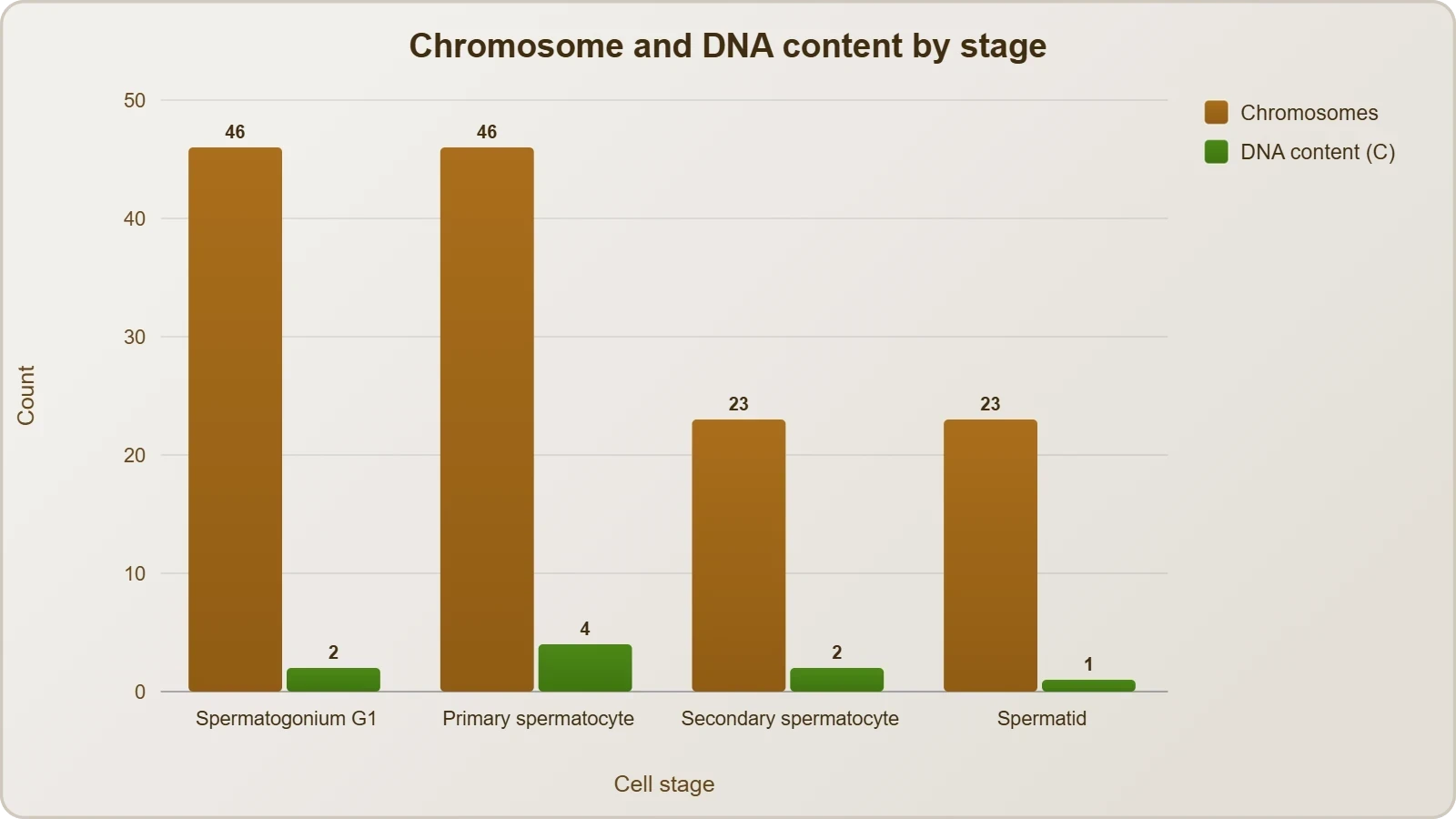
Examiners frequently ask for the chromosome number and DNA content at each stage, and the safest way to answer is to track both quantities explicitly. A spermatogonium in the G1 phase has 46 chromosomes and a DNA content conventionally denoted 2C. After S-phase replication it still has 46 chromosomes but 4C of DNA, because each chromosome now consists of two sister chromatids. The primary spermatocyte that enters meiosis therefore carries 46 chromosomes and 4C DNA, and this is the state in which it remains through prophase I, metaphase I and anaphase I.
At the completion of meiosis I, the two secondary spermatocytes each receive 23 chromosomes, but each chromosome still comprises two chromatids, so the DNA content is 2C. The second meiotic division separates the chromatids, and each spermatid ends with 23 chromosomes and 1C of DNA. The following relationship summarises the whole trajectory, where ##n## is the haploid number and ##C## the haploid DNA content:
Why the Divisions Are Equal
In the male, cytoplasmic division at both meiotic stages is essentially equal, so four functional spermatids emerge from each primary spermatocyte. This is a deliberate contrast with oogenesis, where the first meiotic division is markedly unequal, producing one large secondary oocyte and a tiny first polar body so that the future embryo retains the bulk of the nutrient-rich cytoplasm. The male strategy is numerical: maximise gamete count, since each sperm is a small, disposable delivery vehicle. The female strategy is provisioning: concentrate resources in a single cell that must sustain early development.
Textbook diagrams often compress the process into a neat vertical cascade, but the reality is a wave. Spermatogenesis proceeds in coordinated stages along the length of each tubule, so that cross-sections at different points show different combinations of cell types. This spatial organisation, together with the supporting Sertoli cells, creates the blood–testis barrier that shelters developing germ cells from immune attack and from many circulating toxins. Recognising this architecture explains why certain drugs and infections damage fertility in characteristic patterns rather than uniformly.
Spermiogenesis and Spermiation: Building and Releasing the Sperm
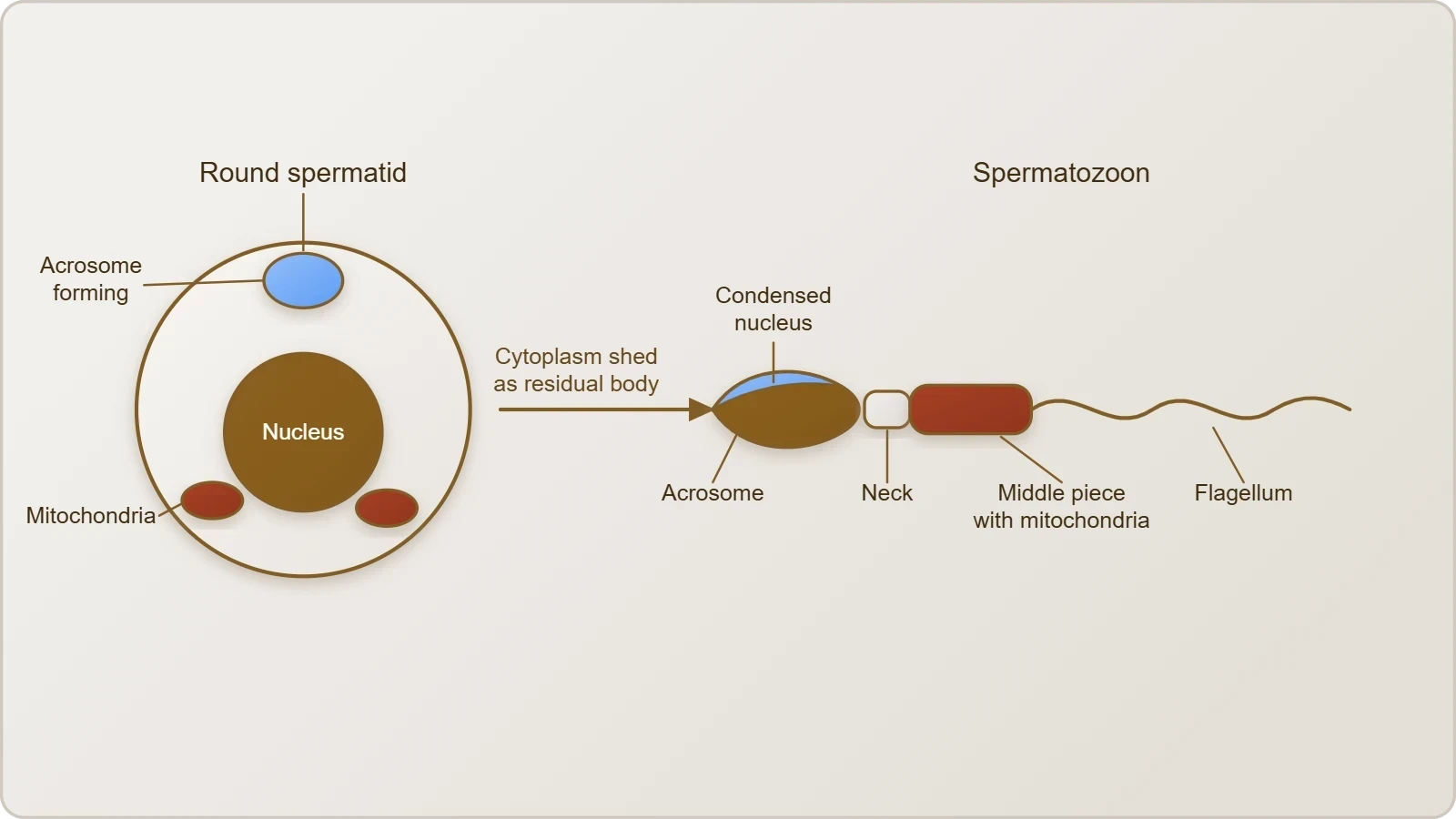
The haploid spermatids produced by meiosis are round, unremarkable cells. Their transformation into spermatozoa is a separate morphological process called spermiogenesis, and it is here that most of the structural drama occurs. The nucleus condenses dramatically as histones are largely replaced by protamines, the acrosome is assembled over the anterior nucleus from Golgi-derived vesicles, the flagellum grows from the distal centriole, and superfluous cytoplasm is discarded as a residual body. Mitochondria migrate to form a helical sheath around the proximal flagellum, and the cell elongates into the familiar streamlined profile.
Once spermiogenesis is complete, the sperm heads become embedded in the Sertoli cells, which nurse and orient the maturing cells throughout their development. The final release from the seminiferous tubule into the lumen is called spermiation. The released spermatozoa are morphologically mature but functionally immature: they lack vigorous progressive motility and cannot yet fertilise an oocyte. Their maturation continues during transit through the epididymis, where secretions modify the plasma membrane and stabilise the flagellum, a point that connects directly to the accessory ducts covered earlier in the course.
The Acrosome and the Acrosome Reaction
The acrosome is a cap-like structure covering the anterior portion of the sperm head, filled with hydrolytic enzymes including hyaluronidase and acrosin. Its purpose becomes clear at fertilisation. When a sperm binds to the zona pellucida of the oocyte, the binding triggers the acrosome reaction, an exocytotic event in which the outer acrosomal membrane fuses with the overlying plasma membrane and releases its enzymes. These enzymes digest a path through the zona pellucida, allowing the sperm to reach the oolemma and fuse with it. Only a sperm that has undergone capacitation in the female tract can execute this reaction.
The acrosome reaction is therefore a precisely timed event, not a continuous leak. Premature release of acrosomal enzymes would leave the sperm unable to penetrate the oocyte investments, while failure to react would leave it trapped at the zona. This is one reason why sperm quality is assessed not only by count and motility but also by morphology: an abnormally shaped acrosome may be functionally incompetent even when the sperm swims well. It also explains why the acrosome is described as the sperm's chemical key, matched to the lock presented by the oocyte's extracellular coat.
Mitochondria, ATP and the Middle Piece
The middle piece of the sperm possesses numerous mitochondria, and their arrangement is no accident. The flagellum is driven by dynein motor proteins that consume ATP, and the mitochondrion is the organelle that produces it through oxidative phosphorylation. The helical mitochondrial sheath sits immediately behind the neck, close to the base of the axoneme, minimising diffusion distance between ATP generation and ATP consumption. This local energy supply is what sustains the vigorous motility required for the sperm to traverse the cervix, uterus and oviduct.
Energy metabolism in sperm is more nuanced than a simple mitochondrion-equals-motility equation. Sperm can also generate ATP by glycolysis along the fibrous sheath of the principal piece, and different species and even different sperm populations rely on these pathways to differing degrees. Nevertheless, the mitochondrial contribution is central, and mitochondrial dysfunction is associated with reduced motility. The following expression captures the overall energy demand in qualitative terms, where ##v## is swimming velocity and ##\eta## an effective drag coefficient:
Hormonal Control: The Hypothalamic–Pituitary–Testicular Axis
Spermatogenesis does not run on its own; it is switched on and sustained by a three-tier endocrine loop. At puberty, secretion of gonadotropin-releasing hormone (GnRH) from the hypothalamus increases significantly. GnRH travels in the hypophyseal portal blood to the anterior pituitary, where it stimulates the secretion of two gonadotropins: luteinising hormone (LH) and follicle stimulating hormone (FSH). These two hormones then act on different testicular cell types, and their combined action drives germ cell development. The elegance of the system lies in its division of labour between a steroid signal and a peptide signal.
LH acts on the Leydig cells, the interstitial cells lying between the seminiferous tubules, and stimulates the synthesis and secretion of androgens, principally testosterone. Androgens in turn stimulate spermatogenesis, acting on both the germ cells and the somatic support cells. FSH acts on the Sertoli cells and stimulates the secretion of factors that support spermiogenesis, including androgen-binding protein, which concentrates testosterone within the tubule, and a range of paracrine signals. Neither hormone alone is sufficient; the process requires the coordinated steroid and peptide input, which is why hypogonadotropic states impair fertility.
Negative Feedback and Its Clinical Meaning
The axis is restrained by negative feedback. Testosterone and its metabolite dihydrotestosterone act on the hypothalamus and pituitary to suppress GnRH and LH secretion, while inhibin, produced by Sertoli cells, selectively suppresses FSH. This feedback architecture explains a familiar clinical pattern: exogenous anabolic steroids suppress LH and FSH, shrink the testes and reduce sperm production, sometimes to zero, even though the user's muscles grow. The body reads the exogenous androgen as a signal that production is adequate and shuts down the endogenous drive.
Conversely, failure of the testes to produce testosterone removes negative feedback, so LH and FSH rise, producing the hormonal signature of primary testicular failure. Failure at the hypothalamic or pituitary level produces low gonadotropins with low testosterone, the signature of secondary failure. Distinguishing these patterns is a routine part of infertility investigation, and it follows directly from the feedback logic taught here. The table below summarises the principal hormones, their sources and their testicular targets.
Timing: Sixty-Four to Seventy-Four Days and a Lifetime of Output
In humans, the journey from spermatogonium to released spermatozoon takes approximately 64 to 74 days, with the epididymal transit adding roughly another 10 to 14 days before the sperm is fully mature and motile. Because the process is continuous and asynchronous along the tubule, sperm are produced without interruption rather than in seasonal or monthly batches. This is a fundamental difference from the female cycle, where a single ovum is released roughly every 28 days. The continuous output means that a temporary insult, such as fever or a course of medication, may depress sperm counts for weeks before recovery.
That delay has practical consequences. A man starting a medication that might affect fertility will not see the effect in a semen analysis immediately, because the cells currently in the ejaculate were already well advanced in development two to three months earlier. Similarly, recovery after stopping a harmful exposure takes weeks to months. Clinicians therefore repeat semen analyses after an interval of roughly two to three months when assessing change. Understanding the timeline turns a memorised number into a usable piece of clinical reasoning.
The Structure of a Spermatozoon
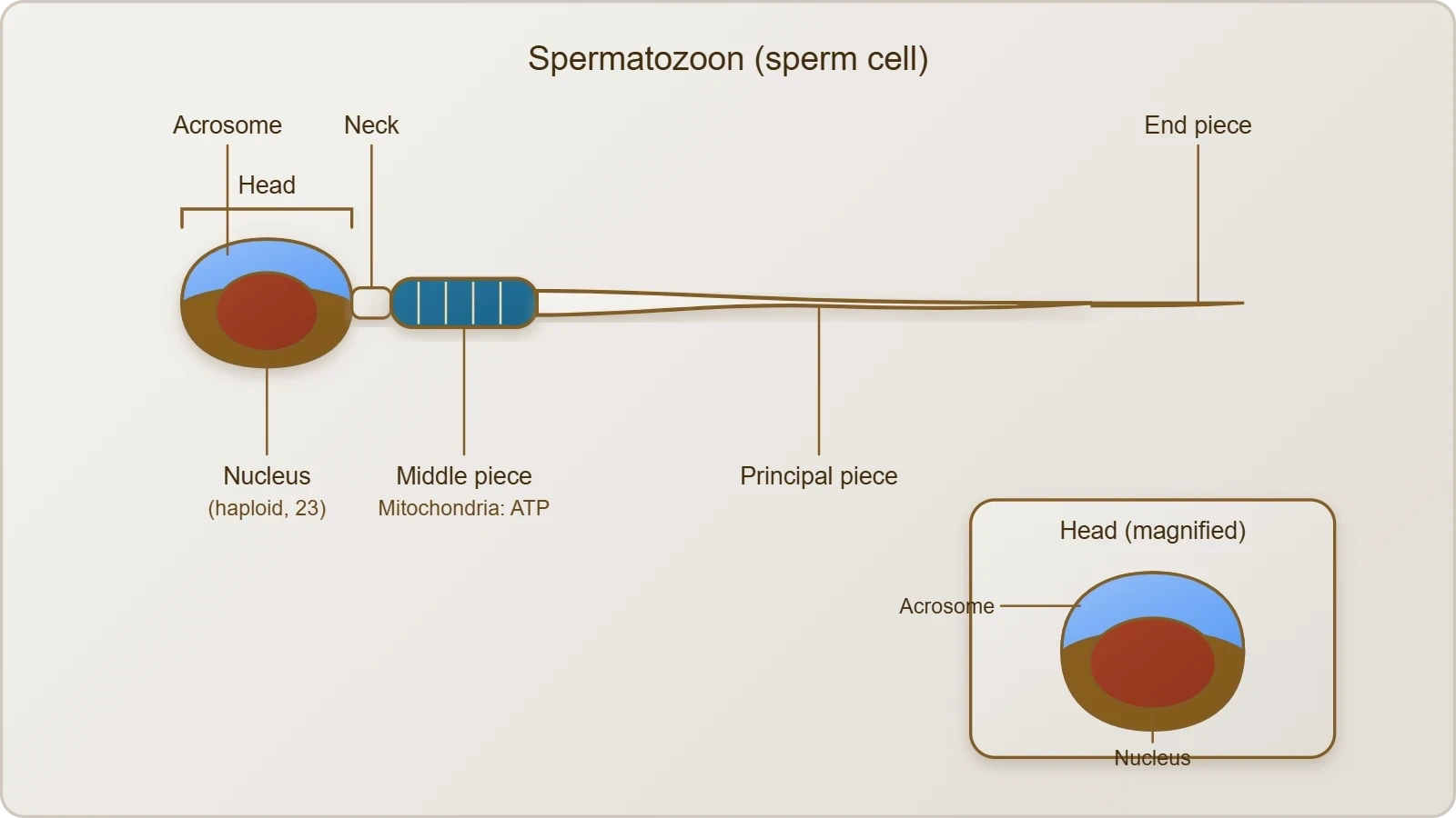
The mature sperm is a microscopic structure composed of a head, a neck, a middle piece and a tail, all enveloped by a plasma membrane. The head contains an elongated haploid nucleus, the anterior portion of which is covered by the acrosome, the enzyme-filled cap that will later permit penetration of the oocyte. The neck contains the centriole that organises the flagellar axoneme. The middle piece carries the mitochondrial sheath that supplies ATP, and the tail is the flagellum proper, whose beating propels the cell. Each compartment maps onto a specific function, which is why morphology matters as much as count.
Beyond the textbook description, the sperm is a highly specialised cell that has jettisoned almost everything it does not need. It carries virtually no cytoplasm, no endoplasmic reticulum of consequence and no ribosomes capable of protein synthesis; it is transcriptionally silent. Its genome is packaged with protamines rather than histones, giving the nucleus exceptional compactness and protecting the DNA during transit. The residual body discarded during spermiogenesis removes excess cytoplasm, reducing the cell's mass and improving its hydrodynamic efficiency. The design is that of a delivery vehicle, not a self-sufficient organism.
Head, Neck, Middle Piece and Tail in Functional Terms
The head's acrosome is a modified lysosome-like vesicle, and its enzymes are the tools of penetration. The nucleus within is haploid, carrying 23 chromosomes, and its condensation is so extreme that the DNA is almost crystalline. The neck is a short segment containing the proximal centriole, which is delivered to the oocyte at fertilisation and helps organise the first mitotic spindle of the zygote. The middle piece is the power plant, its mitochondria arranged helically around the axoneme. The tail, or principal piece, contains the fibrous sheath that provides elastic recoil and supports glycolysis.
This division of labour is reflected in the way abnormalities present. A sperm with a defective acrosome may swim perfectly but fail to penetrate. A sperm with a defective mitochondrial sheath may have normal morphology but poor motility. A sperm with an abnormal head shape may carry chromatin damage. This is why modern semen analysis reports several parameters rather than a single number, and why morphology is graded strictly. The table below sets out the compartments and their functions for rapid revision.
Numbers, Motility and the Limits of Simplified Criteria
The reference figures state that a human male ejaculates about 200 to 300 million sperms during a coitus, and that for normal fertility at least 60 per cent must have normal shape and size while at least 40 per cent must show vigorous motility. These numbers are useful for examination purposes and capture the essential idea that both morphology and motility matter. They should, however, be recognised as simplified criteria. Modern laboratory practice follows the World Health Organization reference values, which are more detailed and are based on the lower fifth percentile of men who have fathered children within twelve months.
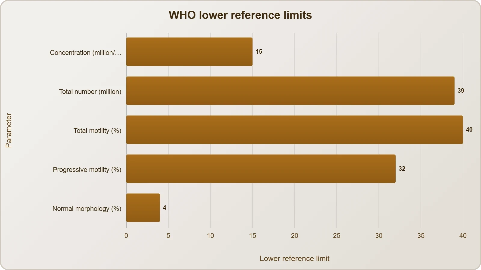
The current WHO framework sets a lower reference limit for sperm concentration of 15 million per millilitre, total sperm number of 39 million per ejaculate, total motility of 40 per cent, progressive motility of 32 per cent, and normal morphology of 4 per cent using strict criteria. The apparent discrepancy with the older 60 per cent figure arises because strict morphology grading counts even minor deviations as abnormal, so a lower percentage of strictly normal forms is compatible with fertility. The lesson is not that one figure is wrong but that clinical thresholds are statistical and context-dependent.
Transport, Maturation and the Making of Semen
Spermatozoa released from the seminiferous tubules are transported by the accessory ducts, beginning with the rete testis and the efferent ductules and continuing into the epididymis. Along this route they are not merely conveyed but modified. Secretions of the epididymis, vas deferens, seminal vesicle and prostate are essential for maturation and motility of sperms. The epididymis in particular provides a milieu in which the sperm plasma membrane is remodelled, surface proteins are altered and the flagellum acquires the capacity for coordinated beating. Sperm leaving the testis are immature; sperm leaving the epididymis are competent.
The seminal plasma along with the sperms constitutes the semen. Seminal vesicle secretion is rich in fructose, which serves as a glycolytic substrate for the sperm, together with prostaglandins and coagulating proteins. Prostatic secretion contributes proteolytic enzymes that liquefy the coagulum, plus zinc and citrate. Bulbourethral secretions add lubricating mucus. The result is a complex fluid that nourishes, buffers and protects the sperm and modulates the female tract. The functions of the male sex accessory ducts and glands are maintained by the testicular hormones, the androgens, which is why androgen deficiency affects fertility and glandular function together.
Capacitation and the Journey Through the Female Tract
Ejaculated sperm cannot fertilise an oocyte immediately. In the female tract they undergo capacitation, a series of membrane and biochemical changes that remove cholesterol from the plasma membrane, alter ion channel activity and increase intracellular signalling. Capacitation destabilises the acrosomal region so that it becomes responsive to the zona pellucida, and it enhances the flagellar beat. Only capacitated sperm can execute the acrosome reaction on contact with the oocyte's coat. This delay is a safety mechanism, preventing premature enzyme release before the sperm has reached the egg.
The journey itself is formidable. Of the hundreds of millions of sperm deposited, only a few hundred reach the ampulla of the oviduct, where fertilisation normally occurs. Many are lost through the cervix, destroyed by the acidic vaginal environment or engulfed by immune cells. The survivors are guided by thermotaxis and chemotaxis toward the oocyte. This numerical attrition is the evolutionary rationale for the enormous sperm count: the system is designed for redundancy, and a reduction in count or motility narrows the margin of success. The table below traces the maturation journey.
Common Misconceptions Worth Correcting
Several errors recur in examinations and in popular discussion. The first is to treat spermatogenesis and spermiogenesis as synonyms; the former is the whole process from spermatogonium to spermatozoon, the latter only the morphological transformation of spermatid to sperm. The second is to believe that meiosis in the male is completed before birth, as it is in the female; in the male, meiosis begins only at puberty and continues throughout life. The third is to assume that the presence of many sperm guarantees fertility, ignoring that morphology and motility are equally important. The fourth is to imagine that sperm are produced in a monthly cycle rather than continuously.
A fifth misconception concerns the hymen and virginity, which belongs to the previous lesson but is worth a brief reminder because it is so persistent: the presence or absence of the hymen is not a reliable indicator of virginity or sexual experience. A sixth is the belief that sperm count is fixed and cannot be influenced by lifestyle, when in fact heat, smoking, obesity, some medications and infections can all depress production, usually with a delay of weeks to months. Correcting these errors is as much a part of the lesson as learning the positive facts.
Worked Examples, Comparisons and Revision
Quantitative questions on spermatogenesis reward a systematic approach. The key is to identify the starting cell, apply the division rule, and track chromosome number and DNA content separately. A useful habit is to write the ploidy and the chromatid status beside each cell name before attempting arithmetic. The examples below illustrate the common question types, and the programming snippet that follows models the divisions so that the logic can be checked computationally. The comparison table then sets spermatogenesis against oogenesis, preparing the ground for the next lesson.
Worked Calculations
Consider a question asking how many spermatozoa arise from 100 primary spermatocytes. Each primary spermatocyte completes meiosis I to give two secondary spermatocytes and meiosis II to give four spermatids, so 100 primary spermatocytes yield 400 spermatids and, after spermiogenesis, 400 spermatozoa. If the question instead begins with 100 spermatogonia, the answer depends on how many of them commit to meiosis, since some continue mitotic division to replenish the stem population. Examiners usually specify the committed fraction, and the calculation then follows the same fourfold rule.
A second common question asks for the chromosome number in a secondary spermatocyte. Since the reduction division has already occurred, the answer is 23, but each chromosome still has two chromatids, so the DNA content is 2C. A third asks how many chromatids are present in a primary spermatocyte at metaphase I: 46 chromosomes each with two chromatids gives 92 chromatids. These questions are simple once the distinction between chromosome number and DNA content is secure, and they are the most frequent source of lost marks in this topic.
def spermatogenesis(primary_spermatocytes):
    """Model the meiotic yield from primary spermatocytes."""
    secondary = primary_spermatocytes * 2   # meiosis I: reduction division
    spermatids = secondary * 2              # meiosis II: chromatid separation
    spermatozoa = spermatids                # spermiogenesis does not change number
    return {
        "primary_spermatocytes": primary_spermatocytes,
        "secondary_spermatocytes": secondary,
        "spermatids": spermatids,
        "spermatozoa": spermatozoa,
        "chromosomes_per_spermatid": 23,
    }

result = spermatogenesis(100)
print(result)
# {'primary_spermatocytes': 100, 'secondary_spermatocytes': 200,
#  'spermatids': 400, 'spermatozoa': 400, 'chromosomes_per_spermatid': 23}
Spermatogenesis Versus Oogenesis
The contrast with oogenesis is instructive and prepares the next lesson. Spermatogenesis begins at puberty and continues throughout life, whereas oogenesis begins during embryonic development, when a couple of million oogonia are formed in each fetal ovary, and no more are added after birth. Spermatogenesis produces four functional gametes from each primary cell, whereas oogenesis produces one large secondary oocyte and a tiny first polar body from each primary oocyte, because the first meiotic division is unequal. Spermatogenesis is continuous and asynchronous; oogenesis is cyclical, culminating in ovulation roughly every 28 days.
The timing of meiotic arrest also differs. In the male, primary spermatocytes proceed through meiosis without prolonged arrest. In the female, primary oocytes enter prophase I during fetal life and remain arrested there for years, sometimes decades, until the surge of luteinising hormone triggers completion of the first meiotic division just before ovulation. The secondary oocyte is then arrested at metaphase II and completes the second division only if fertilisation occurs. These differences reflect the different biological strategies of the two gametes and explain why maternal age affects oocyte quality more than paternal age affects sperm production.
Clinical Connections and Exam-Style Practice
Spermatogenesis is a sensitive indicator of general health, and its disruption produces recognisable clinical patterns. Varicocele, a dilated vein in the scrotum, raises testicular temperature and impairs production. Cryptorchidism, in which a testis fails to descend, exposes germ cells to abdominal temperature and causes progressive damage unless corrected early. Infections such as mumps orchitis can destroy seminiferous tubules. Chemotherapy and radiotherapy are deliberately toxic to dividing cells and therefore hit spermatogenesis hard, which is why sperm banking is offered before treatment. Heat, smoking, heavy alcohol use and obesity all exert smaller but measurable effects.
The diagnostic pathway follows the physiology. A semen analysis is the first test, interpreted against WHO reference values. Hormone measurements then distinguish primary from secondary failure using the feedback logic described earlier. Genetic testing may follow when counts are very low. Treatment depends on the cause: surgical correction of varicocele, hormone replacement in hypogonadotropic states, or assisted reproduction where no correctable cause is found. Each step makes sense only if the underlying biology of spermatogenesis, its hormonal control and its timeline are understood, which is precisely why this lesson matters beyond the examination hall.
Practice Questions with Model Answers
Question one: state the chromosome number in a human spermatid and explain your reasoning. Model answer: 23, because the first meiotic division reduces the diploid number of 46 to a haploid 23 in each secondary spermatocyte, and the second division separates sister chromatids without changing the chromosome number. Question two: explain why FSH is required for spermatogenesis even though testosterone is the principal androgen. Model answer: FSH acts on Sertoli cells to stimulate secretion of factors that support spermiogenesis and to produce androgen-binding protein, which concentrates testosterone within the tubule; without this support, maturation is impaired.
Question three: a man taking exogenous anabolic steroids develops testicular shrinkage and a very low sperm count. Explain the mechanism. Model answer: the exogenous androgen raises circulating androgen levels, which exert negative feedback on the hypothalamus and pituitary, suppressing GnRH, LH and FSH. Without LH, Leydig cells stop producing testosterone locally, and without FSH, Sertoli cell support declines, so spermatogenesis is impaired and the testes shrink. Question four: why does a febrile illness cause a fall in sperm count that appears weeks later? Model answer: because spermatogenesis takes about 64 to 74 days, the cells affected by the illness are still maturing and only reach the ejaculate weeks afterwards.
Key Takeaways for Revision
The essential sequence is spermatogonium, primary spermatocyte, secondary spermatocyte, spermatid and spermatozoon, with meiosis reducing 46 chromosomes to 23 and one primary spermatocyte yielding four spermatozoa. Spermiogenesis is the morphological transformation, and spermiation is the release. GnRH from the hypothalamus drives LH and FSH from the anterior pituitary; LH acts on Leydig cells to make androgens, and FSH acts on Sertoli cells to support spermiogenesis. The sperm has a head with acrosome and haploid nucleus, a neck, a mitochondrial middle piece and a tail. The process takes roughly 64 to 74 days and continues throughout life.
Beyond the core, remember that the acrosome reaction releases enzymes to penetrate the zona pellucida, that mitochondrial ATP powers the flagellum, that capacitation in the female tract is a prerequisite for fertilisation, and that the textbook fertility figures are simplified compared with WHO reference values. Remember also the common misconceptions: spermatogenesis is not spermiogenesis, male meiosis is not completed before birth, and sperm production is continuous rather than monthly. With these points secure, the transition to oogenesis in the next lesson becomes a matter of comparison rather than confusion, and the whole architecture of human reproduction falls into place.
Also Read
- No links available.
From our network :
- 10 Physics Numerical Problems with Solutions for IIT JEEPrepare for IIT JEE with 10 physics numerical problems, complete with detailed solutions and conceptual explanations. Ideal for mastering kinematics, thermodynamics, electromagnetism, and optics.
- 98% of Global MBA Programs Now Prefer GRE Over GMAT Focus EditionExplore why 98% of global MBA programs now prefer the GRE over GMAT Focus. Learn about the shorter GRE format, verbal logic importance, and strategic versatility for students.
- How to Print Cheaply: Cutting the Cost per PageA practical, measured guide to lowering what each printed page costs — duplex and draft modes, third-party ink, yield mathematics, and knowing when a new printer beats refills.
- EV 2.0: The Solid-State Battery Breakthrough and Global Factory ExpansionDiscover the impact of the Solid-State Battery Breakthrough on the EV market. Learn about CATL and BYD stock growth, global factory expansion in Europe, and the future of EV 2.0 technology.
- AI-Powered 'Precision Diagnostic' Replaces Standard GRE Score ReportsDiscover the new GRE Precision Diagnostic. Learn how AI-powered behavioral analytics and time-to-solve tracking are replacing standard GRE score reports to empower students.
- Analyzing Trump Deportation Numbers Insights Into The 2026 Immigration Crackdown
- Do You Need a New Router or Is Your Internet Plan the Problem?A practical comparison of the signs that point to router limits versus signs your broadband speed or provider is the bottleneck, so you can decide what to replace before spending money.
- Where to Put Your Wireless Router for Better CoverageA practical, physics-grounded guide to router placement: why the centre of the home beats the corner, how walls and metal sap signal, and how to run a simple survey that proves the improvement.
- Trump Political Strategy How Geopolitical Stunts Serve As Media Diversions
RESOURCES
- Endocrinology of the Testis and Spermatogenesis - Endotext - NCBIncbi.nlm.nih.govJan 7, 2026 ... The endocrine regulation of these cells and their role in supporting sperm development and androgen production are also considered. For…
- Hormone Regulation in Testicular Development and Function - PMCpmc.ncbi.nlm.nih.govSpermatogenesis is a complex process that involves the development of male germ cells, known as spermatogonia, into fully mature spermatozoa, or sperm. This ...
- Male Reproductive System: Structure & Function - Cleveland Clinicmy.clevelandclinic.orgMay 8, 2023 ... LH is necessary to continue the process of spermatogenesis. Testosterone. Testosterone is the main male sex hormone. It helps you…
- The Roles of Luteinizing Hormone, Follicle-Stimulating ... - PMC - NIHpmc.ncbi.nlm.nih.gov... process of spermatogenesis. 13. Inference and Summary. The study of ... O'Shaughnessy P.J. Hormonal Control of Germ Cell Development and Spermatogenesis.
- Mechanisms of Hormonal, Genetic, and Temperature Regulation of ...mdpi.comMar 29, 2025 ... Spermatogenesis is a complex and highly regulated process involving the proliferation, differentiation, and apoptosis of germ cells.
- Regulation of spermatogenesis: An evolutionary biologist's ...sciencedirect.comThe regulatory mechanisms controlling sperm output act primarily on spermatogonia. •. Differentiation of germ cells follows a highly coordinated and strictly ...
- Anatomy & Physiology Reproductive System Study Guide | Notespearson.comStructure of Sperm. Head: Contains ... Progesterone prepares endometrium (days 15-28). Female reproductive cycle phases Hormonal regulation of female cycle ...
- Single-cell RNA sequencing uncovers dynamic roadmap and cell ...cell.comDec 6, 2022 ... ... sperm structural assembly already being expressed in the lP–D spermatocytes stage. ... hormonal control of spermatogenesis. Ann. N. Y.…
- Metabolic Reprogramming in Male Infertility: Mechanistic Integration ...onlinelibrary.wiley.comJun 12, 2026 ... ... hormonal, energy-sensing and epigenetic regulation during spermatogenesis. ... regulation, spermatogenesis, sperm motility and capacitation [148].
- High fat diet-induced obesity prolongs critical stages of the ... - Naturenature.comJan 11, 2022 ... Deleterious effects of obesity upon the hormonal and molecular mechanisms controlling spermatogenesis and male fertility. Hum. Fertil. 18 ...
- 27.1 Anatomy and Physiology of the Testicular Reproductive Systemopenstax.orgApr 20, 2022 ... In this section, we examine each of these different structures, and discuss the process of sperm production and transport. This…
- effects on sperm morphology according to spermatogenesis stagestau.amegroups.orgDec 27, 2019 ... Abnormal sperm morphology and MAI reverted to control values 73 days after cessation of heating. Conclusions: A mild sustained increase…
- In utero and lactational exposure of male rats to 2,3,7,8 ... - PubMedpubmed.ncbi.nlm.nih.gov... stages of development; effects on spermatogenesis and male reproductive capability are reported herein. ... morphology of these sperm appeared to be unaffected.
- Part 2: Male Reproductive System - Normal Physiology and Structureoecd.orgstages of the spermatogenic cycle. During the transition ... Regulation of spermatogenesis relies not only on the classical endocrine control involving.
- Exposure to zinc oxide nanoparticles affects testicular structure ...atm.amegroups.orgJul 15, 2022 ... The method of obtaining the body weights, organ weights, histopathological examination of testis, sperm parameters, and serum sex hormones of ...