Within roughly thirty hours of fertilisation, a single diploid zygote sitting in the ampullary region of the fallopian tube begins one of the most tightly choreographed sequences in all of developmental biology. It divides without growing, then reorganises itself into a fluid-filled sphere, then hatches from its own protective coat and finally burrows into the lining of the uterus. Each of these steps — cleavage, the morula, the blastocyst and implantation — is not merely a stage on a diagram but a precisely timed physiological event, and each one can fail in ways that explain a substantial share of early pregnancy loss in humans.
This lesson follows that trajectory in detail, building directly on the chromosomal logic of sex determination established in the previous session and preparing the ground for the placenta, which is the subject of the next. The emphasis falls on mechanism rather than memorisation: why cleavage produces smaller and smaller cells instead of a bigger embryo, why the blastocyst must differentiate into two distinct cell populations before it can attach, and why the trophoblast is the true architect of the maternal–fetal interface. Along the way we will examine the molecular signals that let a genetically foreign embryo survive inside its mother, the clinical realities of ectopic pregnancy and implantation failure, and the laboratory significance of pluripotent stem cells derived from the inner cell mass.
Because this material sits at the intersection of NCERT syllabus content and genuine clinical embryology, it rewards a learner who asks "how" as well as "what". The numbers matter — 2, 4, 8, 16 blastomeres, a morula of 8 to 16 cells, implantation at six to seven days — but the reasoning behind those numbers matters more. By the end of this lesson you should be able to trace a zygote from the isthmus of the oviduct to its embedded position in the endometrium, explain the endocrine conversation that begins the moment the trophoblast touches uterine tissue, and anticipate the questions an examiner or a clinician would ask next.
On This Page
- Cleavage: Division Without Growth in the Oviduct
- The Morula: A Compact Ball of Blastomeres
- Blastocyst Architecture: Trophoblast and Inner Cell Mass
- Implantation: Embedding in the Endometrium
- The Endocrine Handshake: hCG and the Rescue of the Corpus Luteum
- Quantitative and Laboratory Perspectives
- From Implantation to the Placenta: Setting Up the Next Lesson
- Clinical and Research Frontiers
Cleavage: Division Without Growth in the Oviduct
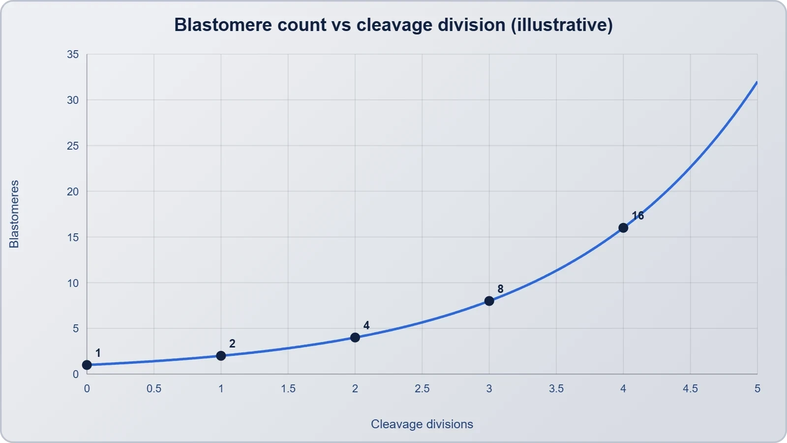
Cleavage is the series of rapid mitotic divisions that begins as the zygote travels through the isthmus of the oviduct toward the uterus. The defining feature of cleavage is that the embryo does not increase in overall volume; the cytoplasm of the zygote is simply partitioned into progressively smaller daughter cells called blastomeres. This is possible because the divisions occur with abbreviated gap phases, bypassing much of the growth that normally separates one mitotic cycle from the next. The zona pellucida, the glycoprotein coat surrounding the oocyte, remains intact throughout and constrains the embryo to a fixed spherical volume, so each division necessarily halves the cytoplasmic allotment of every cell.
The arithmetic of cleavage is deceptively simple and worth stating explicitly. Starting from one cell, successive divisions give 2, then 4, then 8, then 16 blastomeres, so that after ##n## divisions the embryo contains ##2^n## cells. The volume of each blastomere therefore scales as ##V_0 / 2^n##, where ##V_0## is the original zygote volume, which is why blastomeres become visibly smaller with each round. In humans the first division typically occurs about 24 to 30 hours after fertilisation, the second around 40 hours, and the third near 60 hours, so the eight-cell stage is reached on roughly day three.
Timing, Transport and the Zona Pellucida
Cleavage is not a stationary process. The embryo is being propelled along the oviduct by ciliary beating and smooth muscle contraction, and it reaches the uterine cavity at approximately day four to five, by which time it has formed a morula or early blastocyst. The zona pellucida serves two critical functions during this transit: it prevents the embryo from adhering prematurely to the tubal epithelium, and it holds the blastomeres together in a compact mass. Premature hatching or abnormal zona hardening are both associated with implantation failure, which is one reason zona manipulation is treated so carefully in assisted reproduction.
A useful way to remember the relationship between time and cell number is to note that cleavage divisions are asynchronous and not every blastomere divides at once. Real embryos at "day three" may contain anywhere from six to ten cells, and embryologists grade them partly on how uniform and regular those divisions appear. Perfectly synchronous division is the exception rather than the rule, and minor asynchrony is compatible with entirely normal development. What matters clinically is the overall trajectory: steady division, intact zona, and timely arrival in the uterus.
Compaction and the First Differentiation Event
Around the eight-cell stage the embryo undergoes compaction, a process in which the blastomeres flatten against one another and maximise their contact surfaces, transforming a loose cluster into a tight, smooth ball. Compaction is driven by the expression of cell adhesion molecules, principally E-cadherin, which links the cells into a coherent epithelium-like sheet. This is the first morphological sign that the embryo is preparing to differentiate, because cells on the outside of the compacted mass will adopt one fate and cells on the inside another. Compaction is also the point at which the embryo becomes metabolically more active and switches to relying partly on its own genome rather than stored maternal transcripts.
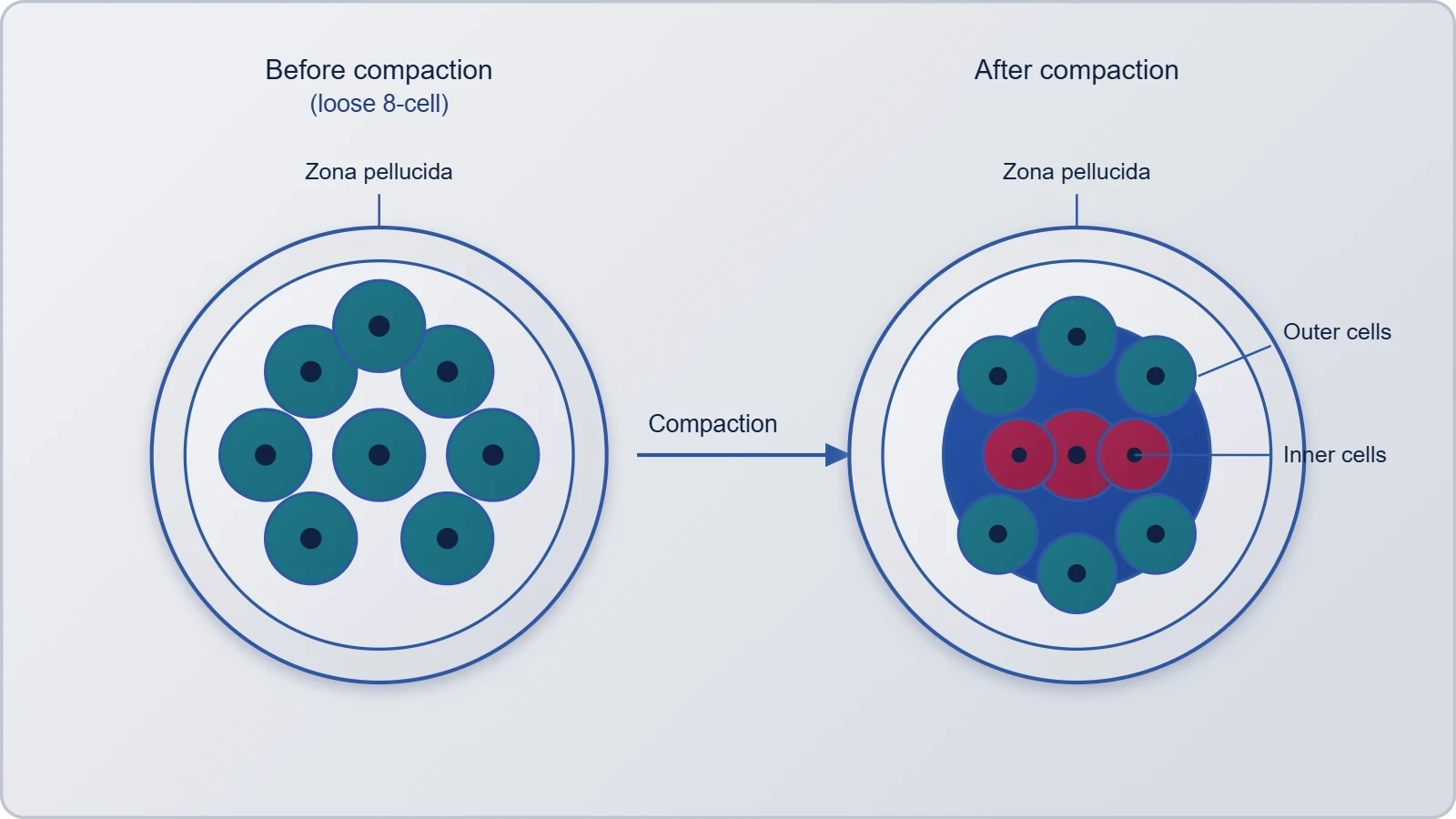
The significance of compaction extends beyond appearance. Tight junctions forming between outer cells create a sealed barrier, which is the prerequisite for the next step: pumping fluid into the interior to create a cavity. Without a functional outer seal, no blastocoel can form, and the embryo cannot become a blastocyst. This is why compaction is regarded as a quality checkpoint in embryo assessment and why embryos that fail to compact rarely progress to successful implantation.
The Morula: A Compact Ball of Blastomeres
The embryo with 8 to 16 blastomeres is called a morula, a term derived from the Latin for mulberry because of its resemblance to the fruit. The morula stage occupies roughly day three to four after fertilisation and represents the transition between the loose cleavage divisions and the organised blastocyst. At this point the embryo is still enclosed within the zona pellucida and has not yet begun to accumulate fluid. It is a stage of preparation rather than of dramatic visible change, but the molecular reorganisation occurring within it determines everything that follows.
It is worth being precise about the cell count, because textbooks and examiners both use the 8-to-16 figure. A morula is not defined by a single number but by a range, and the upper end of that range already contains the beginnings of the outer–inner distinction that will define the blastocyst. Learners who memorise "morula equals sixteen cells" often stumble on questions that present a twelve-cell embryo and ask for its stage; the correct answer is morula, because the defining criterion is the range and the compacted morphology, not an exact count.
From Morula to Blastocyst: Cavitation
The morula continues to divide and transforms into a blastocyst as it moves further into the uterus. The transformation is driven by cavitation: trophoblast cells on the outer surface actively transport sodium ions inward, water follows osmotically, and a fluid-filled cavity called the blastocoel accumulates. As the cavity expands, it pushes the inner cells to one pole, producing the characteristic structure of a hollow sphere with a small cluster of cells attached to its inner wall. This rearrangement is complete by about day five in humans and marks the embryo's arrival as a fully organised pre-implantation structure.
The osmotic logic of cavitation can be expressed simply. If the trophoblast pumps solute into the interior at a rate that maintains a concentration difference ##\Delta C## across the epithelium, the resulting osmotic pressure difference is ##\Delta \pi = RT\,\Delta C##, and water enters until the hydrostatic pressure of the expanding cavity balances it. This is why the integrity of the outer cell layer is essential: a leaky trophoblast cannot sustain the gradient, and the blastocoel fails to expand. The physics of cavitation is thus a direct consequence of the tight junctions established during compaction.
Hatching from the Zona Pellucida
Before implantation can occur, the blastocyst must escape from the zona pellucida, a process called hatching. The expanding blastocoel generates internal pressure, and the trophoblast secretes proteolytic enzymes, including a trypsin-like protease, that locally digest the zona. The embryo squeezes through the resulting opening, typically around day five to six. Hatching is a genuine bottleneck: embryos that fail to hatch cannot implant regardless of how healthy they otherwise appear, and in assisted reproduction a technique called assisted hatching is sometimes used to create a small opening in the zona for selected patients.
Once hatched, the blastocyst is free in the uterine cavity and becomes dependent on the endometrial environment for survival. The window during which the uterus is receptive to a hatched blastocyst is narrow, generally estimated at four to five days within a menstrual cycle of about twenty-eight days, corresponding to days nineteen to twenty-three. An embryo that arrives outside this window, whether too early or too late, faces a lining that is biochemically unprepared to receive it.
Blastocyst Architecture: Trophoblast and Inner Cell Mass
The blastocyst is defined by the arrangement of its blastomeres into two distinct populations. The outer layer is the trophoblast, a single epithelium of cells surrounding the blastocoel, and attached to its inner surface at one pole is a small group of cells called the inner cell mass. These two populations are not interchangeable: the trophoblast gives rise to the placental tissues, while the inner cell mass gives rise to the embryo proper. This division of labour is the first true cellular differentiation in human development and is established by the positional signals generated during compaction.
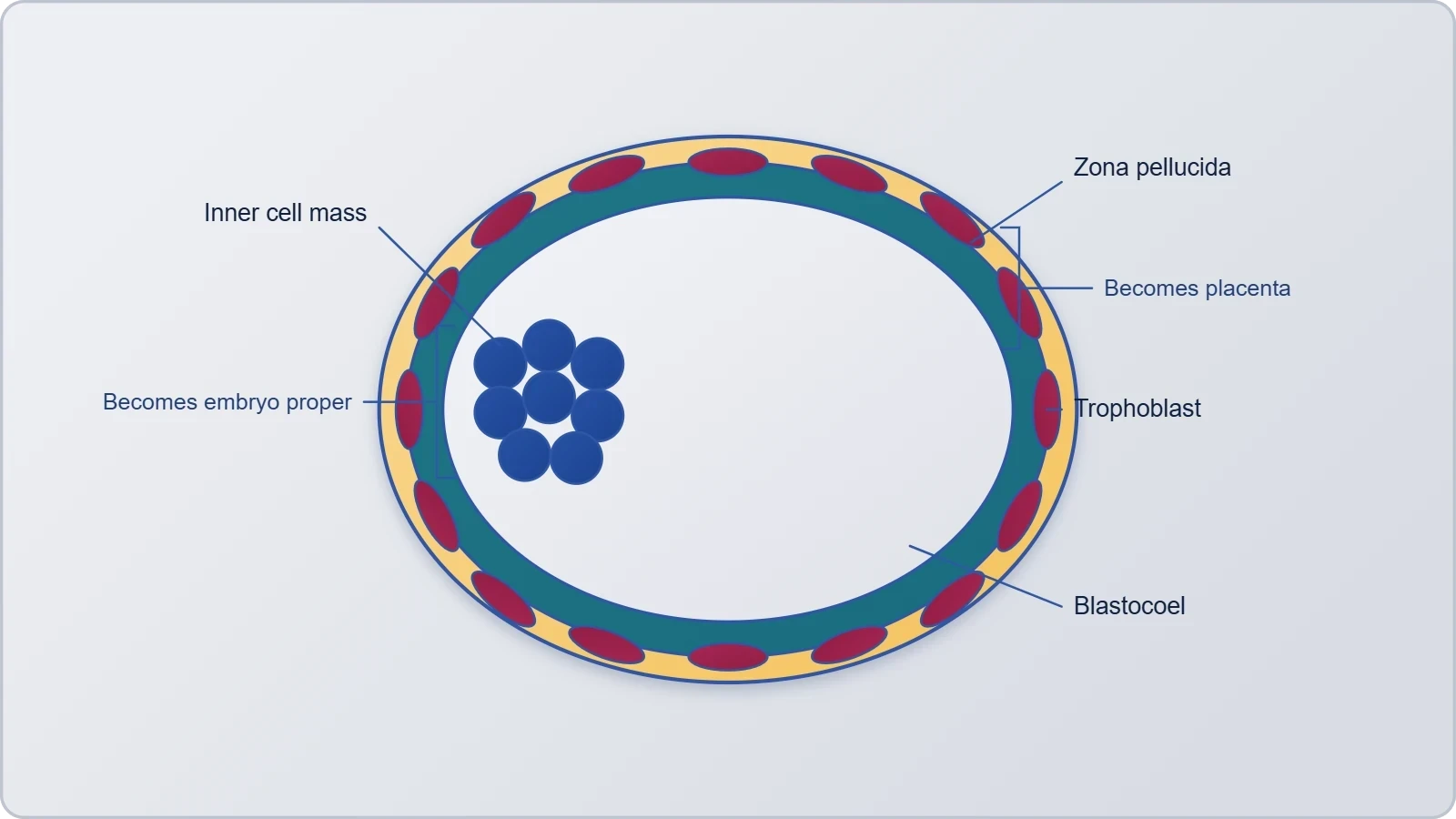
The mechanism behind this fate decision is elegantly simple. Cells on the outside of the compacted morula are exposed to the external environment and express the transcription factor CDX2, which commits them to the trophoblast lineage. Cells on the inside are shielded from the exterior, express OCT4, SOX2 and NANOG, and become the inner cell mass. The Hippo signalling pathway mediates this positional sensing, so that a cell's fate is determined not by lineage history but by where it happens to sit. This is a classic example of conditional specification in embryology.
The Trophoblast: Architect of the Placenta
The trophoblast is the embryo's interface with the mother, and its importance can hardly be overstated. After attachment it proliferates and differentiates into two principal layers: an inner cytotrophoblast of individual dividing cells and an outer syncytiotrophoblast, a multinucleate mass formed by cell fusion that invades the endometrium. The syncytiotrophoblast is the tissue that erodes maternal vessels and, crucially, the tissue that secretes human chorionic gonadotropin (hCG). This hormone rescues the corpus luteum from regression, sustaining progesterone production and thereby maintaining the pregnancy until the placenta can take over endocrine duties.
The appearance of hCG in maternal blood and urine is the basis of every pregnancy test in clinical use. Because the syncytiotrophoblast begins secreting hCG almost immediately upon contact with uterine tissue, detectable levels can appear within a few days of implantation, well before any missed period. The trophoblast therefore does two jobs simultaneously: it physically anchors and invades, and it sends the hormonal signal that tells the mother's body to preserve the pregnancy. Both functions are essential, and both are initiated by the outer cells of the blastocyst.
The Inner Cell Mass and Pluripotency
The inner cell mass gives rise to the embryo proper, differentiating subsequently into the three germ layers — ectoderm, mesoderm and endoderm — from which every tissue of the body derives. Its cells are pluripotent, meaning they can give rise to all cell types of the adult body but not to extraembryonic tissues such as the placenta. This property is the foundation of human embryonic stem cell research, since inner cell mass cells can be isolated and cultured indefinitely while retaining the capacity to differentiate into neurons, cardiomyocytes, hepatocytes and many other cell types.
The distinction between pluripotency and totipotency is a frequent source of confusion. The zygote and the early blastomeres up to the morula stage are totipotent, capable of forming both the embryo and the extraembryonic membranes. Once the blastocyst forms, the inner cell mass is pluripotent and the trophoblast is restricted to placental fates. Understanding this hierarchy clarifies why the blastocyst stage is such a pivotal moment: it is the point at which the embryo surrenders total developmental potential in exchange for organised structure.
Implantation: Embedding in the Endometrium
Implantation is the process by which the blastocyst becomes embedded in the endometrium of the uterus, and it is the event that converts a free-floating embryo into a pregnancy. The trophoblast layer first attaches to the endometrial surface, and the inner cell mass, already positioned against the trophoblast at the embryonic pole, is thereby brought into contact with the maternal tissue. After attachment, the uterine cells divide rapidly and grow over the blastocyst, so that the embryo becomes completely enclosed within the endometrial stroma. This embedding is what the term implantation properly denotes.
Implantation normally occurs about six to seven days after fertilisation, which places it in the mid-luteal phase of the menstrual cycle. The endometrium at this time is in its secretory phase, thickened and rich in glands and blood vessels under the influence of progesterone from the corpus luteum. The blastocyst attaches most often to the posterior wall of the uterine body, near the fundus. Successful implantation requires a synchronised dialogue between a competent blastocyst and a receptive endometrium, and disruption of either partner accounts for a large proportion of early pregnancy failures.
The Sequence of Attachment and Invasion
Implantation proceeds through recognisable phases. The hatched blastocyst first becomes loosely apposed to the endometrial epithelium, then firmly adheres as trophoblast integrins bind to extracellular matrix proteins such as laminin and fibronectin. The trophoblast then penetrates between epithelial cells, and the syncytiotrophoblast advances into the underlying stroma, eroding maternal capillaries and establishing the first maternal–fetal contact. By the end of the second week the blastocyst is fully embedded, and the endometrial defect has healed over it.
The maternal side of this dialogue is equally active. Under progesterone, endometrial stromal cells undergo decidualisation, a transformation in which they enlarge, accumulate glycogen and secrete proteins including prolactin and insulin-like growth factor binding protein-1. Decidualisation both supports the embryo and limits trophoblast invasion, preventing the placenta from burrowing too deeply. The balance between invasion and restraint is delicate; excessive invasion underlies conditions such as placenta accreta, while insufficient invasion is associated with pre-eclampsia and fetal growth restriction.
Clinical Correlates: Ectopic Pregnancy and Implantation Failure
When the blastocyst implants anywhere other than the uterine cavity, the result is an ectopic pregnancy. The most common site is the ampullary region of the fallopian tube, where a retained embryo may implant because tubal cilia or peristalsis have failed to transport it. Because the tubal wall cannot accommodate an expanding pregnancy, ectopic gestation risks rupture and life-threatening haemorrhage, and it is a medical emergency. Risk factors include previous tubal surgery, pelvic inflammatory disease, smoking and assisted reproduction, all of which impair tubal transport.
Implantation failure is the other major clinical concern. Even with chromosomally normal embryos, implantation succeeds in only a fraction of natural and assisted cycles, reflecting the narrow window of receptivity and the complexity of the molecular dialogue. Recurrent implantation failure in IVF has prompted the development of endometrial receptivity testing and personalised embryo transfer timing. Understanding the normal sequence — apposition, adhesion, invasion, decidualisation — gives clinicians a framework for diagnosing where the process has gone wrong.
The Endocrine Handshake: hCG and the Rescue of the Corpus Luteum
The moment the syncytiotrophoblast contacts maternal blood, it begins secreting human chorionic gonadotropin, and this single hormone is the reason a pregnancy survives its first weeks. The corpus luteum, which formed after ovulation, would normally regress around day twenty-two to twenty-four of the cycle, withdrawing progesterone and triggering menstruation. hCG is structurally similar to luteinising hormone and binds the same receptor on luteal cells, sustaining progesterone secretion and preventing endometrial breakdown. Without this rescue, the implanted embryo would be shed along with the uterine lining.
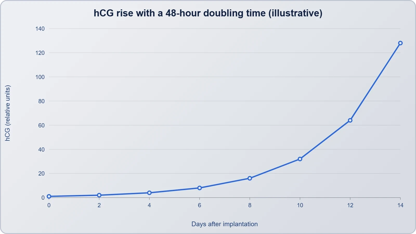
The kinetics of hCG are clinically informative. Levels roughly double every forty-eight to seventy-two hours in early pregnancy, and the rate of rise is used to assess viability; a suboptimal rise may indicate an ectopic or failing pregnancy. Because hCG appears in maternal urine, it forms the basis of home pregnancy tests, which typically detect the hormone around the time of the missed period. The trophoblast thus functions as an endocrine organ from the earliest days of implantation, well before the placenta itself is formed.
Why the Embryo Is Not Rejected
Half of the embryo's genetic material comes from the father, making it a semi-allograft that ought, in principle, to provoke maternal immune rejection. That it does not is one of the more remarkable features of mammalian reproduction. The trophoblast expresses an unusual combination of HLA molecules, notably HLA-G, which interacts with maternal natural killer cells to promote tolerance rather than attack. The syncytiotrophoblast, being a continuous multinucleate layer without intercellular gaps, presents no classical transplantation antigens to maternal lymphocytes at the interface.
Maternal immune adaptation extends beyond the interface. Decidual natural killer cells, macrophages and regulatory T cells accumulate in the endometrium and shift the local environment toward tolerance and vascular remodelling. Progesterone contributes directly by promoting a Th2-biased immune profile. This is not a failure of immunity but a highly regulated local modulation, and disturbances in it are implicated in recurrent miscarriage and pre-eclampsia. The embryo, in other words, negotiates its survival rather than simply evading detection.
Common Misconceptions Worth Correcting
Several errors recur in examinations and in popular writing. The first is that the zygote grows during cleavage; it does not, because the zona pellucida constrains the volume and the divisions lack growth phases. The second is that implantation occurs immediately after fertilisation; in reality there is a delay of about six to seven days while cleavage, morula formation, blastocyst formation and hatching occur. The third is that the inner cell mass forms the placenta; it is the trophoblast that does so, while the inner cell mass forms the embryo proper.
A fourth misconception concerns the timing of sex determination. As established in the previous lesson, the chromosomal sex of the zygote is fixed at fertilisation, but the gonads do not differentiate until several weeks later. A fifth error is to describe the morula as a fluid-filled structure; the cavity appears only at the blastocyst stage. Finally, learners often assume that hCG is produced by the placenta; in early pregnancy it is produced by the trophoblast, and placental production follows later. Precision on these points distinguishes a secure understanding from a superficial one.
Quantitative and Laboratory Perspectives
Beyond the descriptive account, several quantitative relationships sharpen understanding of early development. The exponential decline in blastomere volume during cleavage, the osmotic basis of cavitation, and the doubling kinetics of hCG all lend themselves to simple calculation. Working through these relationships builds the kind of numerical fluency that examination questions and clinical reasoning both demand. The following sections present a worked example and a short computational illustration, followed by a summary of the laboratory techniques that allow embryologists to observe these stages directly.
It is worth emphasising that these calculations are models rather than exact descriptions. Real embryos vary in division timing, blastomere size and hCG trajectory, and clinical interpretation always accounts for that variability. Nevertheless, the models capture the underlying logic and provide a scaffold for interpreting deviations. A learner who can compute expected values and then explain why an observed value differs is far better prepared than one who has memorised the numbers alone.
A Worked Example: Blastomere Volume and Cell Number
Consider a zygote of diameter approximately 120 micrometres, giving a volume of roughly ##V_0 = \dfrac{4}{3}\pi r^3 \approx 9.0 \times 10^5\ \mu m^3##. After three cleavage divisions the embryo has eight blastomeres, so each has a volume of about ##1.1 \times 10^5\ \mu m^3##, corresponding to a diameter of roughly 60 micrometres. The total volume is unchanged, which is precisely the point of cleavage: the embryo redistributes its cytoplasm rather than accumulating more of it.
The second expression follows from the first because volume scales with the cube of radius. It shows that radius declines more slowly than volume, since ##2^{-n/3}## falls less steeply than ##2^{-n}##. This is why blastomeres at the eight-cell stage still look substantial under a microscope even though each contains only one-eighth of the original cytoplasm. The relationship also explains why the morula, despite having more cells than the zygote, occupies essentially the same space within the zona pellucida.
Modelling hCG Rise and a Short Code Illustration
Early hCG concentrations approximate exponential growth with a doubling time of about two days, so that ##C(t) = C_0 \cdot 2^{t/t_d}##, where ##t_d \approx 48## hours. If a sensitive assay first detects hCG at ##C_0 = 5\ \text{mIU/mL}## on day seven after fertilisation, then by day thirteen the predicted concentration is ##5 \times 2^{3} = 40\ \text{mIU/mL}##, comfortably above the detection threshold of most urine tests. This arithmetic explains why home testing is recommended around the time of the missed period rather than immediately after conception.
import math

def hcg_level(c0, days, doubling_time=2.0):
    """Predict hCG concentration assuming exponential rise."""
    return c0 * (2 ** (days / doubling_time))

def blastomere_volume(v0, divisions):
    """Volume of each blastomere after n cleavage divisions."""
    return v0 / (2 ** divisions)

print(round(hcg_level(5, 6), 1))          # ~40.0 mIU/mL on day 13
print(round(blastomere_volume(9.0e5, 3))) # ~112500 cubic micrometres
Such simple models are used in teaching and in the design of clinical algorithms, though real hCG trajectories are fitted with more elaborate curves that account for a plateau and eventual decline. The value of the exercise lies in connecting a biological process to a mathematical form, which is exactly the kind of reasoning that distinguishes a strong student. The same exponential logic applies to bacterial growth and radioactive decay, making it a genuinely transferable skill.
From Implantation to the Placenta: Setting Up the Next Lesson
Implantation is not an endpoint but a beginning. Once the blastocyst is embedded, the trophoblast begins to form the finger-like chorionic villi that interdigitate with uterine tissue and maternal blood, jointly creating the placenta. That structure will supply oxygen and nutrients, remove carbon dioxide and waste, and act as an endocrine organ producing hCG, human placental lactogen, estrogens and progestogens. The foundations for all of this are laid in the first week, which is why a secure grasp of cleavage, morula, blastocyst and implantation is indispensable before studying the placenta.
The endocrine groundwork deserves particular attention as a bridge. The syncytiotrophoblast's early hCG secretion rescues the corpus luteum, and the progesterone it sustains maintains the decidualised endometrium. As the placenta matures, it gradually assumes the steroidogenic role, and the corpus luteum becomes dispensable after roughly the tenth week. This handover is a recurring theme in reproductive endocrinology and will be examined in detail in the next lesson, where the structure and functions of the placenta take centre stage.
Consolidation: Key Points to Carry Forward
Cleavage partitions the zygote into progressively smaller blastomeres without net growth, producing 2, 4, 8 and 16 cells within the confines of the zona pellucida. The 8-to-16-cell embryo is the morula, which compacts and then cavitates to become a blastocyst with an outer trophoblast and an inner cell mass. The trophoblast mediates attachment, invasion and hCG secretion, while the inner cell mass, whose cells are pluripotent, gives rise to the embryo proper. Implantation occurs about six to seven days after fertilisation and establishes the pregnancy.
These facts are best retained as a narrative rather than a list. A zygote is carried through the oviduct, divides without growing, compacts into a mulberry-like morula, hollows out into a blastocyst, hatches from its coat, and burrows into a prepared endometrium that has been waiting under progesterone. Each step depends on the one before, and each offers a point of clinical vulnerability. Holding the sequence in mind makes the details easier to retrieve and far easier to apply.
Practice Questions and Self-Assessment
Attempt the following before moving on. First, explain why the overall size of the embryo does not increase during cleavage, and identify the structure responsible. Second, distinguish between the trophoblast and the inner cell mass in terms of position, potency and derivatives. Third, state the normal timing of implantation and describe the hormonal signal that prevents regression of the corpus luteum. Fourth, explain why an embryo that fails to hatch cannot implant, and identify the clinical situation in which assisted hatching might be considered.
More advanced questions follow. Explain how positional signalling during compaction determines whether a cell becomes trophoblast or inner cell mass, naming the transcription factors involved. Discuss the immunological paradox of the semi-allograft embryo and the role of HLA-G. Finally, calculate the expected hCG concentration twelve days after fertilisation if the initial detectable level was 4 mIU/mL and the doubling time is 48 hours. Working through these questions consolidates the lesson and prepares you for the placental biology that follows.
Clinical and Research Frontiers
Understanding early development has direct consequences for reproductive medicine and for basic research. Pre-implantation genetic testing, in which a few trophectoderm cells are biopsied from a blastocyst, relies on the fact that the trophoblast is representative of the embryo's chromosomal constitution while leaving the inner cell mass undisturbed. Time-lapse imaging of cleavage patterns has refined embryo selection, and endometrial receptivity arrays now allow transfer timing to be individualised. Each of these technologies rests on the developmental biology covered in this lesson.
Research on the inner cell mass has also reshaped regenerative medicine. Human embryonic stem cells, first derived from blastocysts in 1998, are pluripotent and can be directed to form virtually any adult cell type. More recently, induced pluripotent stem cells have been generated by reprogramming adult cells with defined factors, reducing reliance on embryos. The ethical debates surrounding these technologies turn partly on the moral status of the blastocyst, which is why precise understanding of its biology matters well beyond the laboratory.
Assisted Reproduction and Embryo Assessment
In IVF, embryos are cultured to the blastocyst stage before transfer, which allows selection of those that have successfully cavitated and hatched. Blastocyst transfer is associated with higher implantation rates per embryo transferred and reduces the risk of multiple pregnancy when single embryo transfer is used. Grading systems assess the expansion of the blastocoel, the quality of the inner cell mass and the cohesiveness of the trophectoderm, all of which are direct readouts of the processes described in this lesson.
Vitrification, the ultra-rapid cooling technique used to cryopreserve blastocysts, has made elective single embryo transfer practical because surplus embryos can be stored with high survival rates. The ability to freeze and later thaw a blastocyst depends on its having a small, intact inner cell mass and a resilient trophectoderm. Thus the same structural features that predict natural implantation also predict cryosurvival, a neat illustration of how developmental biology informs clinical practice.
Ethical and Educational Dimensions
The blastocyst stage occupies a contested position in bioethics because it is the earliest point at which the embryo is a structured entity with distinct cell lineages, yet it is not yet implanted and has no nervous system. Different legal frameworks set different limits on research using human embryos, typically defined in terms of days of development. The fourteen-day rule, widely adopted though not universal, reflects a judgement about when meaningful development begins, and it is informed directly by the biology of implantation.
For learners, the educational value lies in seeing how a sequence of apparently simple divisions generates the complexity required for a pregnancy. Cleavage, compaction, cavitation, hatching and implantation are not arbitrary vocabulary items but a chain of causal events. Mastery of this chain prepares the ground for the placenta, for fetal development, and for the clinical reasoning that underpins obstetrics. The next lesson takes up the placenta, the organ that makes the whole enterprise possible.
Also Read
- No links available.
From our network :
- Do You Need a New Router or Is Your Internet Plan the Problem?A practical comparison of the signs that point to router limits versus signs your broadband speed or provider is the bottleneck, so you can decide what to replace before spending money.
- 10 Physics Numerical Problems with Solutions for IIT JEEPrepare for IIT JEE with 10 physics numerical problems, complete with detailed solutions and conceptual explanations. Ideal for mastering kinematics, thermodynamics, electromagnetism, and optics.
- Where to Put Your Wireless Router for Better CoverageA practical, physics-grounded guide to router placement: why the centre of the home beats the corner, how walls and metal sap signal, and how to run a simple survey that proves the improvement.
- Trump Political Strategy How Geopolitical Stunts Serve As Media Diversions
- EV 2.0: The Solid-State Battery Breakthrough and Global Factory ExpansionDiscover the impact of the Solid-State Battery Breakthrough on the EV market. Learn about CATL and BYD stock growth, global factory expansion in Europe, and the future of EV 2.0 technology.
- How to Print Cheaply: Cutting the Cost per PageA practical, measured guide to lowering what each printed page costs — duplex and draft modes, third-party ink, yield mathematics, and knowing when a new printer beats refills.
- Analyzing Trump Deportation Numbers Insights Into The 2026 Immigration Crackdown
- AI-Powered 'Precision Diagnostic' Replaces Standard GRE Score ReportsDiscover the new GRE Precision Diagnostic. Learn how AI-powered behavioral analytics and time-to-solve tracking are replacing standard GRE score reports to empower students.
- 98% of Global MBA Programs Now Prefer GRE Over GMAT Focus EditionExplore why 98% of global MBA programs now prefer the GRE over GMAT Focus. Learn about the shorter GRE format, verbal logic importance, and strategic versatility for students.
RESOURCES
- Cleavage pattern, morula compaction and blastocyst morphology as ...frontiersin.orgLimited implantation success of direct-cleaved human zygotes: a time-lapse study. Fertil Steril. (2012) 98(6):1458–63. 10.1016/j.fertnstert.2012.07.1135.
- Association of early cleavage, morula compaction and blastocysts ...nature.comJan 7, 2024 ... ... human assisted reproduction cycles. Reprod. Biomed. Online 9, 511 ... Comparison of single euploid blastocyst transfer cycle outcome derived…
- Prediction of blastocyst development and implantation potential in ...pmc.ncbi.nlm.nih.gov... cleavage do not always reach morula and blastocyst ... Early cleavage predicts the viability of human embryos in elective single embryo transfer procedures.
- MicroRNAs in spent blastocyst culture medium are ... - PubMedpubmed.ncbi.nlm.nih.govMicroRNAs in spent blastocyst culture medium are derived from trophectoderm cells and can be explored for human embryo reproductive competence assessment.
- Elective single morula transfer achieved better clinical outcomes ...link.springer.comOct 21, 2025 ... ... morula transfer or double cleavage-stage embryo transfer. The ... reproduction technology success rates after euploid embryo transfer ...
- Embryology, Week 1 - StatPearls - NCBI Bookshelfncbi.nlm.nih.govApr 17, 2023 ... ... , mitotic cleavage of the blastomere, morula formation, blastocyst formation, and implantation of the blastocyst.
- Clinical outcomes following selection of human preimplantation ...pubmed.ncbi.nlm.nih.govJun 2, 2014 ... ... morula, blastocyst and hatching (n = 4). Five studies used combined ... implantation, prior to routine adoption in the…
- MicroRNAs in spent blastocyst culture medium are derived from ...sciencedirect.comSpent Culture Medium from Single-embryo Transfer Cycles. The spent culture medium was collected from embryos at the cleavage and morula stages or from expanded ...
- P-231 Artificial Intelligence predicts conversion to utilisable ...academic.oup.comJun 23, 2025 ... P-231 Artificial Intelligence predicts conversion to utilisable blastocysts, from cleavage or morula and early blastocyst stages. E Gallagher ...
- Stages of Embryo Development: Understanding the Journey of Lifecarolinasfertilityinstitute.comJan 31, 2025 ... Monitoring Cleavage and Morula Stages: ... The stages of embryo development showcase human reproduction's extraordinary complexity and precision.
- Istanbul consensus update: a revised ESHRE/ALPHA consensus on ...academic.oup.comApr 27, 2025 ... ... cleavage stage embryo, morula, blastocyst ... R. Cleavage kinetics analysis of human embryos predicts development to blastocyst and implantation.
- Biology #HumanReproduction #PostFertilisation #Fertilisation ...facebook.comAug 18, 2026 ... OCR: ZYGOTE CLEAVAGE MORULA POST FERTILISATION EVENTS THE JOURNEY FROM ZYGOTE TO DEVELOPING EMBRYO BLASTOCYST CLEAVAGE ZYGOTE IMPLANTATION ...
- Morula Stage Transfer | Definition, Related Videos & FAQs - JoVEjove.comMorula stage transfer is an assisted-reproduction technique in which a compacted early embryo is placed into the uterus, typically before it forms a blastocyst.