Muscle tissue is not a single uniform material, and the body does not treat it as one. When you hold a heavy bag for a minute, sprint up a staircase, or simply keep your posture while reading, different populations of fibres are recruited to do the work, and they pay for that work in different currencies. Some fibres burn oxygen steadily and resist exhaustion for long periods; others fire powerfully and briefly, then fade. Understanding this division is the natural next step after the sliding filament theory, because contraction explains how force is generated, while fibre type explains how long that force can be sustained and what metabolic cost it carries.
This lesson examines two connected ideas that the chapter places side by side. The first is muscle fatigue, described as the consequence of repeated activation, where lactic acid accumulates because glycogen is broken down anaerobically inside the muscle. The second is the classification of fibres into red and white types, distinguished by myoglobin content, mitochondrial density, sarcoplasmic reticulum volume, and the metabolic route each prefers. Together these ideas explain why some muscles look darker than others, why endurance and explosive strength rarely peak in the same tissue, and why a muscle that has worked hard refuses to keep working at the same intensity.
The treatment below stays close to the chapter's own terminology and sequence of ideas, building on the contraction mechanism already covered and preparing the ground for the skeletal framework that follows. Each concept is paired with structured reference tables, a short mathematical treatment of fatigue onset, and a clearly separated "Going further" note where established background is added without disturbing the source material. By the end, the distinction between aerobic red fibres and anaerobic white fibres should read not as a list to memorise but as a logical solution to two different physiological problems.
On This Page
Fatigue as a Consequence of Repeated Activation
The chapter introduces fatigue in the middle of its account of contraction, immediately after describing relaxation. The sequence matters: calcium ions are pumped back into the sarcoplasmic cisternae, actin filaments are masked again, and the Z lines return to their original position. That is relaxation. But when activation is repeated without adequate recovery, the chemistry inside the fibre begins to shift, and the muscle's capacity to keep producing the same output declines. Fatigue, in this account, is not a failure of the nerve or a loosening of the tendon; it is a metabolic event inside the fibre itself.
The stated cause is the accumulation of lactic acid arising from the anaerobic breakdown of glycogen. Glycogen is the stored carbohydrate fuel within muscle, and when oxygen supply cannot keep pace with demand, its breakdown follows an anaerobic route that leaves lactic acid behind. The chapter also notes that the reaction time of fibres can vary in different muscles, which is a useful reminder that fatigue is not a single clock ticking at one rate everywhere. Different muscles, with different fibre compositions and different demands placed on them, will tire on different schedules.
The Metabolic Chain Behind Fatigue
To read the chapter's sentence carefully is to see a chain rather than a single event. Repeated activation raises demand for ATP; ATP production outpaces oxygen delivery; glycogen is broken down anaerobically; lactic acid accumulates; and the muscle's performance falls. Each link is conditional on the one before it, which is why rest restores function: it allows oxygen delivery to catch up and the accumulated products to be cleared. The chapter does not elaborate on clearance mechanisms, and this lesson does not invent them, but the logic of accumulation and recovery is implicit in the description.
It is worth being precise about what the chapter does and does not claim. It attributes fatigue to lactic acid accumulation due to anaerobic glycogen breakdown, and it frames this as a consequence of repeated activation. It does not specify a threshold concentration, a particular fibre type as uniquely vulnerable, or a molecular receptor involved. Those details belong to specialised literature. For this lesson, the essential claim is causal and directional: repeated use under anaerobic conditions produces a chemical environment in which continued contraction becomes progressively harder.
Modelling the Onset of Fatigue
A simple quantitative picture helps fix the idea without contradicting it. If a muscle consumes ATP at a rate ##R## and regenerates it aerobically at a maximum rate ##R_{a}##, then the anaerobic shortfall is the difference between them whenever demand exceeds aerobic capacity. The accumulated lactic acid, written ##L(t)##, grows in proportion to that shortfall over time. This is a deliberately minimal model, but it captures the chapter's causal structure: fatigue onset depends on how far demand outruns oxygen-supported supply, and on how long that gap persists.
Two practical readings follow. First, a muscle with greater aerobic capacity, meaning more mitochondria and more stored oxygen, keeps ##R_{a}## high and delays the point at which the integral grows quickly. Second, intermittent demand with recovery intervals allows clearance between bouts, so the integral does not climb monotonically. Both readings point directly toward the red and white fibre distinction that the chapter develops next, since the two fibre types differ precisely in the machinery that sets ##R_{a}##.
Myoglobin and the Colour of Muscle
Muscle contains a red coloured oxygen storing pigment called myoglobin, and this single sentence carries much of the chapter's explanatory weight. Myoglobin is the reason muscle tissue is not uniformly pale. Where the pigment is abundant, the tissue takes on a reddish appearance; where it is scarce, the tissue looks pale or whitish. Colour, in other words, is not decorative but diagnostic, a visible signal of how much oxygen-storing pigment a fibre holds and, by extension, how that fibre is likely to generate energy.
The chapter is careful to link pigment to function rather than to appearance alone. Myoglobin stores oxygen, and stored oxygen is only useful if there is machinery to use it. That is why the description of red fibres immediately adds that they contain plenty of mitochondria, the organelles that can utilise the large amount of oxygen stored in them for ATP production. Pigment and organelle are presented as a matched pair: the store and the consumer. A fibre with abundant myoglobin but few mitochondria would be poorly equipped, and the chapter avoids implying otherwise.
Why Stored Oxygen Matters
Oxygen delivery through the bloodstream is continuous but not instantaneous, and during intense activity the gap between delivery and demand widens. A local oxygen store inside the fibre buffers that gap. Myoglobin holds oxygen close to the contractile apparatus, and mitochondria convert it into ATP on site. The chapter's phrase, that these muscles can utilise the large amount of oxygen stored in them for ATP production, captures this arrangement precisely. The store is not a reserve for emergencies alone; it is part of the routine supply line that keeps aerobic metabolism running.
This is also why the chapter can call red fibres aerobic muscles without hesitation. The label follows from the combination of stored oxygen and mitochondrial capacity, not from colour as such. Colour is the outward sign of an inward arrangement. When you look at a dark red muscle, you are seeing, in effect, the visible trace of a metabolic strategy built around oxygen. The next section turns to the fibres that adopt the opposite strategy, and the contrast is sharpest when the two are placed side by side.
Reading Fibre Colour as a Functional Signal
In practice, the colour cue is a useful first approximation for students encountering muscle tissue in dissection or in diagrams. A deep red appearance suggests high myoglobin and therefore a strong aerobic inclination; a pale appearance suggests the reverse. The chapter does not claim that colour alone determines function, and it does not need to. It offers colour as a reliable correlate, then supplies the underlying reasons: myoglobin content, mitochondrial number, and the metabolic route on which the fibre depends. The correlate and the cause are kept distinct, which is exactly the discipline a careful reader should maintain.
It is also worth noting what the chapter leaves open. It does not state that every muscle is purely red or purely white, and the phrasing, that myoglobin content is high in some of the muscles, implies variation rather than a strict binary. Real muscles are mixtures, and the chapter's language accommodates that. For examination purposes, the two categories are what matter, but the underlying idea is a spectrum of aerobic dependence, with red and white as the instructive extremes.
Red Fibres: The Aerobic Machinery
Red fibres are defined by a cluster of features that reinforce one another. Myoglobin content is high, giving the reddish appearance from which the name derives. Mitochondria are plentiful, allowing the stored oxygen to be used for ATP production. Because oxygen is both stored and consumed efficiently, the chapter calls these aerobic muscles. The designation is functional rather than anatomical: it describes how the fibre meets its energy needs, not where it sits in the body or which bone it moves. A single muscle can contain red fibres alongside others, and the proportion shapes what that muscle is good at.
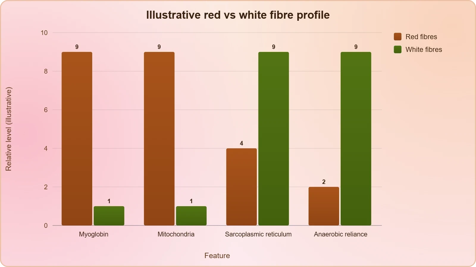
The logic of the arrangement deserves attention. Storing oxygen without mitochondria would be wasteful; possessing mitochondria without a local oxygen supply would leave them underused during intense activity. Red fibres avoid both problems by pairing the store with the consumer. This is why the chapter presents myoglobin and mitochondria in the same breath, and why the term aerobic muscles follows so naturally. The fibre is built around the expectation that oxygen will be available and that ATP will be generated steadily rather than in sudden bursts.
Endurance as a Structural Property
Steady aerobic ATP production translates into resistance to fatigue. A fibre that can keep generating ATP with oxygen does not need to rely on anaerobic glycogen breakdown to the same degree, so lactic acid accumulation is slower and the fatigue pathway described earlier is delayed. This is a structural property, not a matter of willpower. The fibre's endurance is written into its pigment content and organelle density before any contraction begins. When the chapter links red fibres to aerobic metabolism, it is effectively explaining why certain muscles can work for long periods while others cannot.
There is a further implication worth drawing out. If fatigue arises from anaerobic breakdown of glycogen, then a fibre that minimises anaerobic dependence also minimises its exposure to the chemical conditions that produce fatigue. Red fibres are therefore not merely capable of endurance; they are constructed to postpone the very process that ends sustained activity. The chapter does not spell out this inference in so many words, but it follows directly from the two descriptions placed side by side, and it is the kind of connection that turns separate facts into a coherent account.
Where Red Fibres Serve the Body
Postural muscles, which must hold the body upright for hours, illustrate the principle well. Their task is low in intensity but long in duration, exactly the profile that aerobic fibres handle efficiently. The chapter does not name specific postural muscles in this section, and this lesson does not attribute claims to it that it does not make. The point is illustrative: wherever sustained, moderate force is required, a high proportion of red fibres is functionally sensible. The same reasoning explains why muscles used for continuous breathing or maintained stance are typically darker than those reserved for brief, forceful actions.
Students should resist the temptation to treat red fibres as simply better. They are better at endurance, but the chapter's contrast implies a trade-off rather than a hierarchy. A fibre optimised for steady aerobic output is not automatically optimised for rapid, powerful contraction, and the next section describes the fibres that take the opposite approach. The two categories are best understood as complementary specialisations, each solving a different problem, and the body's use of both reflects the range of demands it must meet.
White Fibres: Speed Without Oxygen
White fibres possess very little myoglobin and therefore appear pale or whitish. The number of mitochondria is also few in them, so the machinery for aerobic ATP production is limited. Yet the chapter adds a detail that prevents the picture from becoming a simple story of deficiency: the amount of sarcoplasmic reticulum is high. This matters because the sarcoplasmic reticulum is the calcium store whose release and reuptake drive contraction and relaxation. A fibre with abundant sarcoplasmic reticulum is equipped for rapid, repeated calcium cycling, which is precisely what fast, forceful contraction requires.
Energy for such contraction comes from anaerobic processes, as the chapter states directly. White fibres depend on anaerobic breakdown of glycogen rather than on oxygen-supported metabolism. This is the same pathway that produces lactic acid and, when repeated activation continues, contributes to fatigue. The white fibre is therefore powerful but self-limiting: it can deliver force quickly, yet the metabolic route it relies on generates the very products associated with declining performance. Speed and stamina are traded against each other, and the white fibre sits firmly on the speed side of that trade.
Sarcoplasmic Reticulum and Rapid Cycling
The emphasis on sarcoplasmic reticulum is easy to overlook but central to the contrast. In the contraction account, calcium release triggers sliding and calcium reuptake produces relaxation. A fibre with a large reticulum can cycle calcium quickly, supporting rapid alternation between contraction and relaxation. This is a structural advantage for fast movement, and it explains why white fibres are not simply weaker versions of red ones. They are built for a different tempo. The chapter's inclusion of this detail signals that the red-white distinction involves the calcium handling system, not only the energy system.
Combining high reticulum volume with low myoglobin and few mitochondria yields a coherent profile. The fibre is set up to release and recapture calcium swiftly, and to fuel that activity through anaerobic glycogen breakdown. It does not store much oxygen because it does not depend on oxygen. The features are mutually consistent, and reading them together is more instructive than memorising them separately. Each component supports the same underlying strategy: rapid force generation sustained by non-oxygen-dependent fuel.
The Cost of Anaerobic Reliance
Dependence on anaerobic processes carries an inherent cost, and the chapter's earlier description of fatigue supplies the terms. Anaerobic breakdown of glycogen leaves lactic acid, and repeated activation allows it to accumulate. A muscle rich in white fibres is therefore well suited to brief, intense efforts and less suited to prolonged ones. This is not a defect but a design consequence. The same metabolic route that frees the fibre from oxygen dependence also ties it to a fatigue-prone chemistry. Recognising this connection unifies the lesson's two halves: fibre type and fatigue are two views of the same metabolic arrangement.
For learners, the practical takeaway is that fibre composition predicts fatigue behaviour. A muscle with a high proportion of white fibres will tire quickly under sustained load, while one with more red fibres will last longer at lower intensity. The chapter does not quantify these proportions, and this lesson does not invent figures. What it does provide is the reasoning that makes such predictions sensible, grounded in myoglobin content, mitochondrial number, sarcoplasmic reticulum volume, and the choice between aerobic and anaerobic energy supply.
Integrating Fibre Type with the Contraction Cycle
The previous lesson established that contraction proceeds by sliding filaments, driven by calcium release and ended by calcium reuptake into the sarcoplasmic cisternae. Fibre type now appears as a set of variations on that shared mechanism. Every fibre contracts by the same sliding process, but fibres differ in how quickly they cycle calcium, how much oxygen they store, and how they generate ATP. The sliding filament account is the common grammar; red and white fibres are different dialects of it. Keeping this relationship clear prevents the two topics from becoming disconnected lists.
Fatigue, too, belongs to the same integrated picture. It arises when repeated activation outpaces recovery, and the fibre's metabolic route determines how quickly that happens. A red fibre, with stored oxygen and abundant mitochondria, delays the shift toward anaerobic glycogen breakdown. A white fibre, with little myoglobin and few mitochondria, reaches that shift sooner. The contraction cycle supplies the events; the fibre type supplies the metabolic context; fatigue is the outcome when the context can no longer support the events at the required rate.
A Unified View of Structure and Stamina
Presented this way, the lesson's content forms a single explanatory structure rather than three separate topics. Structure, meaning pigment and organelle content, sets the metabolic route. The metabolic route sets the rate at which lactic acid accumulates under repeated activation. Accumulation sets the onset of fatigue. Each step follows from the one before, and each is stated or directly implied in the chapter. The value of the structure is that it allows a learner to reason from a fibre's appearance to its likely endurance without memorising every pairing independently.
This reasoning also clarifies why the chapter places fatigue before the fibre classification. The fatigue paragraph establishes the anaerobic pathway and its product; the fibre paragraphs then show that some muscles lean heavily on that pathway while others avoid it. The order is pedagogical, moving from a general metabolic fact to its structural consequences. Reading the sections in sequence, rather than as isolated definitions, reveals the argument the chapter is making about how muscle design matches muscle demand.
Preparing for the Skeletal Framework
With the muscular side of movement established, attention turns naturally to the framework that muscles act upon. The next lesson examines the skeletal system, described as a framework of bones and a few cartilages, with bone and cartilage as specialised connective tissues distinguished by their matrices. The human system comprises 206 bones and a few cartilages, grouped into the axial and appendicular divisions. Muscles generate force, but bones and joints give that force direction and purpose, converting contraction into organised movement of the body.
The fibre-type distinction carries forward usefully. Different skeletal regions place different demands on the muscles attached to them, and the proportion of red and white fibres shapes how those demands are met. A framework built for sustained posture and a framework built for rapid limb movement call for different muscular specialisations. The skeletal lesson will supply the anatomical map; this lesson has supplied the physiological vocabulary. Together they explain how a body both holds itself steady and moves through space, which is the central concern of the chapter as a whole.
Going Further: Pigment Chemistry and Fatigue Chemistry
Two pieces of established background help place the chapter's statements in a wider context, and both are offered here as additions rather than corrections. The first concerns myoglobin's relationship to haemoglobin. Myoglobin belongs to the same family of oxygen-binding proteins as haemoglobin, the pigment that carries oxygen in the blood, and both owe their colour to a haem group bound to iron. This is why muscle rich in myoglobin looks red in the same way that blood does. The chapter's description of myoglobin as a red coloured oxygen storing pigment is entirely consistent with this broader biochemical picture.
The second concerns the fate of lactic acid. The chapter associates it with fatigue following anaerobic breakdown of glycogen, and this association is well established in physiology. What the wider literature adds is detail about clearance and transport, which the chapter does not address. For the purposes of this lesson, the essential point is that the chapter's causal claim, repeated activation leading to lactic acid accumulation and hence fatigue, stands as stated. The additional background enriches understanding without altering the sequence of ideas the chapter presents.
Myoglobin, Haemoglobin and the Red Colour
Recognising the shared chemistry clarifies why the red-white distinction is visible at all. Both pigments bind oxygen reversibly, and both depend on iron within a haem group. Myoglobin's role is local storage within the fibre, while haemoglobin's role is transport through the circulation. The two work in sequence: haemoglobin delivers, myoglobin stores, mitochondria consume. Seen this way, the colour of muscle is a visible marker of a supply chain that begins in the blood and ends in the organelle, and the chapter's emphasis on stored oxygen fits neatly into that chain.
This perspective also explains why fibres with little myoglobin appear pale. Without the pigment, there is no local store and no red colour, and the fibre must rely on anaerobic routes when demand rises. The visual contrast between red and white tissue is therefore a contrast between two positions in the oxygen supply chain, one richly provisioned and one minimally so. The chapter's terminology, red fibres and white fibres, captures this contrast in language that is both memorable and biochemically grounded.
Lactic Acid and the Experience of Fatigue
The association between lactic acid and fatigue is one of the most familiar ideas in exercise physiology, and the chapter states it plainly. Anaerobic breakdown of glycogen produces lactic acid, and its accumulation accompanies the decline in muscular performance that follows repeated activation. What learners should take from this is a causal sequence rather than a vague sensation. Fatigue is not merely a feeling; it is the observable consequence of a chemical accumulation within the tissue, and it resolves when the conditions that produced it are reversed through rest and restored oxygen supply.
It is worth noting that the chapter presents this as a general mechanism rather than a complete account. It does not claim that lactic acid is the only factor in fatigue, and this lesson does not either. The value of the statement lies in its clarity: repeated activation, anaerobic glycogen breakdown, lactic acid accumulation, fatigue. That chain is sufficient to explain the difference between fibres that resist fatigue and those that do not, and it provides the conceptual bridge between the contraction mechanism of the previous lesson and the skeletal framework of the next.
Consolidating the Lesson Through Practice
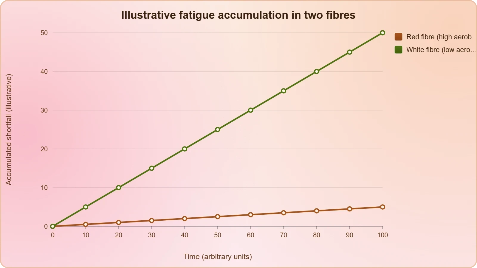
A short programming exercise can make the fatigue model concrete without adding biological claims beyond the chapter. The function below computes the accumulated anaerobic shortfall over a series of time steps, given a demand rate and an aerobic capacity. It is a teaching aid, not a physiological simulation, and its purpose is to let learners see how changing the aerobic capacity alters the accumulation curve. The logic mirrors the integral presented earlier, discretised into steps for clarity and easy experimentation.
def lactic_accumulation(demand, aerobic_capacity, steps, dt):
    """Approximate accumulated anaerobic shortfall over time."""
    total = 0.0
    history = []
    for _ in range(steps):
        shortfall = max(demand - aerobic_capacity, 0.0)
        total += shortfall * dt
        history.append(total)
    return history

# Red fibre: high aerobic capacity keeps the shortfall small
red = lactic_accumulation(demand=10.0, aerobic_capacity=9.0, steps=5, dt=1.0)
# White fibre: low aerobic capacity widens the shortfall
white = lactic_accumulation(demand=10.0, aerobic_capacity=3.0, steps=5, dt=1.0)

print("Red fibre accumulation:", red)
print("White fibre accumulation:", white)
Running the example shows the red fibre accumulating slowly because its aerobic capacity nearly matches demand, while the white fibre accumulates rapidly because the gap is wide. The comparison is deliberately simple, yet it captures the chapter's central contrast: fibres differ in aerobic capacity, and that difference governs how quickly the products of anaerobic metabolism build up. Learners can vary the parameters to explore intermediate cases, which reinforces the idea that real muscles occupy positions along a spectrum rather than falling into two neat boxes.
Self-Check Questions
Before moving to the skeletal system, test understanding with a few targeted questions. What pigment gives red muscle its colour, and what does it store? Which fibre type has more mitochondria, and why does that matter for ATP production? What metabolic route do white fibres depend on, and what product accumulates when that route is used repeatedly? Why does the chapter describe red fibres as aerobic muscles? Answering these from memory, without consulting the tables, is a reliable way to confirm that the causal chain from contraction to fatigue has been absorbed rather than merely read.
A further question invites synthesis: why would a muscle that must hold a posture for hours benefit from a high proportion of red fibres, while a muscle that must produce a brief, forceful movement might benefit from more white fibres? The answer draws on myoglobin content, mitochondrial density, sarcoplasmic reticulum volume, and the trade-off between aerobic endurance and anaerobic power. Framing the answer in these terms demonstrates that the lesson's concepts have been integrated, and it sets up the skeletal discussion by showing how structure at the tissue level serves function at the level of the whole body.
Key Terms at a Glance
The vocabulary of this lesson is compact but precise, and a final review of the terms ensures nothing is carried forward loosely. Myoglobin, lactic acid, glycogen, mitochondria, sarcoplasmic reticulum, red fibres, white fibres, aerobic muscles, and fatigue each carry a specific meaning in the chapter's account. Keeping these definitions sharp matters because the next lesson introduces a new set of terms, bones and cartilages, axial and appendicular divisions, and the vocabulary of joints. A firm grasp of the muscular terms prevents confusion when the skeletal framework is introduced.
Finally, remember that the chapter's order of ideas is itself instructive. Contraction, relaxation, fatigue, myoglobin, red fibres, white fibres, then the skeletal system. Each step answers a question raised by the previous one. How does a muscle relax? Calcium returns to the cisternae. What happens with repeated activation? Lactic acid accumulates and fatigue follows. Why do some muscles resist this? Because they store oxygen and possess mitochondria. Why do others not? Because they depend on anaerobic processes. The sequence is a chain of reasoning, and following it is the surest way to retain the material.
Also Read
- No links available.
From our network :
- EV 2.0: The Solid-State Battery Breakthrough and Global Factory ExpansionDiscover the impact of the Solid-State Battery Breakthrough on the EV market. Learn about CATL and BYD stock growth, global factory expansion in Europe, and the future of EV 2.0 technology.
- 10 Physics Numerical Problems with Solutions for IIT JEEPrepare for IIT JEE with 10 physics numerical problems, complete with detailed solutions and conceptual explanations. Ideal for mastering kinematics, thermodynamics, electromagnetism, and optics.
- Vite 6/7 'Cold Start' Regression in Massive Module GraphsIn-depth analysis of Vite 6/7 cold start regressions in massive module graphs. Learn about barrel file issues, pre-bundling optimizations, and how to improve Vite dev server performance.
- Trump Political Strategy How Geopolitical Stunts Serve As Media Diversions
- 98% of Global MBA Programs Now Prefer GRE Over GMAT Focus EditionExplore why 98% of global MBA programs now prefer the GRE over GMAT Focus. Learn about the shorter GRE format, verbal logic importance, and strategic versatility for students.
- Mastering DB2 LUW v12 Tables: A Comprehensive Technical GuideComprehensive guide on DB2 LUW v12 tables. Explore CREATE TABLE syntax, BLU Acceleration, range partitioning, data types, and security features for IBM DB2 LUW v12.
- Analyzing Trump Deportation Numbers Insights Into The 2026 Immigration Crackdown
- AI-Powered 'Precision Diagnostic' Replaces Standard GRE Score ReportsDiscover the new GRE Precision Diagnostic. Learn how AI-powered behavioral analytics and time-to-solve tracking are replacing standard GRE score reports to empower students.
- Mastering DB2 12.1 Instance Design: A Technical Deep Dive into Modern Database ArchitectureComprehensive guide to DB2 12.1 Instance Design. Explore architectural components, HADR setup, buffer pool tuning, and security policies for IBM DB2 12.1. Includes 10 functional SQL and Shell samples.
RESOURCES
- Analysis of Titin in Red and White Muscles: Crucial Role on ... - PMCpmc.ncbi.nlm.nih.govNov 18, 2018 ... ... fatigue than are white muscle fibers due to their ability to oxidatively regenerate ATP. In contrast, ATP generation in…
- Skeletal Muscle Fiber Type: Influence on Contractile and Metabolic ...ncbi.nlm.nih.govOct 12, 2004 ... ... muscles of the same animal. These early observations formed the basis of the classical terminology of red and white…
- High-frequency fatigue in rat skeletal muscle: role of extracellular ion ...pubmed.ncbi.nlm.nih.gov... muscle fibers. HFF was slower in slow-twitch soleus fibers than in fast-twitch red or white sternomastoid fibers; denervation accelerated fatigue …
- Anatomy of Skeletal Muscle and Its Vascular Supply - NCBI - NIHncbi.nlm.nih.govThese type I fibers are resistant to fatigue, relying on oxidative ... Comparison of capillary network anatomy in red and white muscle indicates ...
- Difference Between Red And White Muscle - BYJU'Sbyjus.comRed Muscle Fibers. White Muscle Fibers ; Definition ; Red muscles are a type of skeletal muscle which are dense with capillaries and is…
- Muscle Fibre Types - Physiopediaphysio-pedia.comFG fibers use anaerobic metabolism to produce powerful, high-tension contractions but fatigue quickly. Most muscles possess a mixture of each fiber type. The ...
- Differentiation of muscle fiber types in the teleost Brachydanio reriolink.springer.com... white fibers, which are fast-contracting and fatigue rapidly. ... Nag, A.C., Nursall, J.R.: Histogenesis of white and red muscle fibres of trunk muscles…
- Differences in Sizes of Red and White Muscle Fibres in Vastus ...tandfonline.comThe weight lifters with high isometric muscular strength had significantly larger white fibres than the others. The endurance events athletes with high aerobic ...
- Slow Muscle Fiber - an overview | ScienceDirect Topicssciencedirect.comWhite muscle fibers containing little myoglobin and mitochondria are fast-twitch fibers that fatigue rapidly. Table 3.7.4 compares some of the different ...
- Significance of calcium: its correlation with red and white muscle ...pesquisa.bvsalud.org... red and white muscle contraction, fatigue and potential. Castro, A. A.; Perez ... Calcium plays a crucial role in both red and white…
- its correlation with red and white muscle contraction, fatigue and ...msjonline.orgMar 14, 2024 ... Conversely, in red (slow-twitch) muscle fibers, the release of calcium occurs in smaller amounts and over a prolonged period, leading…
- Understanding Fast Twitch vs Slow Twitch Muscle Fibers - NASMnasm.orgApr 1, 2025 ... ... fibers are sometimes called red fibers ... Because slow-twitch muscle fibers use oxygen to produce energy, they are more…
- Significance of calcium: its correlation with red and white muscle ...msjonline.orgMar 21, 2024 ... Hortemo KH, Munkvik M, Lunde PK, Sejersted OM. Multiple causes of fatigue during shortening contractions in rat slow twitch skeletal…
- Difference Between Red and White Muscle Fibers - Biology - Vedantuvedantu.comWhite Muscles perform anaerobic oxidation and in the process, they accumulate more lactic acid than the Red Muscles. This leads to fatigue in the…
- Muscles - Fast and slow twitch - BBCbbc.co.ukFast twitch muscle fibres: Contract quickly, but rapidly get tired. Endurance ... Muscles that contain a lot of slow twitch fibres are red, because…