Muscle contraction is often imagined as a single event, yet it depends on an intricate molecular architecture in which two distinct protein filaments cooperate with extraordinary precision. Within each sarcomere, the functional unit of contraction established in the preceding lesson, actin and myosin are arranged as rod-like structures running parallel to one another and to the longitudinal axis of the myofibrils. Their relative thickness gives them their familiar names: actin filaments are thinner and are therefore called thin filaments, while myosin filaments are thicker and are known as thick filaments. Understanding how these two proteins are built is the essential bridge between the static anatomy of the sarcomere and the dynamic sliding that produces movement.
This lesson examines the structure of contractile proteins at the molecular level, following the sequence of ideas in the chapter. It begins with the thin filament, resolving the helical arrangement of F actin, the monomeric G actin units that compose it, and the regulatory strands of tropomyosin and troponin distributed along its length. It then turns to the thick filament, where meromyosin monomers assemble into a polymerised protein with projecting cross arms, heavy and light meromyosin regions, and a globular head that functions as an active ATPase enzyme. Each structural detail is presented with its functional significance, so that the machinery is understood before the sliding filament theory is introduced in the next lesson.
The material deliberately avoids the mechanism of contraction itself, which belongs to the following lesson. Instead, it establishes the vocabulary and spatial relationships that the sliding filament theory will later rely upon: binding sites for myosin on actin, binding sites for ATP on the myosin head, and the regulatory proteins that mask those sites in the resting state. Learners who grasp these structures will find the sliding filament theory far easier to follow, because every step of that theory refers back to the molecular features described here. The lesson closes with a short "Going further" block that adds well-established background on regulatory proteins and ATPase enzymes without departing from the chapter's terminology.
On This Page
Thin Filaments: The Architecture of Actin
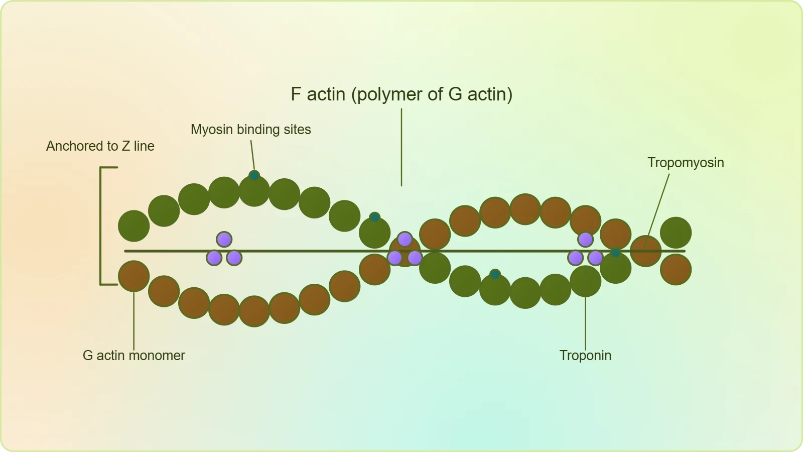
Each actin filament is a finely organised polymer, and its construction follows a clear hierarchy that the chapter describes with precision. The filament is made of two F (filamentous) actins helically wound around each other, giving the thin filament its characteristic twisted, rope-like appearance. Each F actin is itself a polymer of monomeric G (globular) actins, so the thin filament is a polymer of polymers, built from small globular subunits assembled into long fibrous strands. This two-tier organisation explains both the flexibility and the tensile strength of the thin filament, and it provides the repeating surface along which myosin heads will later engage.
The helical winding of the two F actins is not merely decorative. It creates a periodic structure with repeating grooves and ridges, and it is along this structure that the regulatory proteins are positioned. Because the two F actins twist around one another, the filament presents a continuously repeating pattern of binding surfaces, each of which must be controlled so that contraction occurs only when appropriate. The thin filament is therefore both a structural rod and a regulated track, and its geometry is central to the way muscle activity is switched on and off at the molecular level.
Tropomyosin and Troponin Along the Filament
Two filaments of another protein, tropomyosin, run close to the F actins throughout the length of the thin filament. They lie in the grooves formed by the helical winding, following the same twisted path as the actin strands. A complex protein called troponin is distributed at regular intervals on the tropomyosin. Together these two proteins form the regulatory system of the thin filament, and their regular, repeating placement ensures that control is exerted uniformly along the entire length of the actin polymer rather than at isolated points.
The chapter states a crucial functional detail about the resting state: a subunit of troponin masks the active binding sites for myosin on the actin filaments. In other words, when the muscle is relaxed, the sites where myosin heads would otherwise attach are physically covered. This masking is the reason a resting muscle does not contract spontaneously. The thin filament is fully assembled and ready, but its binding sites are concealed until the appropriate signal arrives, at which point the regulatory proteins shift and expose the sites for interaction.
Why the Thin Filament Matters Structurally
The thin filament's firm attachment to the Z line, described in the previous lesson, gives it a fixed anchor at each end of the sarcomere. Because the filament is anchored rather than free-floating, any movement of the thin filament relative to the thick filament translates directly into a change in sarcomere length. The molecular structure described here therefore has mechanical consequences at the level of the whole fibre. The helical actin strands, the tropomyosin filaments, and the periodically distributed troponin together form a structure that is anchored, regulated, and ready to slide.
It is worth appreciating how economically the thin filament is designed. A single repeating geometry serves three purposes at once: it provides the binding surface for myosin, it accommodates the tropomyosin strands within its grooves, and it presents troponin at regular intervals so that regulation is evenly distributed. This economy of design is a recurring theme in muscle anatomy, where structure and function are so tightly integrated that describing one inevitably describes the other. The thin filament is thus a compact solution to the problem of building a regulated, anchorable, sliding track.
Thick Filaments: Meromyosin and Its Parts
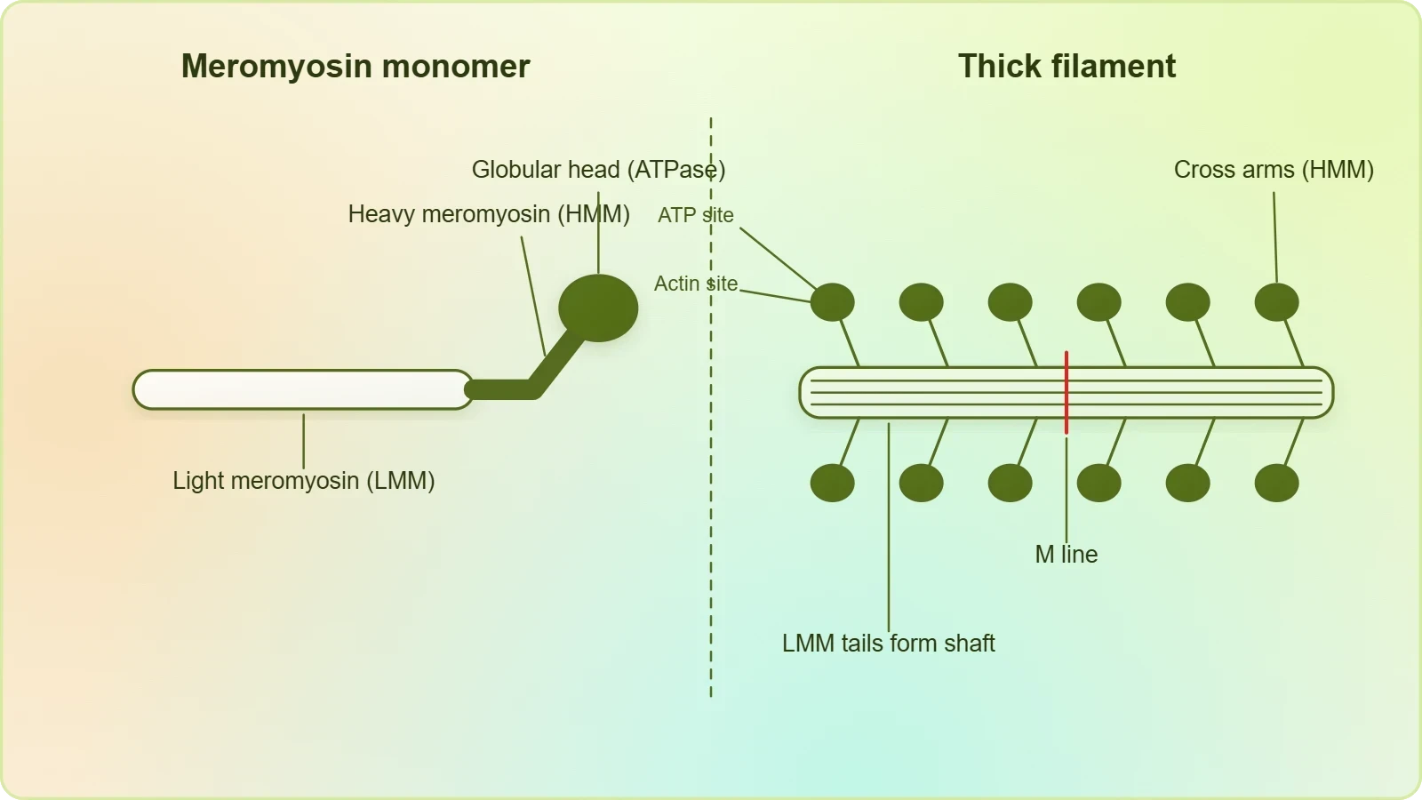
Each myosin filament is also a polymerised protein, and its building block is the meromyosin monomer. Many monomeric proteins called meromyosins constitute one thick filament, so the thick filament is an assembly of many identical units arranged in a regular pattern. This modular construction is what allows the thick filament to present a dense array of projecting heads along its length, each capable of interacting with a neighbouring thin filament. The meromyosin monomer is therefore the fundamental unit of the thick filament, just as G actin is the fundamental unit of the thin filament.
Each meromyosin has two important parts: a globular head with a short arm, and a tail. The former, the head and short arm together, is called the heavy meromyosin (HMM), while the latter, the tail, is called the light meromyosin (LMM). This division is not arbitrary; it reflects a genuine functional separation within the molecule. The tail region is responsible for assembly into the filament, while the head region carries the machinery for interaction with actin and for the chemical activity that powers contraction. The naming of the two regions thus encodes their distinct roles.
The Cross Arm and Its Projection
The HMM component, that is the head and short arm, projects outwards at regular distances and angles from each other from the surface of the polymerised myosin filament, and this projecting structure is known as the cross arm. The regularity of the projection is significant: it means the thick filament presents its heads in an ordered array rather than at random, so that many heads can engage the thin filament simultaneously. The angle of projection also matters, because the geometry of the cross arm determines how the head can reach across to the adjacent thin filament.
Visualising the thick filament as a central shaft studded with regularly spaced cross arms helps clarify why the filament is described as thick. The shaft is formed by the tails of the meromyosin monomers, packed together, while the heads project outward like bristles. This arrangement maximises the number of potential contact points between thick and thin filaments while keeping the filament itself compact. The cross arm is therefore the structural feature that makes the thick filament an effective partner for the thin filament during contraction.
The Globular Head as an ATPase Enzyme
The globular head is described as an active ATPase enzyme, and it has binding sites for ATP and active sites for actin. This dual capability is the chemical heart of the contractile machinery. The head can attach to actin through its active site, and it can bind and split ATP through its ATP-binding site. The energy released by the splitting of ATP is what the head uses to perform its cycle of attachment, movement, and release, a cycle that the next lesson will describe in detail as the sliding filament theory.
The fact that the head carries both an actin-binding site and an ATP-binding site means that the two functions are physically coupled within a single small structure. This coupling is what allows chemical energy to be converted into mechanical work at the molecular scale. The head is not merely a passive hook; it is an enzyme and a motor combined. Recognising this dual nature is essential for understanding why the thick filament is described as the active, energy-consuming partner in the contractile apparatus.
How the Two Filaments Are Arranged Together
The structure of the individual filaments only becomes meaningful when their arrangement within the sarcomere is considered. Both actin and myosin are arranged as rod-like structures, parallel to each other and also to the longitudinal axis of the myofibrils. This parallel alignment ensures that the thin and thick filaments lie side by side along the full length of the contractile unit, ready to interact along their overlapping regions. The longitudinal orientation is what allows sliding to shorten the sarcomere rather than distort it sideways.
The thin filaments are firmly attached to the Z line, the elastic fibre at the centre of each I band that bisects it. The thick filaments in the A band are held together in the middle of that band by a thin fibrous membrane called the M line. These two anchoring structures, Z line and M line, define the geometry of the sarcomere and keep the filaments in their correct relative positions. The A and I bands are arranged alternately throughout the length of the myofibrils, producing the characteristic banded appearance of skeletal muscle under the microscope.
Overlap and the H Zone
In a resting state, the edges of the thin filaments on either side of the thick filaments partially overlap the free ends of the thick filaments, leaving the central part of the thick filaments uncovered. This central part of the thick filament, not overlapped by thin filaments, is called the H zone. The H zone is therefore a direct visual indicator of the degree of overlap between the two filament types, and its width changes as the sarcomere changes length. Recognising the H zone helps connect the molecular structure to the banded pattern seen in the previous lesson.
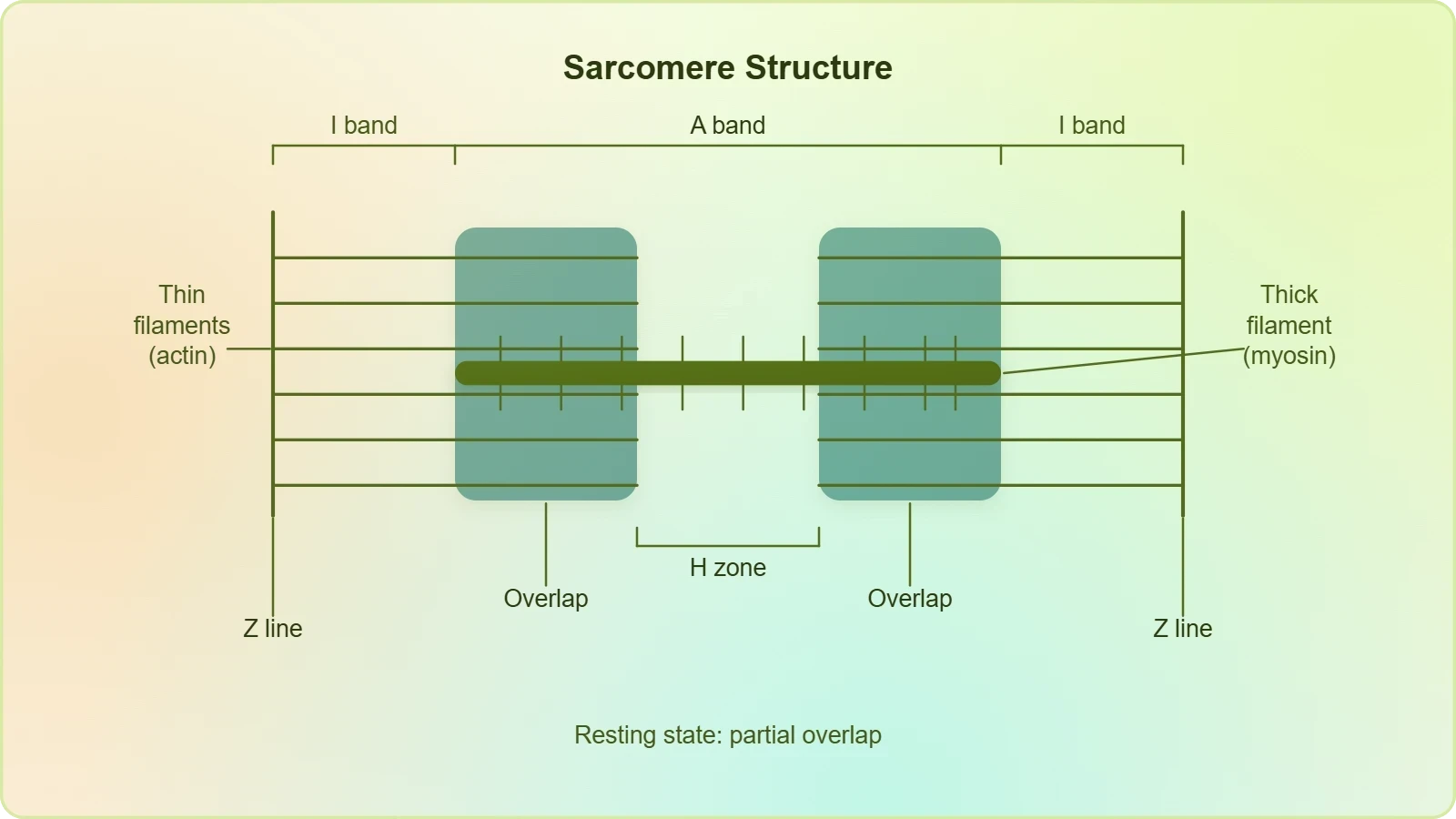
The partial overlap in the resting state is precisely what makes contraction possible. Because the filaments already lie alongside one another, a small relative sliding movement can immediately increase the overlap without any need for the filaments to find each other first. The resting arrangement is thus a poised configuration, with the machinery assembled and the binding sites masked. When the regulatory proteins later expose those sites, the cross arms are already positioned to engage the thin filaments and begin the cycle of attachment and movement.
From Structure to Function
The structural details covered in this lesson all point forward to the mechanism of contraction. The myosin head's ATPase activity, its actin-binding site, the regular projection of cross arms, and the masking of actin's binding sites by troponin are not isolated facts; each is a component of a coordinated system. When the sliding filament theory is presented in the next lesson, it will describe how these components act in sequence: the exposure of binding sites, the attachment of cross arms, the power stroke, and the splitting of ATP that resets the cycle.
Keeping the structural vocabulary clear is therefore the best preparation for what follows. A learner who can distinguish F actin from G actin, tropomyosin from troponin, and heavy from light meromyosin will be able to follow the mechanism without confusion. The chapter's order of ideas is deliberate: structure is established first, in precise molecular terms, and only then is the dynamic process explained. This lesson completes that structural foundation, leaving the contractile cycle ready to be described in full.
Regulatory Control and the Resting State
The resting state of the muscle is an active condition maintained by the regulatory proteins, not a passive absence of activity. The chapter is explicit that a subunit of troponin masks the active binding sites for myosin on the actin filaments. This masking is the molecular basis of relaxation. The thin filament is fully formed and the thick filament is fully assembled, yet no cross-bridge can form because the sites where the myosin heads would attach are covered. Relaxation is therefore a state of readiness held in check by a specific molecular mechanism.
The distribution of troponin at regular intervals on the tropomyosin is what makes this control effective along the whole filament. Because tropomyosin runs the full length of the F actins and troponin is spaced regularly along it, the masking is not confined to one spot but is repeated throughout the thin filament. A single regulatory arrangement thus governs the entire actin surface. This is an elegant solution to the problem of controlling a long polymer: rather than regulating each binding site individually, the filament uses a continuous strand with periodic control points.
Binding Sites and Their Significance
The chapter identifies two distinct sets of sites that matter for contraction. On the actin filament, there are active binding sites for myosin, which are masked by troponin in the resting state. On the myosin head, there are binding sites for ATP and active sites for actin. The interaction between these two sets of sites is the physical basis of cross-bridge formation. Understanding which site is on which filament, and which is masked or exposed, is essential for following the sequence of events in the next lesson.
It is useful to think of the system as a lock and key arrangement with a safety cover. The myosin head carries the key, in the form of its actin-binding site, and the actin filament carries the lock, in the form of its binding sites. In the resting state, the safety cover, provided by troponin and tropomyosin, keeps the lock inaccessible. When the cover is removed, the key can engage. This simple analogy captures the essential logic of the regulatory system without distorting the chapter's terminology.
Energy and the ATPase Head
The description of the globular head as an active ATPase enzyme introduces the energy dimension of contraction. The head has binding sites for ATP, and as an ATPase it is capable of splitting ATP. The energy released by this splitting is what powers the cycle of cross-bridge formation and release. Without an active ATPase head, the thick filament could bind actin but could not cycle, and sustained contraction would be impossible. The enzymatic nature of the head is therefore as important as its structural role as a cross arm.
This enzymatic capability also explains why muscle tissue is so metabolically demanding. Every cycle of attachment and release requires the splitting of ATP by the myosin head, and contraction involves many heads cycling repeatedly. The structure described in this lesson thus connects directly to the energy metabolism of muscle, a topic that extends beyond the present chapter but follows naturally from the observation that the globular head is an active ATPase enzyme with binding sites for ATP.
Going Further: Regulatory Proteins and ATPase Enzymes
Troponin and tropomyosin are best understood as regulatory proteins whose role is to control when myosin can bind to actin. The chapter describes their positions and the masking effect in the resting state; the broader principle is that these proteins act as a switch. When the appropriate signal reaches the thin filament, the regulatory proteins shift so that the binding sites are exposed, allowing cross-bridges to form. This switch-like behaviour is what makes muscle contraction responsive rather than continuous, and it explains why the thin filament is described as regulated rather than merely structural.
ATPase enzymes are a broad class of proteins that split ATP, releasing energy that cells can use. The myosin head belongs to this class, and its ATPase activity is the specific instance described in the chapter. Recognising the general principle helps place the myosin head within a wider biological context: many cellular processes depend on enzymes that harvest the energy stored in ATP. In muscle, that harvested energy is converted into mechanical movement, but the underlying chemistry of splitting ATP is shared with other energy-consuming systems throughout the cell.
Consolidating the Structural Picture
Bringing the two filaments together, the picture is one of complementary design. The thin filament provides a regulated track with binding sites that can be exposed or concealed, anchored at the Z line. The thick filament provides a shaft studded with cross arms, each carrying an ATPase head with sites for both actin and ATP, held in the middle of the A band by the M line. The partial overlap in the resting state, with the H zone at the centre of the thick filament, sets the stage for sliding. Every element described in this lesson has a counterpart in the other filament.
This complementarity is the reason the chapter treats the two filaments in a single section. Neither is meaningful in isolation: actin without myosin has nothing to slide against, and myosin without actin has nothing to pull. The contractile proteins are a partnership, and their structures are matched to one another in scale, spacing, and chemistry. Holding this paired picture in mind is the best preparation for the sliding filament theory, which will describe how the partnership operates dynamically to shorten the sarcomere and produce movement.
Key Terms and a Worked Check
A short consolidation of terminology helps fix the material before moving on. The thin filament is built from F actins, each a polymer of G actins, with tropomyosin strands and troponin at regular intervals. The thick filament is built from meromyosins, each divided into heavy meromyosin (head and short arm) and light meromyosin (tail), with the head projecting as a cross arm and acting as an ATPase enzyme. These terms recur throughout the remaining lessons, so fluency with them pays dividends.
It is also worth noting the numerical and spatial relationships the chapter supplies. The thin filaments are attached to the Z line, the thick filaments are held at the M line, and the A and I bands alternate along the myofibril. The H zone is the central region of the thick filament not overlapped by thin filaments in the resting state. These positional facts are simple but precise, and they allow the structure of the sarcomere to be reconstructed from memory, which is exactly the kind of recall that examination questions reward.
A Simple Structural Check
One way to test understanding is to trace a single myosin head from its origin to its target. The head is part of the heavy meromyosin region of a meromyosin monomer, which is one of many monomers constituting a thick filament. The head projects outward as a cross arm at a regular distance and angle from its neighbours. Its active site for actin faces the adjacent thin filament, where the corresponding binding sites are masked by troponin in the resting state. Following this path confirms that every structural term has a clear place in the overall arrangement.
A second check is to compare the two filaments feature by feature. Actin is thinner, myosin thicker; actin is anchored at the Z line, myosin at the M line; actin carries the masked binding sites, myosin carries the ATPase head. If these contrasts can be stated without hesitation, the structural foundation is secure. The next lesson will then add the dynamic dimension, showing how the cross arms engage the exposed sites and how the splitting of ATP drives the sliding that shortens the sarcomere.
Preparing for the Sliding Filament Theory
The sliding filament theory states that contraction of a muscle fibre takes place by the sliding of the thin filaments over the thick filaments. Everything in this lesson supports that statement. The parallel arrangement of the filaments, the partial overlap in the resting state, the cross arms projecting from the thick filament, and the binding sites on actin all describe a system built for sliding. When the theory is presented, it will not introduce new structures but will explain how the existing ones move relative to one another.
Learners who have followed the molecular detail will recognise that the theory is a description of motion, not of shortening of the filaments themselves. The filaments slide, and the sarcomere shortens as a consequence. The H zone narrows as overlap increases, and the bands shift in their apparent width. These changes follow directly from the structures described here, and the next lesson will trace them step by step, from the exposure of binding sites to the completion of the contractile cycle.
Also Read
- No links available.
From our network :
- 98% of Global MBA Programs Now Prefer GRE Over GMAT Focus EditionExplore why 98% of global MBA programs now prefer the GRE over GMAT Focus. Learn about the shorter GRE format, verbal logic importance, and strategic versatility for students.
- Analyzing Trump Deportation Numbers Insights Into The 2026 Immigration Crackdown
- Vite 6/7 'Cold Start' Regression in Massive Module GraphsIn-depth analysis of Vite 6/7 cold start regressions in massive module graphs. Learn about barrel file issues, pre-bundling optimizations, and how to improve Vite dev server performance.
- 10 Physics Numerical Problems with Solutions for IIT JEEPrepare for IIT JEE with 10 physics numerical problems, complete with detailed solutions and conceptual explanations. Ideal for mastering kinematics, thermodynamics, electromagnetism, and optics.
- AI-Powered 'Precision Diagnostic' Replaces Standard GRE Score ReportsDiscover the new GRE Precision Diagnostic. Learn how AI-powered behavioral analytics and time-to-solve tracking are replacing standard GRE score reports to empower students.
- Mastering DB2 LUW v12 Tables: A Comprehensive Technical GuideComprehensive guide on DB2 LUW v12 tables. Explore CREATE TABLE syntax, BLU Acceleration, range partitioning, data types, and security features for IBM DB2 LUW v12.
- Mastering DB2 12.1 Instance Design: A Technical Deep Dive into Modern Database ArchitectureComprehensive guide to DB2 12.1 Instance Design. Explore architectural components, HADR setup, buffer pool tuning, and security policies for IBM DB2 12.1. Includes 10 functional SQL and Shell samples.
- EV 2.0: The Solid-State Battery Breakthrough and Global Factory ExpansionDiscover the impact of the Solid-State Battery Breakthrough on the EV market. Learn about CATL and BYD stock growth, global factory expansion in Europe, and the future of EV 2.0 technology.
- Trump Political Strategy How Geopolitical Stunts Serve As Media Diversions
RESOURCES
- Muscle Contraction - PMC - NIHpmc.ncbi.nlm.nih.govThe two types of striated muscle—skeletal and cardiac—organize their contractile filaments of actin and myosin into regular arrays called sarcomeres. These ...
- Physiology, Skeletal Muscle - StatPearls - NCBI Bookshelf - NIHncbi.nlm.nih.govJul 30, 2023 ... Actin filaments are double-helical structures ... They are the largest fibers in diameter due to their high density of actin…
- Special Issue: The Actin-Myosin Interaction in Muscle - PMC - NIHpmc.ncbi.nlm.nih.govSuperimposed on these two types of A-band structure, there are subtle variations in contractile protein isoforms, M-band structure, Z-band structure and energy ...
- Actin, Myosin, and Cell Movement - The Cell - NCBI Bookshelfncbi.nlm.nih.govStructure of muscle cells. Muscles are composed of bundles of single large cells (called muscle fibers) that form by cell fusion and contain multiple…
- Contractile Protein - an overview | ScienceDirect Topicssciencedirect.comThick filaments are made up of myosin and the structural protein titin. The ... Lying between the thin actin filaments are thick myosin filaments…
- Altered expression of titin and contractile proteins in failing human ...pubmed.ncbi.nlm.nih.gov... template for myosin and actin filaments in sarcomerogenesis. Human cardiac tissue obtained at the time of transplantation surgery was investigated using ...
- The effect of muscle ultrastructure on the force, displacement and ...royalsocietypublishing.orgMay 22, 2024 ... ... contractile proteins, actin and myosin. Structural variation contributes. ... contractile protein filaments: actin, myosin and titin. Considerable ...
- The actin and myosin filaments of human and bovine blood plateletspubmed.ncbi.nlm.nih.gov... structures which may subserve this contractile function. In order to identify actin, the ability of the filaments to react with heavy meromyosin (HMM)…
- Structure of Contractile Proteins | CK-12 Foundationflexbooks.ck12.orgThe two main types, actin (thin filaments) and myosin (thick filaments), are ... Contractile proteins in muscle, chiefly actin (thin filament) and ...
- The ultrastructural organization of actin and myosin II filaments in the ...molbiolcell.orgJan 5, 2017 ... The CR contains a suite of characteristic protein constituents, including three essential filament systems consisting of the contractile ...
- Smooth muscle ultrastructure – still shrouded in mystery?link.springer.comOct 14, 2026 ... ... contractile proteins actin and myosin ... To form a contractile structure, smooth muscle myosin filaments are surrounded by actin…
- Sarcomere - Physiopediaphysio-pedia.comA sarcomere is the basic contractile unit of a myocyte (muscle fiber). A sarcomere comprises two main protein filaments (thin actin and thick myosin ...
- Cardiac Myocytes and Sarcomeres - CV Physiologycvphysiology.comThe interactions between actin and myosin serve as the basis for the sliding filament theory of muscle contraction. Myosin and actin Myosin is a…