A skeletal muscle is not a single uniform tissue but an ordered hierarchy of nested structures, each wrapped in its own connective sheath and each serving a distinct mechanical purpose. When you examine a muscle at increasing magnification, the organisation reveals itself in stages: the whole muscle, the bundles within it, the individual fibre, the fibrils inside the fibre, and finally the repeating protein arrays that actually shorten. Understanding this ladder of scale is the foundation for everything that follows about contraction, because the sliding of filaments only makes sense once the architecture that holds those filaments in register is clear.
This lesson examines that architecture in detail, following the sequence in which the structures nest inside one another. It begins with the fascia that binds muscle bundles, moves inward through the sarcolemma and sarcoplasm of the muscle fibre, and then descends to the myofibrils with their alternating dark A bands and light I bands. From there it defines the sarcomere together with the Z line, M line and H zone, the landmarks that give the contractile machinery its geometry. Each term is introduced where it belongs in the physical hierarchy, so the vocabulary accumulates in a logical order rather than as a list to be memorised.
Because the previous lesson established the types of muscle and their properties, this lesson can concentrate entirely on internal structure. It deliberately stops short of the molecular detail of actin and myosin, which belongs to the next lesson on contractile proteins, and it does not yet explain the sliding filament mechanism. What it does provide is the structural map: the compartments, the membranes, the bands and the boundaries. With that map in hand, the behaviour of the contractile proteins becomes a question of mechanics rather than of unfamiliar anatomy.
On This Page
The Connective Framework: Fascicles and Fascia
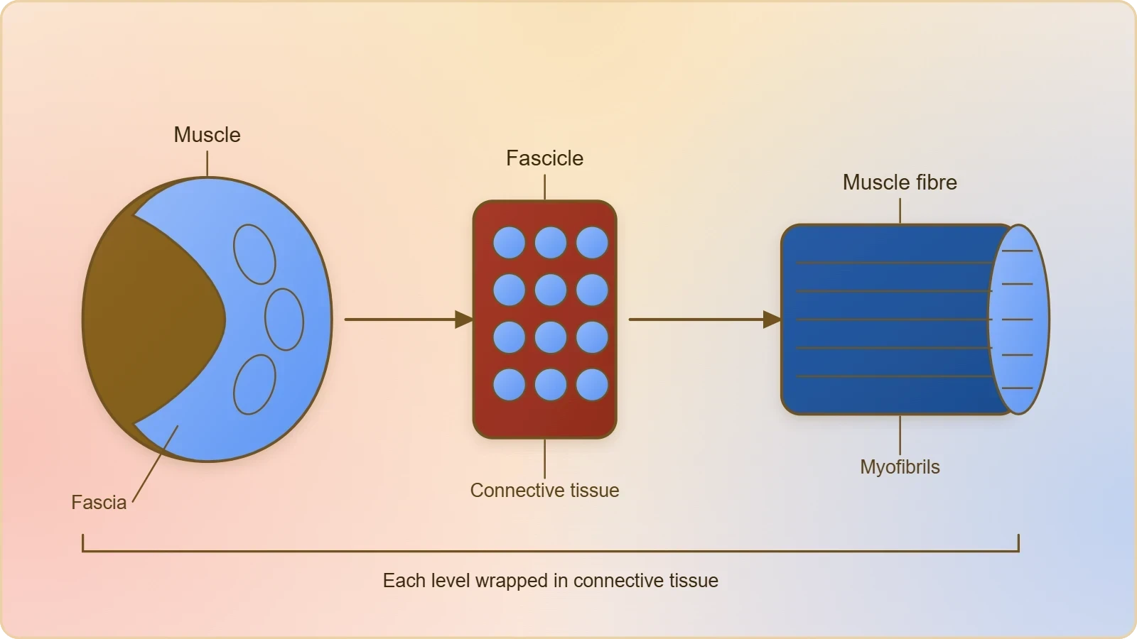
Every organised skeletal muscle in the body is built from a number of muscle bundles, or fascicles, held together by a common collagenous connective tissue layer called fascia. The fascia is the outermost binding material, a tough sheet of collagen that invests the muscle as a whole and continues into the tendons that attach it to bone. Within this envelope, the fascicles lie roughly parallel to one another, and it is this parallel packing that allows the forces generated by individual fibres to sum into a single directed pull. The arrangement is not incidental; it is the reason a muscle can act as one coordinated unit rather than as a loose collection of independent cells.
The fascicular plan also explains why a muscle can be dissected into visible bundles with little more than a blunt probe. Each bundle is a discrete package, separated from its neighbours by connective tissue that carries blood vessels and nerves into the interior. This vascular supply is essential, because a contracting muscle has a high and continuous demand for oxygen and fuel, and the connective partitions provide the routes by which capillaries reach every fibre. The fascia therefore performs a dual role: it is a mechanical binder and a conduit for the vessels and nerves that keep the tissue alive and responsive.
From Whole Muscle to Individual Fibre
Each muscle bundle contains a number of muscle fibres, and the fibre is the individual muscle cell. In a diagrammatic cross-sectional view of a muscle, the fascicles appear as rounded groups, and within each group the fibres are seen as closely packed circular or polygonal profiles. The fibre is the unit that actually contracts; the fascicle is a collection of such units, and the whole muscle is a collection of fascicles. This three-tier arrangement, muscle, fascicle and fibre, is the first level of the hierarchy that the lesson asks you to hold in mind.
The size of a muscle's effect depends on how many fibres it contains and how they are arranged, not on any difference in the internal machinery of the fibres themselves. A fibre from a small hand muscle and a fibre from a large thigh muscle share the same basic internal design. What varies is the number of fibres recruited and the mechanical advantage of their arrangement. Recognising this uniformity is important, because it means the structural principles learned from one fibre apply across the entire skeletal system.
Blood Supply and the Living Bundle
The cross-sectional view shows blood capillaries running between the fibres, threading through the connective tissue that separates them. These capillaries are not decorative additions to the diagram; they represent the dense microcirculation that sustains aerobic metabolism during sustained activity. A muscle fibre is metabolically expensive to operate, and the capillary network must deliver oxygen and glucose while removing carbon dioxide and heat. The proximity of capillaries to individual fibres reflects how finely the vascular supply is divided, matching the fine division of the muscle into contractile units.
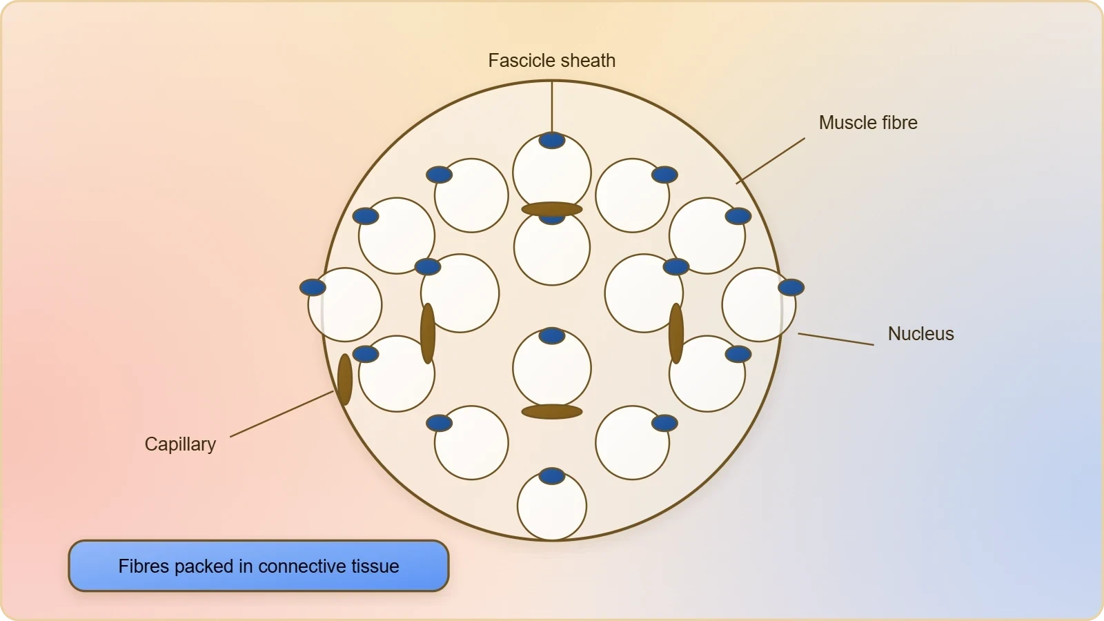
This vascular intimacy also has clinical meaning. When muscle tissue is injured, the bleeding and swelling that follow arise from this rich capillary bed, and the fascia that binds the bundles can confine the swelling, producing the tight, painful quality of a muscle tear. The connective framework described here is thus not merely a passive wrapper; it shapes how injury presents and how repair proceeds. The structural picture, in other words, carries directly over into the disorders that a later lesson will consider.
Inside the Fibre: Sarcolemma, Sarcoplasm and Nuclei
The muscle fibre is lined by the plasma membrane, called the sarcolemma, which encloses the sarcoplasm, the fibre's cytoplasm. The sarcolemma is more than a simple boundary; it is the excitable surface across which the signal to contract travels, and its continuity around the entire fibre ensures that excitation reaches every part of the cell. The sarcoplasm within holds the usual cellular constituents together with the specialised machinery of contraction, and its composition is tailored to the demands of rapid, repeated shortening. These two terms, sarcolemma and sarcoplasm, name the fibre's outer membrane and inner substance, and they anchor all subsequent description.
A characteristic feature of the muscle fibre is the presence of a large number of parallelly arranged filaments in the sarcoplasm, called myofilaments or myofibrils. These filaments dominate the interior, occupying most of the fibre's volume and running lengthwise along its axis. Their parallel alignment is what gives skeletal muscle its ordered, striated appearance under the microscope, and it is the reason a single fibre can produce a coherent directional force. The fibre is, in effect, a cylinder packed with longitudinal cables, each cable itself composed of the protein arrays that will be examined shortly.
The Fibre as a Syncytium
The muscle fibre is described as a syncytium because the sarcoplasm contains many nuclei. During development, precursor cells fuse to form the fibre, and the nuclei of the fused cells persist within a shared cytoplasm rather than being reduced to a single nucleus. The result is a large, multinucleated cell in which many nuclei govern a common expanse of sarcoplasm. This arrangement suits a cell of exceptional length, since each nucleus can direct protein synthesis for the region around it, ensuring that the far reaches of a long fibre are adequately supplied with newly made contractile proteins.
The syncytial nature of the fibre also has functional consequences for signalling and repair. Because the cytoplasm is continuous, a signal arriving at one point on the sarcolemma can spread through the shared interior, and damage to one region does not isolate the rest of the fibre in the way it would in a partitioned tissue. The many nuclei provide a reserve of genetic control that supports the fibre's capacity to rebuild and adapt. The term syncytium, then, is not a curiosity of terminology but a description of a design that solves the problem of maintaining a very large cell.
The Sarcoplasmic Reticulum as Calcium Store
The endoplasmic reticulum of the muscle fibre, called the sarcoplasmic reticulum, is the storehouse of calcium ions. This membrane system threads through the sarcoplasm around the myofibrils, and its role as a calcium reservoir is central to the control of contraction. Calcium is released from this store when the fibre is stimulated and is pumped back when the fibre relaxes, so the sarcoplasmic reticulum acts as the switch that turns contraction on and off. The structural point to retain here is that the calcium store is a specialised, membrane-bound compartment intimately associated with the contractile filaments.
The proximity of the sarcoplasmic reticulum to the myofibrils is what makes rapid control possible. A store located far from the filaments would be useless for the split-second timing that movement demands; a store wrapped around them can flood the contractile apparatus almost instantly. This is a clear example of how structure serves function at the subcellular level, and it prepares the ground for the next lesson, where the calcium-sensitive proteins on the filaments themselves will be described. For now, the sarcoplasmic reticulum is simply the fibre's calcium warehouse, positioned exactly where its contents are needed.
Myofibrils and the Alternating Bands
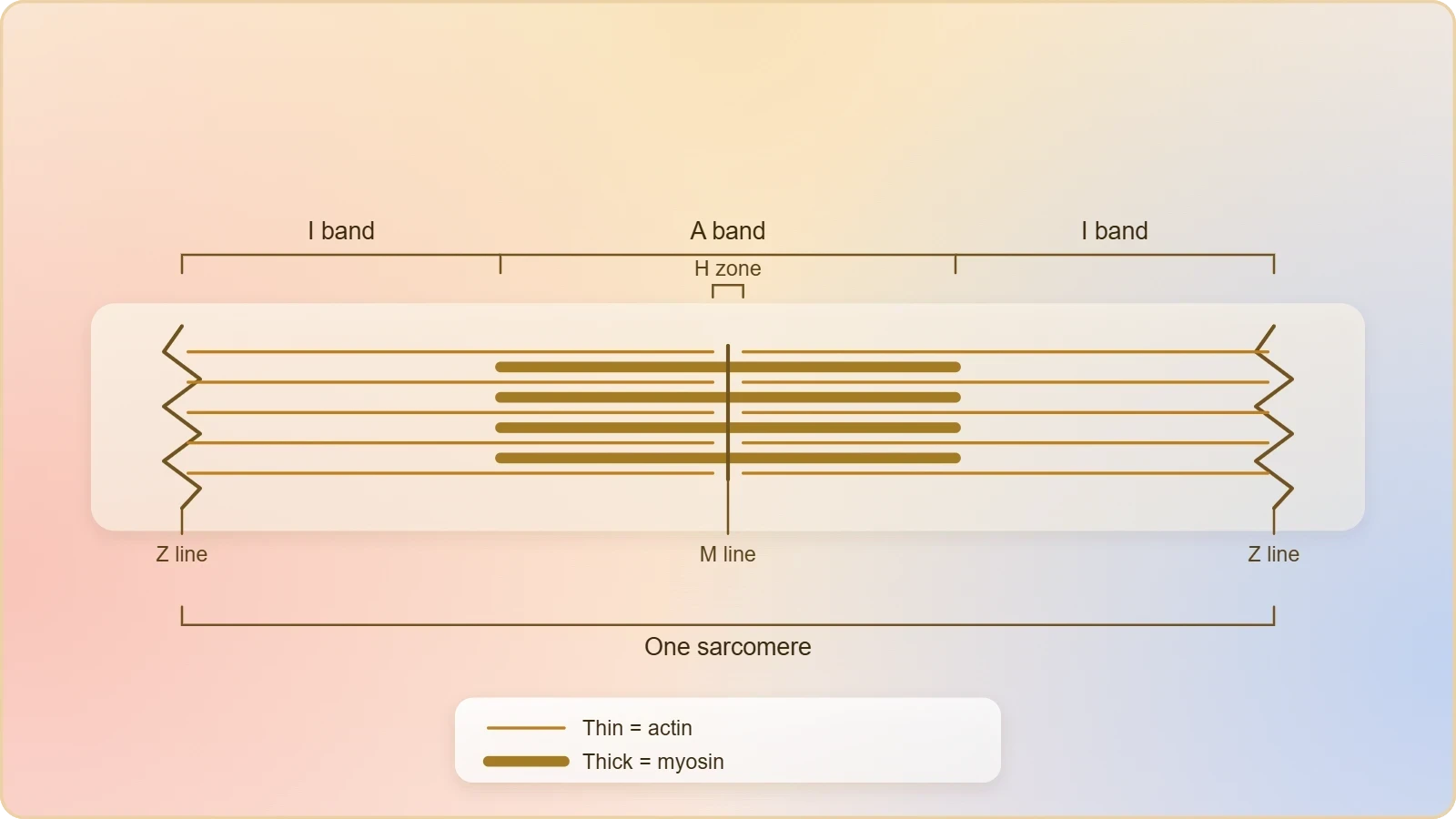
Each myofibril carries alternate dark and light bands along its length, and this banding is the visible signature of the contractile machinery. A detailed study of the myofibril has established that the striated appearance is due to the distribution pattern of two important proteins, actin and myosin. The light bands contain actin and are called I bands, or isotropic bands, while the dark band, called the A or anisotropic band, contains myosin. The two proteins are arranged as rod-like structures, parallel to each other and to the longitudinal axis of the myofibril, and their regular alternation produces the striped pattern seen in striated muscle.
The two filament types differ in thickness as well as in composition. Actin filaments are thinner than myosin filaments and are therefore commonly called thin filaments, while myosin filaments are called thick filaments. This simple size distinction is a useful handle for reading the band pattern: the light I band is the region where thin filaments predominate, and the dark A band is the region occupied by thick filaments. Because the bands repeat along the entire length of the myofibril, any short segment contains a complete sample of the machinery, which is why a small section can represent the whole.
Reading the Band Pattern
The alternation of A and I bands is regular and continuous throughout the length of the myofibril, and this regularity is what allows the structure to be described in repeating units. The dark A band marks the extent of the thick filaments, while the light I band marks the zone where only thin filaments are present. Where the bands meet, the two filament systems overlap, and it is this overlap region that will prove decisive when contraction is explained. For the present lesson, the essential skill is to recognise the bands and to know which protein each contains.
The terminology rewards a moment's attention because it encodes the protein composition directly. The I band is the actin band and the A band is the myosin band; the letters are not arbitrary labels but shorthand for the chemistry of each zone. Once this correspondence is fixed, the diagram becomes readable at a glance, and the positions of the Z line, M line and H zone can be located relative to known filament types. The band pattern is thus the grammar of the myofibril, and fluency in it is the prerequisite for the next lesson's account of the contractile proteins.
Thin and Thick Filaments in Parallel
Both actin and myosin are arranged as rod-like structures lying parallel to one another and to the long axis of the myofibril. This parallel disposition is what converts the chemical activity of the proteins into a linear mechanical event. If the filaments were randomly oriented, their interaction could produce no directed shortening; because they are aligned, the relative sliding of one set past the other shortens the fibril along its axis. The geometry of the array is therefore as important as the chemistry of the proteins, and the band pattern is the visible expression of that geometry.
The parallel arrangement also explains why the myofibril behaves as a coherent unit rather than as a tangle of independent filaments. Each thin filament is positioned to interact with the thick filaments adjacent to it, and each thick filament is positioned to engage the thin filaments on either side. The result is a coordinated system in which many individual interactions sum into a single contraction. This principle of summed parallel action recurs at every level of muscle organisation, from the filament to the fascicle, and it is one of the unifying themes of the subject.
The Sarcomere: Functional Unit of Contraction
The portion of the myofibril between two successive Z lines is considered the functional unit of contraction and is called a sarcomere. This definition is the pivot of the entire lesson, because it identifies the smallest repeating segment that contains a complete set of interacting parts. Within one sarcomere lie the structures needed for shortening: thin filaments anchored at the Z lines, thick filaments centred in the A band, and the overlap regions where the two systems meet. Because the sarcomere repeats along the fibril, the behaviour of one unit describes the behaviour of the whole.
The sarcomere is bounded at each end by a Z line, and the distance between successive Z lines is therefore the length of the unit. In the centre of each I band lies the Z line, an elastic fibre that bisects the light band, and the thin filaments are firmly attached to it. This attachment is what allows the Z line to serve as an anchor: when the thin filaments move, they pull on the Z line, and the Z lines of adjacent sarcomeres transmit the resulting tension along the fibril. The sarcomere is thus a mechanically continuous element, not a merely descriptive region.
The Z Line as Boundary and Anchor
The Z line is described as an elastic fibre situated at the centre of each I band, bisecting it. Its elasticity matters, because a structure that anchors the thin filaments must also accommodate the small changes in spacing that occur as the fibril works. The thin filaments are firmly attached to the Z line, so the line is both a boundary marker and a point of force transmission. When you count sarcomeres along a myofibril, you are counting intervals between successive Z lines, and each interval is a complete contractile module.
Because the Z line defines the sarcomere's limits, it also defines what lies inside and what lies outside. Everything between two Z lines belongs to one sarcomere; the next segment begins immediately beyond the second Z line. This clear demarcation is what makes the sarcomere a convenient unit for study, since its contents can be specified exactly. The Z line is therefore more than a landmark in a diagram; it is the structural boundary that gives the functional unit its identity and its mechanical coherence.
The M Line and the H Zone
The thick filaments in the A band are held together in the middle of the band by a thin fibrous membrane called the M line. This membrane acts as a central brace, keeping the thick filaments in register so that their arrangement remains orderly during contraction. In a resting state, the edges of the thin filaments on either side of the thick filaments partially overlap the free ends of the thick filaments, leaving the central part of the thick filaments uncovered. This central part, not overlapped by thin filaments, is called the H zone.
The H zone is therefore a region defined by absence of overlap rather than by presence of a distinct material. It marks the middle of the A band where only thick filaments are found, flanked on both sides by zones of overlap. The M line lies within this central region, holding the thick filaments together. Together, the Z line, M line and H zone provide a set of reference points that make the sarcomere's internal geography precise, and each will change in a characteristic way when the filament systems slide relative to one another.
From Structure to Function: Preparing for Contraction
The structural account given so far is deliberately static, but every element in it exists to serve movement. The fascia transmits force, the sarcolemma carries excitation, the sarcoplasmic reticulum stores the calcium that will trigger contraction, and the sarcomere organises the filaments whose relative sliding produces shortening. The mechanism of muscle contraction is best explained by the sliding filament theory, which states that contraction of a muscle fibre takes place by the sliding of the thin filaments over the thick filaments. That theory is the destination toward which this lesson's anatomy points, and the anatomy is what makes the theory intelligible.
It is worth noting how the resting arrangement already anticipates the sliding model. The partial overlap of thin and thick filaments at the edges of the A band, and the clear H zone in the centre, describe a system poised for relative movement. If the thin filaments were to slide inward over the thick filaments, the overlap would increase and the H zone would narrow, shortening the distance between Z lines. The static picture therefore contains the seeds of the dynamic one, and the terms defined here are precisely the terms in which the sliding event will be described.
What the Next Lesson Will Add
The present lesson has treated the filaments as structural elements, distinguished by thickness and by the band they occupy. It has not yet described their molecular composition, and that is the task of the next lesson on the structure of contractile proteins. There, actin will be resolved into its filamentous and globular forms, tropomyosin and troponin will be introduced, and myosin will be analysed into its meromyosin components with their heads, arms and tails. Those details belong to the chemistry of the filaments rather than to their arrangement, and separating the two keeps each lesson focused.
The division is pedagogically useful because it prevents the structural map from being obscured by molecular detail. A learner who can already locate the A band, I band, Z line, M line and H zone is in a position to ask how the proteins within those regions are built and how they interact. The questions raised here, about what the thin filaments are made of and how the thick filaments project their cross arms, become the organising questions of the next lesson. Structure first, then composition, then mechanism: that is the sequence this course follows.
Consolidating the Hierarchy
The full hierarchy can now be stated in order: muscle, fascicle, fibre, myofibril, sarcomere, filament. Each level is contained within the one before it, and each is bound or organised by a named structure. The fascia binds the fascicles, the sarcolemma bounds the fibre, the sarcoplasm contains the myofibrils, the Z lines bound the sarcomeres, and the sarcomeres contain the filaments. Holding this ladder in mind is the single most useful outcome of the lesson, because it allows any new term to be placed immediately at its correct level.
This hierarchical habit of thought also guards against a common confusion, in which terms from different levels are treated as if they were interchangeable. A fascicle is not a fibre, and a myofibril is not a sarcomere; each names a distinct tier of organisation with its own boundaries and its own role. Precision about levels is what allows the later discussion of contraction to proceed without ambiguity, since the sliding of filaments occurs at the lowest tier while the shortening of the muscle is observed at the highest.
Going Further
The names isotropic and anisotropic refer to how the bands behave under polarised light. The I band is isotropic because it transmits polarised light equally in all directions, while the A band is anisotropic because its behaviour varies with the direction of the light. This optical distinction is the historical origin of the letters I and A, and it explains why the terminology survives even though modern microscopy describes the bands in terms of filament composition. The names are therefore a reminder that the band pattern was first observed optically, before its molecular basis was understood.
A sarcomere is the repeating unit along a myofibril, which is why it is called the functional unit of contraction. Because the same arrangement recurs from one Z line to the next, a description of one sarcomere applies to every sarcomere along the fibril, and the shortening of the whole fibril is simply the summed shortening of its repeated units. This is the same principle of parallel and serial summation that operates at higher levels of the hierarchy, and it is the reason the sarcomere can stand as a representative of the entire contractile system.
Checking Understanding
A short self-check consolidates the lesson before the next topic begins. The questions below ask only about the structures defined here, so they can be answered from the material already presented. Working through them aloud, or writing brief answers, is more effective than passive re-reading, because it forces the hierarchy to be reconstructed rather than merely recognised. Each question targets a specific level of organisation or a specific landmark, and together they cover the full range of the lesson.
Where an answer feels uncertain, the appropriate response is to return to the relevant section rather than to guess. The band pattern, in particular, rewards repeated inspection, since fluency in reading the A and I bands is the skill on which the next lesson depends. The questions are deliberately phrased to require precise terminology, because precision here is not pedantry but the condition for understanding the mechanism that follows. A learner who can define the sarcomere exactly is ready to learn how it shortens.
Self-Assessment Questions
First, what is a fascicle, and what holds it together? Second, why is the muscle fibre described as a syncytium, and what does the sarcoplasmic reticulum store? Third, which protein occupies the I band and which occupies the A band, and what are the alternative names for these bands? Fourth, define the sarcomere, and state the positions of the Z line, M line and H zone. Finally, explain what the H zone represents in the resting state and why it is defined by the absence of overlap rather than by a distinct material.
These questions can be answered in a few sentences each, and a complete set of correct answers demonstrates mastery of the lesson's structural content. The final question is the most demanding, because it requires the learner to reason about the geometry of overlap rather than simply to recall a definition. That kind of reasoning is exactly what the sliding filament theory will require, and it is the bridge from the static anatomy of this lesson to the dynamic account of contraction that the course will develop.
Looking Ahead
The next lesson takes up the structure of contractile proteins, resolving the thin and thick filaments into their molecular components. Actin will be described as two filamentous actins helically wound together, each a polymer of globular actins, with tropomyosin running alongside and troponin distributed at intervals. Myosin will be described as a polymer of meromyosins, each with a globular head and short arm, the heavy meromyosin, and a tail, the light meromyosin. The cross arms and binding sites introduced there will complete the picture begun here.
With the structural map established and the molecular composition to follow, the stage will be set for the sliding filament theory itself. The terms defined in this lesson, the A band, I band, Z line, M line, H zone and sarcomere, are the vocabulary in which that theory is expressed, and the hierarchy of muscle, fascicle, fibre, myofibril and sarcomere is the framework within which it operates. The present lesson has therefore done its work if the map is clear and the terms are secure.
Also Read
- No links available.
From our network :
- AI-Powered 'Precision Diagnostic' Replaces Standard GRE Score ReportsDiscover the new GRE Precision Diagnostic. Learn how AI-powered behavioral analytics and time-to-solve tracking are replacing standard GRE score reports to empower students.
- Analyzing Trump Deportation Numbers Insights Into The 2026 Immigration Crackdown
- 10 Physics Numerical Problems with Solutions for IIT JEEPrepare for IIT JEE with 10 physics numerical problems, complete with detailed solutions and conceptual explanations. Ideal for mastering kinematics, thermodynamics, electromagnetism, and optics.
- Mastering DB2 12.1 Instance Design: A Technical Deep Dive into Modern Database ArchitectureComprehensive guide to DB2 12.1 Instance Design. Explore architectural components, HADR setup, buffer pool tuning, and security policies for IBM DB2 12.1. Includes 10 functional SQL and Shell samples.
- Vite 6/7 'Cold Start' Regression in Massive Module GraphsIn-depth analysis of Vite 6/7 cold start regressions in massive module graphs. Learn about barrel file issues, pre-bundling optimizations, and how to improve Vite dev server performance.
- EV 2.0: The Solid-State Battery Breakthrough and Global Factory ExpansionDiscover the impact of the Solid-State Battery Breakthrough on the EV market. Learn about CATL and BYD stock growth, global factory expansion in Europe, and the future of EV 2.0 technology.
- Trump Political Strategy How Geopolitical Stunts Serve As Media Diversions
- Mastering DB2 LUW v12 Tables: A Comprehensive Technical GuideComprehensive guide on DB2 LUW v12 tables. Explore CREATE TABLE syntax, BLU Acceleration, range partitioning, data types, and security features for IBM DB2 LUW v12.
- 98% of Global MBA Programs Now Prefer GRE Over GMAT Focus EditionExplore why 98% of global MBA programs now prefer the GRE over GMAT Focus. Learn about the shorter GRE format, verbal logic importance, and strategic versatility for students.
RESOURCES
- Sarcomere-length dependence of myosin filament structure in ...pubmed.ncbi.nlm.nih.govSarcomere-length dependence of myosin filament structure in skeletal muscle fibres of the frog. J Physiol. 2014 Mar 1;592(5):1119-37. doi: 10.1113/jphysiol.
- 10.2 Skeletal Muscle – Anatomy & Physiology 2eopen.oregonstate.educationThe sarcomere is the smallest functional unit of a skeletal muscle fiber and is a highly organized arrangement of contractile, regulatory, and structural ...
- Sarcomere-length dependence of myosin filament structure in ... - PMCpmc.ncbi.nlm.nih.govX-ray diffraction patterns were recorded at beamline ID02 of the European Synchrotron Radiation Facility from small bundles of skeletal muscle fibres from ...
- Human skeletal muscle plasmalemma alters its structure to change ...nature.comFeb 13, 2017 ... The misalignment of sarcomeres at the fibre periphery of healthy muscle ... sarcomere length and intracellular calcium in mouse skeletal…
- Skeletal muscle: A review of molecular structure and function ... - PMCpmc.ncbi.nlm.nih.govFast fibers exhibit shorter twitch parameters, and rapid contraction of the sarcomere. Fast fibers allow for generation of fast and large calcium transients ...
- Sarcomere - Physiopediaphysio-pedia.comA sarcomere is the basic contractile unit of a myocyte (muscle fiber). A sarcomere comprises two main protein filaments (thin actin and thick myosin ...
- Physiology, Skeletal Muscle - StatPearls - NCBI Bookshelf - NIHncbi.nlm.nih.govJul 30, 2023 ... Titin is found at the Z disk and anchors myosin longitudinally within the sarcomere. Muscle fiber types can be broken…
- Skeletal Muscle | Anatomy and Physiology I - Lumen Learningcourses.lumenlearning.comThis figure shows the structure of muscle fibers. The top panel shows a ... The sarcomere is the functional unit of the muscle fiber.…
- Microtubule architecture and detyrosination bidirectionally modulate ...biorxiv.orgJul 23, 2026 ... Microtubule architecture and detyrosination bidirectionally modulate sarcomere shortening in skeletal muscle fibers. Osman Esen, Eva Larose ...
- DES gene: MedlinePlus Geneticsmedlineplus.govSep 30, 2025 ... ... muscle and in the muscles used for movement (skeletal muscle). Within muscle fibers, structures called sarcomeres generate the force…
- The molecular basis for sarcomere organization in vertebrate ...cell.comMar 24, 2021 ... 21. Cantino, M.E. ∙ Brown, L.D. ∙ Chew, M. ... A-band architecture in vertebrate skeletal muscle: polarity of the myosin…
- Ultrastructure of Muscle - Skeletal - Sliding Filamentteachmeanatomy.infoThe functional unit of contraction in a skeletal muscle fibre is the sarcomere, which runs from Z line to Z line. A sarcomere is…
- The vertebrate muscle Z-disc: sarcomere anchor for structure and ...link.springer.comOct 15, 2009 ... Vertebrate muscle Z-bands have precise widths that depend on fibre type (slow and fast) and muscle type (cardiac and skeletal).…
- 5.4: The Sarcomere — Functional Unit of Muscle - Medicine LibreTextsmed.libretexts.orgMar 15, 2026 ... The sarcomere is the functional unit of a skeletal muscle fiber and is responsible for the striated appearance of muscle.
- The M-band: The underestimated part of the sarcomeresciencedirect.comThe structure of the A-band of human skeletal muscle fibres from ultra-thin cryo-sections negatively stained ... Regulation of skeletal muscle sarcomere integrity ...