Movement is one of the significant features of living beings, and the human body expresses it at every scale, from the streaming of protoplasm inside a single cell to the coordinated swing of a limb. Cells of the human body exhibit three main types of movement, namely amoeboid, ciliary and muscular. Each type is produced by a distinct cellular machinery, and each serves a distinct physiological purpose. Understanding these three categories is the foundation on which the study of muscle, skeleton and joints rests, because every larger locomotory act is ultimately assembled from cellular events of this kind.
This lesson examines the three types of movement in the order in which the chapter presents them. It begins with amoeboid movement, effected by pseudopodia formed by the streaming of protoplasm, and notes the involvement of cytoskeletal elements such as microfilaments. It then turns to ciliary movement, which occurs in most internal tubular organs lined by ciliated epithelium, and finally to muscular movement, which depends on the contractile property of muscles and underlies the movement of limbs, jaws and tongue. The treatment stays close to the chapter's terminology and definitions throughout.
Because the previous lesson established the distinction between movement and locomotion, this lesson builds directly on that foundation. All locomotions are movements, but all movements are not locomotions, and the three cellular types illustrate that principle precisely: amoeboid and ciliary movement may relocate a cell or a particle without any change of place for the organism as a whole, while muscular movement supplies the contractile force that locomotion demands. The lesson also prepares the ground for the next topic, the types and properties of muscle, by showing where muscle sits among the body's movement systems.
On This Page
Amoeboid Movement and the Streaming of Protoplasm
Some specialised cells in our body, such as macrophages and leucocytes in blood, exhibit amoeboid movement. The movement is effected by pseudopodia formed by the streaming of protoplasm, exactly as it occurs in the unicellular organism Amoeba. Cytoskeletal elements like microfilaments are also involved in amoeboid movement. This is the simplest of the three types in terms of visible machinery, yet it performs an essential defensive function, since it allows certain white blood cells to migrate through tissue spaces and reach sites of infection or injury.
Pseudopodia as the Motile Apparatus
A pseudopodium is a temporary projection of the cell surface produced when protoplasm streams forward into a region of the cytoplasm. The projection is not a permanent organelle; it is assembled and withdrawn as the cell moves, which is why the chapter describes the process as streaming rather than as the action of a fixed structure. In Amoeba the same mechanism produces the familiar changing outline of the organism, and in human macrophages and leucocytes it produces directed migration through the tissues. The pseudopodium therefore represents a general cellular strategy rather than a peculiarity of one species.
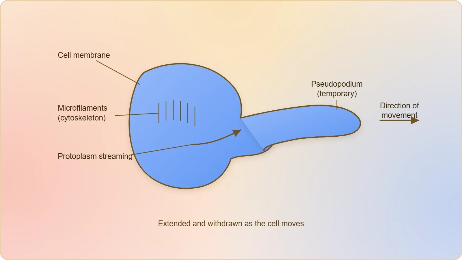
The term amoeboid is used precisely because the human cells that employ this movement resemble Amoeba in behaviour. Macrophages and leucocytes in blood are the named examples, and both belong to the body's defence systems. Their ability to extend pseudopodia lets them squeeze between other cells and pursue material that must be engulfed or destroyed. The chapter places this type first among the three, and the ordering is logical: amoeboid movement requires no specialised tissue, only the capacity of cytoplasm to stream and of the membrane to deform.
The Role of Microfilaments
Cytoskeletal elements like microfilaments are involved in amoeboid movement, and this detail connects the visible behaviour of the cell to its internal architecture. Microfilaments are fine protein threads of the cytoskeleton, and their participation shows that even the apparently simple flowing of protoplasm is an organised, structurally supported event rather than random leakage. The chapter states the involvement without elaborating the molecular mechanism, and that level of detail is sufficient for the present purpose: the student should record that pseudopodia arise from protoplasmic streaming and that microfilaments take part.
Recognising microfilaments here also prepares the mind for the muscular type that follows. Muscle contraction is likewise a property of a specialised protein apparatus, and the contrast between the diffuse, temporary action of microfilaments in amoeboid movement and the highly ordered, forceful action of muscle will become clearer once muscle structure is studied. For now, the essential point is that amoeboid movement is a cellular capacity distributed among particular specialised cells, not a property of the body's muscles or skeleton.
Ciliary Movement and Ciliated Epithelium
Ciliary movement occurs in most of our internal tubular organs, which are lined by ciliated epithelium. The coordinated movements of cilia in the trachea help us in removing dust particles and some of the foreign substances inhaled along with the atmospheric air. Passage of ova through the female reproductive tract is also facilitated by the ciliary movement. This type of movement therefore serves two quite different physiological needs, one protective and one reproductive, using the same basic cellular apparatus in different locations.
Cilia in the Trachea
The trachea is the clearest example of ciliary movement in a protective role. Air entering the respiratory tract carries dust particles and foreign substances, and the ciliated epithelium lining the trachea sweeps these trapped materials away through coordinated beating. The word coordinated is important: the cilia do not act independently but in a synchronised manner, so that their individual strokes combine into a directed current. This is a movement of material across a surface rather than a movement of the organism, which is why it belongs to the category of movement and not to locomotion.
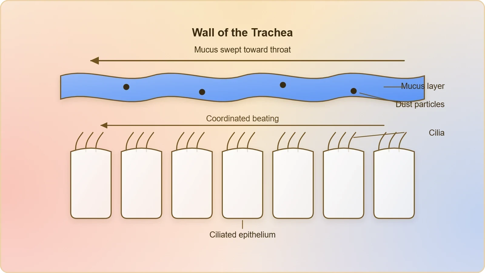
The protective function of tracheal cilia illustrates how a cellular movement can serve the body without producing any visible change of posture. The student should note the precise wording of the chapter: the coordinated movements of cilia help in removing dust particles and some of the foreign substances inhaled along with the atmospheric air. The qualification some is deliberate, since ciliary action is one defence among several. The example also shows that ciliated epithelium is a tissue-level arrangement, with many cells contributing their cilia to a single functional stream.
Cilia and the Passage of Ova
Passage of ova through the female reproductive tract is facilitated by ciliary movement, and this example extends the same mechanism into the reproductive system. The tubular organs of the reproductive tract are lined by ciliated epithelium, and the beating of cilia assists the transport of the ovum along the tract. Here the movement is not defensive but transportive, moving a cell that must reach a particular destination. The chapter presents this alongside the tracheal example to show that ciliary movement is widespread in internal tubular organs rather than confined to the respiratory system.
Placing the two examples together reveals the general principle behind ciliary movement: wherever a tubular organ must move material along its inner surface, ciliated epithelium can provide the motive force. The movement is local, continuous and coordinated, and it requires no muscular contraction. It is worth contrasting this with amoeboid movement, which moves the cell itself, whereas ciliary movement typically moves something past the cell. Both, however, remain movements in the chapter's sense, and neither is locomotion for the human organism as a whole.
Muscular Movement and the Contractile Property of Muscle
Movement of our limbs, jaws, tongue and similar parts requires muscular movement. The contractile property of muscles is effectively used for locomotion and other movements by human beings and the majority of multicellular organisms. Locomotion requires a perfect coordinated activity of muscular, skeletal and neural systems. This third type is therefore the one most directly tied to the locomotory movements introduced in the previous lesson, and it is the type whose structure and mechanism occupy the lessons that follow.
Contractility as the Defining Property
The contractile property of muscles is the source of the force in muscular movement. Muscle is a specialised tissue of mesodermal origin, and about 40 to 50 per cent of the body weight of a human adult is contributed by muscles. They have special properties like excitability, contractility, extensibility and elasticity. Of these, contractility is the one that produces movement, while excitability allows the tissue to respond to stimulation and extensibility and elasticity allow it to lengthen and return. The chapter treats these four properties together because they operate together in every muscular act.
Muscular movement differs from the other two types in scale and in organisation. Amoeboid movement depends on the streaming of protoplasm and ciliary movement on the beating of surface structures, but muscular movement depends on a tissue built specifically for contraction and arranged in masses large enough to move bones. This is why the movement of limbs, jaws and tongue is assigned to the muscular category, and why locomotion, which requires a change of place, is impossible without it in human beings and the majority of multicellular organisms.
Coordination of Muscle, Skeleton and Nerve
Locomotion requires a perfect coordinated activity of muscular, skeletal and neural systems. The muscular system supplies contraction, the skeletal system supplies the levers and supports against which that contraction acts, and the neural system supplies the signals that time and direct the effort. No one of the three can produce locomotion alone. This sentence in the chapter is effectively a map of the remainder of the course, since the sections that follow take up muscle, then the skeletal system, then joints, and finally the disorders that arise when these systems fail.
The word perfect in the chapter's phrasing signals that coordination is not merely helpful but necessary. A muscle that contracts without skeletal attachment produces no useful displacement, and a skeleton without muscular force is a passive framework. The neural system binds the two into a functional unit. Students should carry this three-part scheme forward, because the next lesson examines muscle in detail, and the lessons after that examine the skeleton and joints that complete the locomotory apparatus.
Movement, Locomotion and the Unity of the Three Types
The three cellular types are best understood against the distinction drawn in the previous lesson. Locomotory structures need not be different from those affecting other types of movements. In Paramoecium, cilia help in the movement of food through the cytopharynx and in locomotion as well. Hydra can use its tentacles for capturing prey and also for locomotion. We use limbs for changes in body postures and locomotion as well. These observations suggest that movements and locomotion cannot be studied separately.
Shared Structures, Different Outcomes
The Paramoecium and Hydra examples make the point that a single structure can serve both movement and locomotion depending on the demand of the situation. The chapter links the two by stating that all locomotions are movements but all movements are not locomotions. Applied to the human body, this means that the cilia of the trachea and the pseudopodia of leucocytes are genuine movements even though they never change the body's place, while the muscular movement of limbs can produce either a change of posture or a change of location.
Methods of locomotion performed by animals vary with their habitats and the demand of the situation. Locomotion is generally for search of food, shelter, mate, suitable breeding grounds, favourable climatic conditions, or to escape from enemies and predators. The cellular types described in this lesson supply the raw capability from which these varied locomotory methods are built, and the habitat determines which capability is emphasised. A macrophage migrating through tissue and a runner crossing a field both depend on cellular movement, but only the second is locomotion in the strict sense.
Preparing for Muscle, Skeleton and Joints
The chapter's own sequence moves from the types of movement to muscle, then to the skeletal system, then to joints, and finally to disorders of the muscular and skeletal system. This lesson has covered the first of those sections and has repeatedly touched on the others, because muscular movement cannot be explained without reference to the tissue that produces it and to the skeleton against which it acts. The next lesson takes up muscle directly, examining its types and properties, and will build on the four properties named here: excitability, contractility, extensibility and elasticity.
Students should leave this lesson able to name the three types, state the machinery of each, and give the chapter's examples without hesitation. They should also be able to explain why amoeboid and ciliary movement are movements but not locomotion, and why muscular movement is indispensable to locomotion. These are the load-bearing ideas of the lesson, and everything that follows in the course depends on them being secure.
Going Further: Background That Supports the Lesson
The material above is the backbone of the lesson, and the notes in this section are offered as well-established background that helps the ideas settle. They do not replace or alter anything stated earlier, and nothing here should be read as contradicting the chapter's definitions, examples or order of ideas. Read them as context: they explain why the three types are described as they are and how the study of movement connects to broader biology. The two points below are deliberately brief, and each is clearly separated from the lesson proper.
Cilia as Coordinated Surface Structures
Cilia are hair-like structures on the surface of some cells that can beat in a coordinated way. This background explains why the chapter emphasises coordination in the trachea: the useful effect arises from many cilia beating together, not from any single cilium acting alone. It also clarifies why ciliated epithelium, rather than isolated ciliated cells, is the functional unit in internal tubular organs. The chapter's statement that cilia and flagella are outgrowths of the cell membrane is consistent with this picture, and the flagellar movement of spermatozoa, mentioned in the muscle section, belongs to the same family of surface structures.
Muscle as One of the Four Basic Tissue Types
Muscle tissue is one of the four basic tissue types in the human body, alongside epithelial, connective and nervous tissue. This background places the muscular type of movement in a wider anatomical context and explains why the chapter can describe muscle as a specialised tissue of mesodermal origin with its own set of properties. It also reinforces the point that muscular movement is a tissue-level phenomenon, distinct in kind from the cell-level phenomena of amoeboid and ciliary movement. The remaining lessons of the course develop this tissue-level view through muscle structure, the skeleton, joints and their disorders.
Consolidating the Lesson Before Muscle
A short consolidation is worthwhile before the course moves on, because the three types are easy to confuse when recalled under examination conditions. The reliable way to keep them apart is to ask what actually moves. In amoeboid movement the cell's own protoplasm streams and the cell advances by pseudopodia; in ciliary movement the cilia beat and material passes along a surface; in muscular movement the muscle contracts and a body part is displaced. Each answer points to a different level of organisation, and that difference is the substance of the lesson rather than a mere label.
Common Points of Confusion
Two confusions recur. The first is to treat ciliary movement as locomotion, when in the human body it moves dust, foreign substances and ova rather than the organism. The second is to imagine that amoeboid movement is confined to Amoeba, when the chapter explicitly names macrophages and leucocytes in blood as human examples. A third, subtler confusion is to separate movement from locomotion entirely; the chapter insists they cannot be studied separately, since the same structures often serve both. Keeping these three corrections in mind prevents most errors in later questions on muscle and skeleton.
What the Next Lesson Builds On
The next lesson examines muscle, its types and its properties, and it will assume that the student already knows muscle as the tissue responsible for muscular movement and already recognises the four properties named here. It will also assume familiarity with the statement that locomotion requires coordinated activity of muscular, skeletal and neural systems, since the classification of muscle into skeletal, visceral and cardiac types follows naturally from that scheme. The present lesson has therefore done its work if the three types, their machinery and their examples are secure, and if the relationship between movement and locomotion is clear.
Also Read
- No links available.
From our network :
- Mastering DB2 12.1 Instance Design: A Technical Deep Dive into Modern Database ArchitectureComprehensive guide to DB2 12.1 Instance Design. Explore architectural components, HADR setup, buffer pool tuning, and security policies for IBM DB2 12.1. Includes 10 functional SQL and Shell samples.
- Analyzing Trump Deportation Numbers Insights Into The 2026 Immigration Crackdown
- AI-Powered 'Precision Diagnostic' Replaces Standard GRE Score ReportsDiscover the new GRE Precision Diagnostic. Learn how AI-powered behavioral analytics and time-to-solve tracking are replacing standard GRE score reports to empower students.
- Mastering DB2 LUW v12 Tables: A Comprehensive Technical GuideComprehensive guide on DB2 LUW v12 tables. Explore CREATE TABLE syntax, BLU Acceleration, range partitioning, data types, and security features for IBM DB2 LUW v12.
- Trump Political Strategy How Geopolitical Stunts Serve As Media Diversions
- EV 2.0: The Solid-State Battery Breakthrough and Global Factory ExpansionDiscover the impact of the Solid-State Battery Breakthrough on the EV market. Learn about CATL and BYD stock growth, global factory expansion in Europe, and the future of EV 2.0 technology.
- Vite 6/7 'Cold Start' Regression in Massive Module GraphsIn-depth analysis of Vite 6/7 cold start regressions in massive module graphs. Learn about barrel file issues, pre-bundling optimizations, and how to improve Vite dev server performance.
- 10 Physics Numerical Problems with Solutions for IIT JEEPrepare for IIT JEE with 10 physics numerical problems, complete with detailed solutions and conceptual explanations. Ideal for mastering kinematics, thermodynamics, electromagnetism, and optics.
- 98% of Global MBA Programs Now Prefer GRE Over GMAT Focus EditionExplore why 98% of global MBA programs now prefer the GRE over GMAT Focus. Learn about the shorter GRE format, verbal logic importance, and strategic versatility for students.
RESOURCES
- Types of Movement | CK-12 Foundationflexbooks.ck12.orgNov 1, 2026 ... Human body movements are classified into four types: amoeboid, ciliary, flagellar, and muscular movement. Amoeboid movement occurs through ...
- locomotion and movement chapter 17 - NCERTncert.nic.in17.1 TYPES OF MOVEMENT. Cells of the human body exhibit three main types of movements, namely, amoeboid, ciliary and muscular. LOCOMOTION AND MOVEMENT.
- Types of human movement amoeboid ciliary and muscular - Facebookfacebook.comOct 27, 2026 ... Types of Movement in Animals: Amoeboid, Ciliary & Muscular Movement How do white blood cells crawl through tissues, microscopic organisms ...
- Amoeboid movement - Wikipediaen.wikipedia.org... types of cellular movements in both striated muscle structures and nonmuscle cells. ... Diagram of the three main kinds of amoeboid cell movement.…
- Amoeboid, Ciliary & Muscular Movement How do white blood cells ...facebook.comOct 27, 2026 ... Types of Movement in Animals: Amoeboid, Ciliary & Muscular Movement ... OCR: Mechanisms of Human Movement: Amoeboid, Ciliary, and Muscular ...
- Muscle | Systems, Types, Tissue, & Facts - Britannicabritannica.com6 days ago ... ... amoeboid movement, in which the cell contents flow into extensions, called pseudopodia, from the cell body. Some of the…
- A novel mathematical modeling with solution for movement of fluid ...nature.comOct 18, 2021 ... ... Amoeboid movement (ii) Muscular movement (iii) Ciliary movement. (i) ... Cells in human body which exhibit amoeboid movement are ...
- Types of Body Movements - BYJU'Sbyjus.comDec 1, 2020 ... ... type of ciliary movement. Whenever we move, our limbs are involved. They are the muscular movements. In locomotion and…
- Muscle - Skeletal, Cardiac, Smooth | Britannicabritannica.com6 days ago ... Sperm cells of most higher organisms move using flagella. Many types of unicellular algae and protozoans use flagella in swimming…
- What are the types of movements are shown by the different cells of ...ck12.orgAmoeboid movement: This type of movement is shown by cells that have ... Muscular movement: This type of movement is shown by muscle cells…
- Which of the following type of movements are seen in human body ...allen.inCells of the human body exhibit three main types of movements, namely, amoeboid, ciliary and muscular.
- Introduction to the Muscular System - SEER Training Modulestraining.seer.cancer.govNearly all movement in the body is the result of muscle contraction. ... cells, and amoeboid movement of some white blood cells. The integrated ...
- complete the following paragraph by selecting the correct ... - Allenallen.inCells of the human body exhibit three types of movements namely amoeboid , ciliary , and muscular. Macrophages and leucocytes present in blood exhibit
- 3 Types of Movement in Biology! Living cells and animals use ...instagram.comOct 27, 2026 ... 1️⃣ Amoeboid movement — Macrophages and leucocytes 2️⃣ Ciliary movement — Trachea and female reproductive tract 3️⃣ Muscular movement — ...
- Histology, Respiratory Epithelium - StatPearls - NCBI Bookshelfncbi.nlm.nih.gov... cells for other cell types. The cells that reach the free or apical surface ... Ciliary movement plays a role in many organs…