The gynoecium is the collective term for the female reproductive organs of a flower, and its architecture varies enormously across the angiosperms. A pistil may be built from a single free carpel, described as monocarpellary, or from several carpels, described as multicarpellary. When those carpels are fused into one compound structure the pistil is syncarpous, as in the poppy, Papaver; when they remain distinct and separate the gynoecium is apocarpous, as in Michelia. This distinction between fusion and freedom is not a trivial botanical nicety. It governs how pollen tubes travel, how many stigmas a pollinator must visit, and how the ovary partitions its ovules.
Whatever its carpel count, every pistil resolves into three functional regions: the stigma, the style and the ovary. The stigma is the receptive surface that captures pollen; the style is the elongated conducting tissue that links stigma to ovary; the ovary is the swollen basal chamber that houses the ovules. Each region carries out a distinct task in the chain that leads from pollination to fertilisation, and each shows structural adaptations matched to the plant's breeding system. Understanding this three-part organisation is the necessary prelude to understanding the ovule itself, which is the true site of megasporogenesis and female gametophyte formation.
This lesson examines the pistil and the megasporangium it contains, moving from gross gynoecium architecture to the fine anatomy of a typical angiosperm ovule and then to the cellular events of megasporogenesis. It traces how a single megaspore mother cell, through meiosis, yields four megaspores, and how only one of them survives to build the female gametophyte. The account follows the standard sequence of structures and terminology used in senior secondary biology, and it prepares the ground for the next lesson, where the mature seven-celled, eight-nucleate embryo sac is examined cell by cell.
On This Page
The Gynoecium: Carpel Number and Fusion
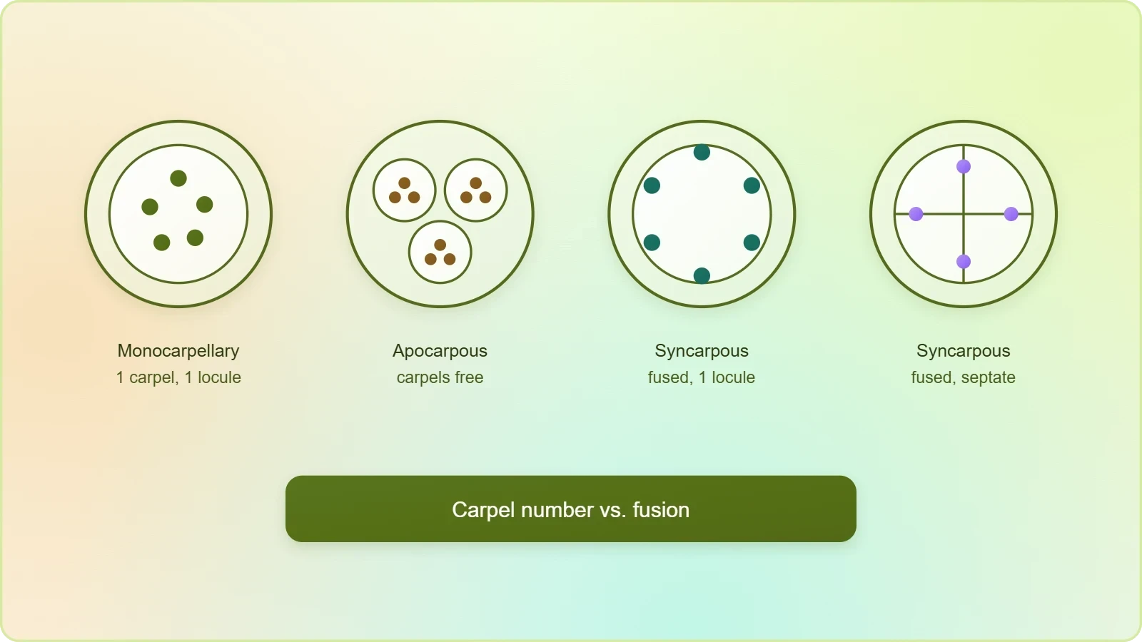
The pistil is the unit of the gynoecium, and its composition can be read from the number of carpels present and from whether those carpels are free or fused. A monocarpellary pistil contains one carpel; a multicarpellary pistil contains several. In a syncarpous gynoecium the carpels are united, often sharing a common ovary and style while retaining separate stigmas, as seen in Papaver. In an apocarpous gynoecium the carpels remain distinct, each with its own ovary, style and stigma, as in Michelia. Recognising these conditions allows a learner to predict where ovules will be borne and how many pollen tubes a single flower must receive.
Monocarpellary and Multicarpellary Conditions
A monocarpellary pistil is the simplest arrangement: one carpel folds to enclose one locule, and the ovules are attached along its inner margin. The legume family offers familiar examples, where a single carpel develops into a pod. Multicarpellary pistils multiply this basic plan. In the syncarpous condition the carpels fuse during development, producing a compound ovary that may be divided internally into chambers by septa. The number of carpels can often be inferred from the number of stigmas, the number of locules, or the pattern of placentation visible in a transverse section of the ovary.
Syncarpous and Apocarpous Gynoecia
Fusion, or syncarpy, is the more common condition among flowering plants, and it carries functional consequences. A single compound stigma presents one large receptive surface, and a single style channels many pollen tubes toward a shared ovary. Apocarpy, by contrast, keeps each carpel independent, so that every carpel must be pollinated separately. The distinction matters for fruit formation as well: a syncarpous ovary typically ripens into one fruit, while an apocarpous gynoecium may yield an aggregate of small fruits, each derived from a separate carpel of the same flower.
The Three Parts of a Pistil
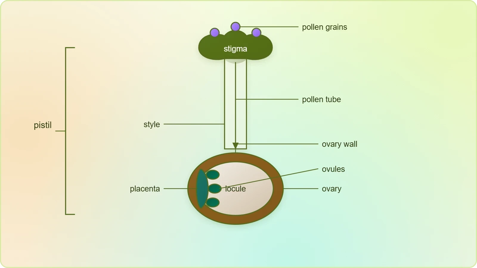
Every pistil, however many carpels it contains, is organised into three regions that work in sequence. The stigma receives the pollen grain; the style conducts the pollen tube downward; the ovary shelters the ovules and later becomes the fruit. These parts are not merely anatomical labels but stages in a functional pipeline. A defect at any one of them, whether a non-receptive stigma, a blocked style or an ovule that fails to differentiate, interrupts reproduction before fertilisation can occur. The three-part plan therefore deserves attention as a coordinated system rather than as a list of separate structures.
Stigma, Style and Ovary
The stigma is the terminal receptive surface, often expanded, lobed or feathery to increase the area available for capturing pollen. Its cells secrete a sticky or moist exudate that holds grains in place and supports their germination. The style is the narrow conducting column beneath it, containing transmitting tissue through which the pollen tube grows toward the ovary. The ovary is the enlarged basal portion, hollow or partitioned, within which the ovules are borne on the placenta. Together these three parts define the pathway that a male gametophyte must traverse before it can deliver its gametes to the embryo sac.
Placenta and Ovule Number
Arising from the placenta are the megasporangia, commonly called ovules. The placenta is the region of the ovary wall where ovules attach, and its arrangement determines the internal geometry of the fruit. The number of ovules in an ovary may be as few as one, as in wheat, paddy and mango, or as many as numerous, as in papaya, water melon and orchids. This numerical range reflects different reproductive strategies: a single ovule concentrates resources into one assured seed, while many ovules allow a plant to set numerous seeds from a single pollination event, provided sufficient pollen arrives.
The Megasporangium: Structure of a Typical Ovule
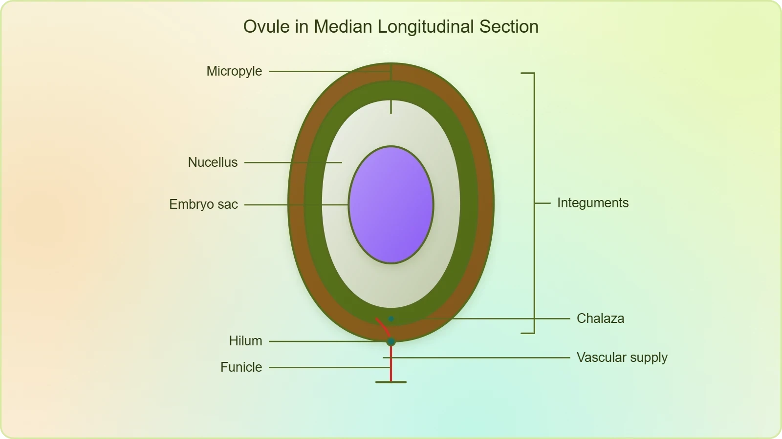
The ovule is a small structure attached to the placenta by means of a stalk called the funicle. Its anatomy is best learned as a series of concentric and longitudinal relationships. The body of the ovule fuses with the funicle in the region called the hilum, so the hilum represents the junction between ovule and funicle. Each ovule has one or two protective envelopes called integuments, which encircle the nucellus except at the tip, where a small opening called the micropyle is organised. Opposite the micropylar end lies the chalaza, representing the basal part of the ovule. Enclosed within the integuments is a mass of cells called the nucellus, whose cells have abundant reserve food materials, and located in the nucellus is the embryo sac, or female gametophyte.
Funicle, Hilum, Integuments and Micropyle
The funicle is the vascularised stalk that suspends the ovule from the placenta and supplies it with water and nutrients. Where the funicle merges into the body of the ovule, the junction is marked as the hilum, a landmark that botanists use to orient the ovule in description. The integuments are the protective coats, one or two in number, that enclose the inner tissues. They leave a small gap at the tip, the micropyle, which is the only opening through the ovule's protective layers and therefore the natural gateway for the pollen tube when it arrives at the embryo sac.
Chalaza, Nucellus and Embryo Sac
The chalaza is the basal part of the ovule, positioned opposite the micropylar end, and it marks the region where the integuments and nucellus converge with the vascular supply. Within the integuments lies the nucellus, a parenchymatous mass whose cells store abundant reserve food materials for the developing gametophyte and, later, the embryo. Embedded in the nucellus is the embryo sac, the female gametophyte. An ovule generally has a single embryo sac formed from a megaspore, so the nucellus typically supports one developing gametophyte rather than several.
Megasporogenesis: From Mother Cell to Megaspores
The process of formation of megaspores from the megaspore mother cell is called megasporogenesis. Ovules generally differentiate a single megaspore mother cell, or MMC, in the micropylar region of the nucellus. The MMC is a large cell containing dense cytoplasm and a prominent nucleus, features that mark it as metabolically active and destined for division. Its position near the micropylar end places the future gametophyte close to the path a pollen tube will take, an arrangement that is both economical and functionally sensible. The MMC then undergoes meiotic division, and meiosis results in the production of four megaspores.
The Megaspore Mother Cell and Meiosis
The importance of the MMC undergoing meiosis lies in the genetic consequences of the process. Meiosis halves the chromosome number and reassorts genetic material through crossing over and independent assortment, so the four megaspores produced are not genetically identical to one another or to the parent cell. This is the central reason why sexual reproduction generates variation. The MMC is diploid, and after meiosis each megaspore carries the haploid chromosome complement. The meiotic division therefore converts a somatic-lineage cell of the nucellus into a set of haploid spores capable of founding a gametophyte.
Ploidy of Nucellus, MMC, Megaspore and Gametophyte
A useful check on understanding is to assign the ploidy of the relevant cells. The cells of the nucellus are diploid, since they belong to the sporophytic tissue of the ovule. The megaspore mother cell is likewise diploid, being a differentiated nucellar cell. The functional megaspore is haploid, the product of meiosis. The female gametophyte derived from that megaspore is also haploid, because it arises by mitotic divisions and no further reduction division occurs. Keeping these four assignments distinct prevents a common confusion between the sporophyte that produces spores and the gametophyte that the spores become.
Monosporic Development of the Female Gametophyte
In a majority of flowering plants, one of the megaspores is functional while the other three degenerate. Only the functional megaspore develops into the female gametophyte, or embryo sac. This method of embryo sac formation from a single megaspore is termed monosporic development. The term is precise: mono refers to one spore, and the entire gametophyte is descended from that single surviving cell. The three degenerating megaspores are not wasted in a metabolic sense, since their contents are typically resorbed and made available to the surviving spore, but they contribute no cells to the mature gametophyte.
Survival of One Megaspore
The tetrad of megaspores produced by meiosis is usually arranged in a linear or T-shaped file within the nucellus. Typically the chalazal-most megaspore becomes the functional one, while the three nearer the micropyle degenerate. The reasons for this asymmetry are developmental rather than arbitrary: the position of the surviving spore relative to the nucellar supply appears to influence its access to nutrients. Whatever the precise mechanism, the outcome is a single founder cell. From this point onward, the female gametophyte is a clonal population of haploid cells derived from one meiotic product, which is why the monosporic pattern is described as a single-spore pathway.
Free Nuclear Divisions and the 2, 4 and 8-Nucleate Stages
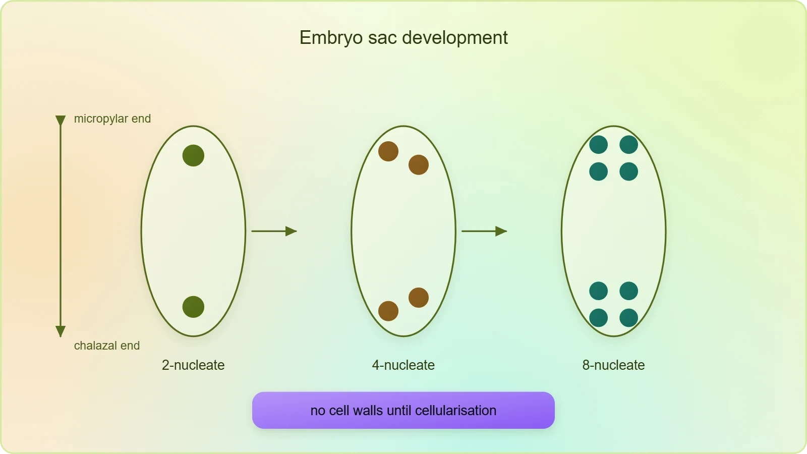
The nucleus of the functional megaspore divides mitotically to form two nuclei, which move to the opposite poles, forming the 2-nucleate embryo sac. Two more sequential mitotic nuclear divisions result in the formation of the 4-nucleate and later the 8-nucleate stages of the embryo sac. It is of interest to note that these mitotic divisions are strictly free nuclear: nuclear divisions are not followed immediately by cell wall formation. The embryo sac therefore passes through a coenocytic phase in which multiple nuclei share a common cytoplasm. Only after the 8-nucleate stage are cell walls laid down, leading to the organisation of the typical female gametophyte.
Organisation of the Embryo Sac and Its Cell Types
After the 8-nucleate stage, cell walls are laid down leading to the organisation of the typical female gametophyte. Observe the distribution of cells inside the embryo sac: six of the eight nuclei are surrounded by cell walls and organised into cells, while the remaining two nuclei, called polar nuclei, are situated below the egg apparatus in the large central cell. There is a characteristic distribution of the cells within the embryo sac. Three cells are grouped together at the micropylar end and constitute the egg apparatus, which consists of two synergids and one egg cell. Three cells are at the chalazal end and are called the antipodals. The large central cell has two polar nuclei. Thus a typical angiosperm embryo sac, at maturity, though 8-nucleate is 7-celled.
Egg Apparatus, Synergids and Filiform Apparatus
The egg apparatus occupies the micropylar end of the embryo sac and comprises two synergids flanking a single egg cell. The synergids have special cellular thickenings at the micropylar tip called the filiform apparatus, which play an important role in guiding the pollen tubes into the synergid. This guidance function makes the synergids far more than passive companions to the egg: they are the entry point through which the male gametophyte is received. The egg cell itself is the female gamete, positioned centrally within the apparatus and ready to fuse with one male gamete after the pollen tube discharges its contents.
Antipodals, Polar Nuclei and the Central Cell
At the chalazal end, opposite the egg apparatus, lie three cells called the antipodals. Their number is constant in the typical sac, though their persistence and function vary among species. The large central cell dominates the interior of the embryo sac and contains the two polar nuclei, which lie below the egg apparatus. These polar nuclei are not enclosed by cell walls at maturity and remain within the central cell's cytoplasm. Their fusion with a male gamete will initiate the triploid endosperm, a topic that belongs to the later account of double fertilisation and seed formation.

Going further
The micropyle is the small opening in the integuments through which the pollen tube enters the ovule. Because the integuments otherwise enclose the nucellus completely, this aperture is the only route by which the male gametophyte can reach the embryo sac. In the organised sac, the synergids lie directly beneath the micropyle, and their filiform apparatus faces the opening, so the geometry of the ovule itself directs the pollen tube toward the receptive cells. This is why the micropylar end is described as the functional front of the ovule and the chalazal end as its base.
The chalaza is the basal part of the ovule opposite the micropylar end. It marks the region where the integuments and nucellus converge and where the vascular supply from the funicle is concentrated. In the mature embryo sac, the antipodals occupy the chalazal pole, mirroring the egg apparatus at the micropylar pole. Recognising this polarity helps in reading diagrams of the ovule: micropyle and egg apparatus at one end, chalaza and antipodals at the other, with the central cell and its two polar nuclei spanning the space between them.
Also Read
- No links available.
From our network :
- Mastering DB2 12.1 Instance Design: A Technical Deep Dive into Modern Database ArchitectureComprehensive guide to DB2 12.1 Instance Design. Explore architectural components, HADR setup, buffer pool tuning, and security policies for IBM DB2 12.1. Includes 10 functional SQL and Shell samples.
- Trump Political Strategy How Geopolitical Stunts Serve As Media Diversions
- 10 Physics Numerical Problems with Solutions for IIT JEEPrepare for IIT JEE with 10 physics numerical problems, complete with detailed solutions and conceptual explanations. Ideal for mastering kinematics, thermodynamics, electromagnetism, and optics.
- 98% of Global MBA Programs Now Prefer GRE Over GMAT Focus EditionExplore why 98% of global MBA programs now prefer the GRE over GMAT Focus. Learn about the shorter GRE format, verbal logic importance, and strategic versatility for students.
- AI-Powered 'Precision Diagnostic' Replaces Standard GRE Score ReportsDiscover the new GRE Precision Diagnostic. Learn how AI-powered behavioral analytics and time-to-solve tracking are replacing standard GRE score reports to empower students.
- Mastering DB2 LUW v12 Tables: A Comprehensive Technical GuideComprehensive guide on DB2 LUW v12 tables. Explore CREATE TABLE syntax, BLU Acceleration, range partitioning, data types, and security features for IBM DB2 LUW v12.
- EV 2.0: The Solid-State Battery Breakthrough and Global Factory ExpansionDiscover the impact of the Solid-State Battery Breakthrough on the EV market. Learn about CATL and BYD stock growth, global factory expansion in Europe, and the future of EV 2.0 technology.
- Analyzing Trump Deportation Numbers Insights Into The 2026 Immigration Crackdown
- Vite 6/7 'Cold Start' Regression in Massive Module GraphsIn-depth analysis of Vite 6/7 cold start regressions in massive module graphs. Learn about barrel file issues, pre-bundling optimizations, and how to improve Vite dev server performance.
RESOURCES
- The Pistil, Megasporangium (ovule) and Embryo sacflexbooks.ck12.orgNov 1, 2026 ... This lesson explores the structure and function of the gynoecium, ovule (megasporangium), megasporogenesis, and development of the female ...
- Structure of Pistil and Embryo Sac | PDF | Plants | Organisms - Scribdscribd.com... Megasporogenesis produces four megaspores from a megaspore mother cell ... 2.2.2 The Pistil, Megasporangium (ovule) and Embryo sac. • The gynoecium ...
- Female gametophyte development, pollen‒pistil interactions and ...pubmed.ncbi.nlm.nih.govJun 25, 2025 ... ... megasporogenesis and megagametogenesis. ... In parallel, for the first time, we characterized both the embryogenesis process and embryo sac ...
- Sexual Reproduction in Flowering Plants: Structured Study Notespearson.comPistil, Megasporangium (Ovule), and Embryo Sac · Megasporogenesis: Formation of megaspores from the megaspore mother cell (MMC) via meiosis. · Embryo Sac: Female ...
- Single-nucleus sequencing deciphers developmental trajectories in ...sciencedirect.comApr 24, 2023 ... ... ovule bearing the embryo sac. The mature structure of a rice pistil contains a flask-like ovary at the base,…
- How flowers make seeds through pistil - Facebookfacebook.comJun 17, 2026 ... OCR: The Pistil and Megasporangium (Ovule) The Female ... pistil to embryo sac. EcoGeneZap Megasporogenesis and the Embryo Sac ...
- Flower Bud Differentiation, Mega-micro Spore Production, and Male ...journals.ashs.org... pistil primordium; ov = ovary; op = ovule primordium. ... According to the observations based on consecutive sections of the embryo sac, the…
- Heat Stress During Gametogenesis Irreversibly Damages Female ...pmc.ncbi.nlm.nih.govJun 28, 2022 ... ... pistil development (megasporogenesis) and ... The abnormalities occurring at the nucleate embryo sac stage, resulted in mature embryo sac ...
- Growth and Development of the Pistillate Flower and Nut in ...journals.ashs.orgDiagram of ovule showing route of secondary pollen tube from resting structure in apex of ovary to embryo sac. a, resting structure in ovary…
- The Ovule and the Embryo Sac - PMCpmc.ncbi.nlm.nih.govAbnormal megasporogenesis in ovm3 and sinl ovules and aborted megagametogenesis in bell and wm2 ... Pistil development. Plant. Cell 5, 1231-1239. Gasser ...
- Heat Stress During Gametogenesis Irreversibly Damages Female ...ttu-ir.tdl.orgAnatomical observations of ovule revealed greater disappearance of megaspore mother cell and nuclei at early stages, and during later stages mature embryo sac ...
- Mutation of DEFECTIVE EMBRYO SAC1 results in a low seed ...academic.oup.comThe embryo sac is embedded in sporophytic tissues of the ovule, making it ... Previous results indicated that LOG regulates the development of the…
- Ovule and pollen development in Camelina sativa provides ... - Naturenature.comNormal seed development in angiosperms is described as functional in the production of male (pollen grain or microgametophyte) and female (embryo sac or ...
- lebo101.pdf - NCERTncert.nic.in1.2.2 The Pistil, Megasporangium (ovule) and Embryo sac. The gynoecium ... Differentiate between microsporogenesis and megasporogenesis. Which type of ...