The mature embryo sac is the quiet culmination of one of the most economical developmental sequences in the plant kingdom: a single surviving spore, through three rounds of free-nuclear mitosis and one act of cellular organisation, becomes a seven-celled, eight-nucleate structure competent to receive a pollen tube. This lesson traces that sequence from the functional megaspore through the 2-nucleate, 4-nucleate and 8-nucleate stages, and then examines how the eight nuclei are partitioned into the egg apparatus, the polar nuclei and the antipodals. The emphasis throughout is on the arithmetic of the divisions and the geometry of the final arrangement, because the familiar description "7-celled and 8-nucleate" only makes sense once both are held together.
The megaspore mother cell has already completed meiosis, and of the four haploid products typically produced, three degenerate while one — usually the chalazal-most — enlarges to become the functional megaspore. Everything that follows is mitotic and, crucially, free nuclear: the nuclei divide without accompanying wall formation, so the embryo sac passes through a coenocytic phase before cellular organisation begins. Recognising this single fact resolves most of the confusion students encounter when counting cells and nuclei in the mature structure. The divisions are also synchronous in the usual account, which is why the stages are named for the number of nuclei rather than the number of cells.
By the end of this lesson you should be able to narrate the transition from functional megaspore to mature embryo sac in order, explain why the mitoses are described as free nuclear, and justify the statement that the mature embryo sac is 8-nucleate but 7-celled. You should also be able to place each of the three cell types — egg apparatus, central cell with its polar nuclei, and antipodals — at the correct pole or centre of the sac. This prepares directly for the next stage of the course, where the pollen tube arrives at the micropylar end and the interaction between male gametes and this precisely organised female structure begins.
On This Page
The Functional Megaspore and the Onset of Free-Nuclear Division
Development of the female gametophyte begins with a single cell set apart from its sisters. Within the nucellus of the ovule, one hypodermal cell enlarges and becomes the megaspore mother cell, which then undergoes meiosis to yield four haploid megaspores arranged in a linear tetrad. In the common monosporic pattern, the three micropylar megaspores degenerate while the chalazal one survives and functions. This surviving cell is the functional megaspore, and its enlargement marks the beginning of the gametophytic phase proper. The degeneration of the other three is not incidental: it concentrates the entire developmental programme of the female gametophyte into one cell, and it explains why the resulting embryo sac is described as monosporic in origin.
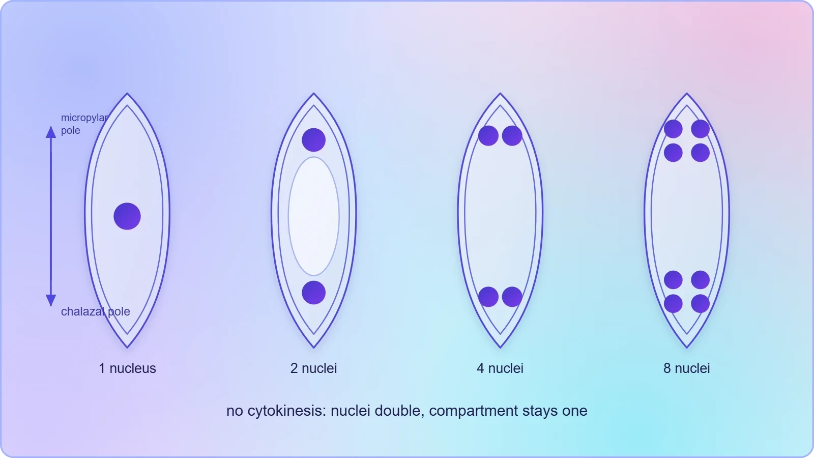
The first mitotic division of the functional megaspore produces two nuclei that remain within the same enlarging cell, separated only by a large central vacuole rather than by a wall. This is the 2-nucleate stage. The absence of cytokinesis is the defining feature of the entire sequence, and it is worth pausing on why it matters. Because no walls are laid down, the nuclei are free to migrate and to occupy positions determined by the vacuolar architecture of the sac, and the subsequent divisions can proceed without the geometric constraints that walled cells would impose. The embryo sac is, in effect, a coenocyte during this phase, and the term free-nuclear mitosis records exactly that condition.
The 2-Nucleate and 4-Nucleate Stages
Each of the two nuclei now divides, giving four nuclei in the same coenocytic sac — the 4-nucleate stage. In the usual account these divisions are synchronous, so the sac passes through a clean doubling sequence: one nucleus, two, four, eight. The four nuclei are distributed, one toward each pole, and a large central vacuole develops between the two groups. This polarisation is the first sign that the sac is acquiring an axis, with a micropylar end facing the opening of the ovule and a chalazal end directed toward the base. The axis established here will govern where the egg apparatus and the antipodals eventually sit, and therefore where the pollen tube must deliver its contents.
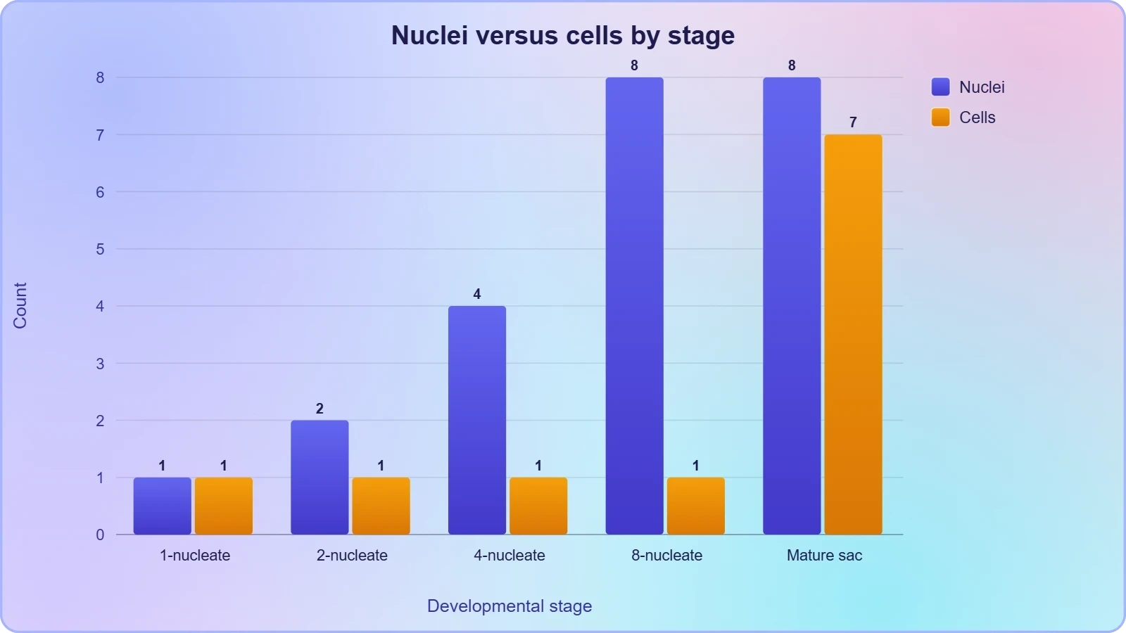
It is useful to keep the arithmetic explicit, because the numbers in the stage names are counts of nuclei, not of cells. At the 4-nucleate stage there are four nuclei and still effectively one compartment. Students who instinctively count cells at this point will find the later "7-celled, 8-nucleate" description puzzling, whereas those who track nuclei separately from walls will find it obvious. The vacuole is not a passive space either: its expansion pushes the nuclei toward the poles and helps to set up the two polar groups that will later contribute the polar nuclei of the central cell.
The Third Division and the 8-Nucleate Condition
A third round of free-nuclear mitosis converts the four nuclei into eight, and this is the 8-nucleate stage. Four nuclei lie at the micropylar pole and four at the chalazal pole, still within a single coenocytic sac. No new walls have appeared, so the structure is 8-nucleate but not yet 7-celled. The doubling sequence is now complete, and the sac has reached its full nuclear complement. What remains is organisation: the migration of one nucleus from each pole to the centre, and the cellularisation of the peripheral nuclei into discrete cells. The transition from 8-nucleate to 7-celled is therefore a matter of wall formation and nuclear migration, not of further division.
The synchrony of these divisions is the standard textbook account and is worth stating plainly, because it explains why the stages are named for nuclear number rather than for elapsed time or size. Each division roughly doubles the nuclear count while the sac itself enlarges, so the nuclear density changes far less than the count suggests. The three mitoses together constitute the entire proliferative phase of the female gametophyte; there is no further mitosis after the eighth nucleus is formed. Everything from this point is rearrangement and cellularisation, which is why the mature structure can be described completely by the positions of eight nuclei and the walls that enclose seven of them.
Organisation of the Mature Embryo Sac
Once eight nuclei are in place, the sac reorganises itself into three distinct regions along its micropylar–chalazal axis. At the micropylar end, three nuclei become enclosed in separate walls to form the egg apparatus. At the chalazal end, three nuclei are similarly walled off as the antipodal cells. One nucleus from each pole migrates to the centre, and these two polar nuclei come to lie in the large central cell without fusing immediately. The result is a structure with seven walled cells and eight nuclei, since the central cell contains two. This is the arrangement that the phrase "7-celled and 8-nucleate" compresses into four words, and unpacking it is the central task of this section.

The polarity established during the free-nuclear phase is what makes this organisation possible. Because the nuclei were already grouped at the two poles, cellularisation can proceed independently at each end, and the central cell is simply the large vacuolate region that remains between them. The micropylar end is the one oriented toward the micropyle, the opening through which the pollen tube will enter, so the egg apparatus is positioned exactly where the male gametes will be delivered. The chalazal end faces the base of the ovule and carries the antipodals, whose role is less obviously connected to fertilisation but which complete the symmetry of the structure.
The Egg Apparatus: Egg Cell and Synergids
The egg apparatus consists of three cells grouped at the micropylar pole: one egg cell and two synergids. The egg cell is the female gamete, the cell that will fuse with one male gamete at fertilisation. The two synergids flank it and are the cells that receive the pollen tube, guiding its entry into the sac. All three are enclosed in walls, which is why they count as three of the seven cells. Their arrangement is not random: the synergids sit at the micropylar tip, with the egg cell typically between and slightly behind them, so that the path of the pollen tube passes through the synergid region before reaching the egg.
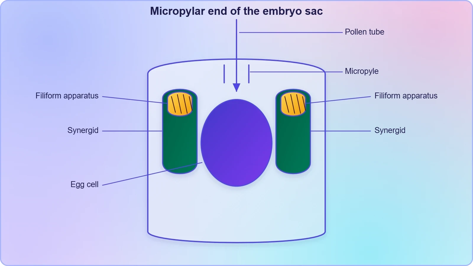
The synergids are often described as short-lived and specialised, and their position at the very entrance of the sac reflects that specialisation. They are the first cells the pollen tube encounters, and their degeneration after the tube has discharged its contents is part of the normal sequence of double fertilisation. For the purposes of this lesson, the essential point is structural: the egg apparatus contributes three cells and three nuclei to the mature sac, and it occupies the micropylar pole. The egg cell itself contributes one of the eight nuclei; the two synergids contribute the other two at that end.
Polar Nuclei and the Central Cell
The central cell is the largest cell of the embryo sac and the only one that contains two nuclei. One polar nucleus arrives from the micropylar pole and one from the chalazal pole, and they come to lie close together in the central region, often below the egg apparatus. They are described as polar nuclei rather than as a fusion nucleus because, in the usual account, they remain distinct until fertilisation, when they fuse with the second male gamete. This is the fusion that produces the triploid primary endosperm nucleus, and it is the reason the central cell is central to the double fertilisation that follows in a later lesson.
Counting carefully here resolves the apparent paradox of the mature sac. The central cell is one cell but holds two nuclei, so the total of seven cells and eight nuclei is consistent: three cells at the micropylar pole, three at the chalazal pole, and one central cell containing two nuclei. The two polar nuclei are not a single diploid nucleus; they are two haploid nuclei sharing a cytoplasm. That distinction matters for the arithmetic of fertilisation, because the second male gamete fuses with both, giving three haploid sets in the primary endosperm nucleus rather than two.
The Antipodals and the Chalazal Pole
At the chalazal end of the sac, the remaining three nuclei are enclosed in walls to form the antipodal cells. They are the least conspicuous of the three regions and, in many species, they degenerate early or vary in number, but in the standard account they are three distinct cells completing the chalazal pole. Their position mirrors the egg apparatus at the opposite end, and together the two groups give the sac its characteristic symmetry. The antipodals contribute three cells and three nuclei to the total, and their presence is what makes the chalazal end a cellular region rather than an empty space.
The functional significance of the antipodals is often described as uncertain or variable, and it is fair to say that their role is less directly tied to fertilisation than that of the egg apparatus or the central cell. What matters for this lesson is their place in the count and in the geometry of the sac. They sit at the pole opposite the micropyle, so they are the last cells the pollen tube's contents would reach, and they are separated from the central cell by the chalazal boundary of that cell. Recognising them as three walled cells at the chalazal pole is sufficient to complete the seven-cell tally.
Why Seven Cells but Eight Nuclei
The apparent contradiction dissolves once the central cell is examined closely. Three cells at the micropylar pole, three at the chalazal pole and one central cell give seven cells. The nuclei are one in the egg, one in each synergid, three in the antipodals and two in the central cell, giving eight. The only cell that departs from the one-cell-one-nucleus rule is the central cell, and it does so because the two polar nuclei have not yet fused. Every other cell in the sac is uninucleate. The description is therefore not a quirk of terminology but a precise statement about the state of the nuclei at maturity.
This arithmetic also explains why the fusion of the polar nuclei at fertilisation changes the count. Once the second male gamete fuses with the two polar nuclei, the central cell contains a single triploid nucleus, and the sac is no longer 8-nucleate in the same sense. The "7-celled and 8-nucleate" description is a snapshot of the mature, unfertilised sac, and it is worth stating that explicitly so that the later events of double fertilisation do not appear to contradict it. The count is a description of a stage, not a permanent property of the structure.
Preparing for the Pollen Tube
The organisation described here is precisely what the pollen tube must negotiate. The tube enters through the micropyle, meets the synergids at the micropylar pole, and discharges its two male gametes into the sac. One gamete fuses with the egg cell and the other with the two polar nuclei, producing the diploid zygote and the triploid primary endosperm nucleus respectively. The antipodals are not directly involved in these fusions. The spatial arrangement of the mature sac — egg apparatus at the entrance, central cell in the middle, antipodals at the far pole — is therefore not decorative but functional, and it is the arrangement that the next stage of the course will assume.
It is also worth noting how much of the later course depends on this structure being correctly understood. The pollen–pistil interaction, the growth of the tube through the style, and the entry of the tube into the ovule all culminate in the delivery of gametes to the cells described here. A student who can place the egg cell, the synergids, the polar nuclei and the antipodals accurately will find the account of double fertilisation straightforward, because each fusion has a defined cellular destination. The mature embryo sac is, in this sense, the map on which the events of fertilisation are plotted.
Going Further
Two details, well established in the wider literature, sharpen the picture without altering the account above. The first concerns the synergids. At their micropylar tip, the synergids develop a specialised thickening known as the filiform apparatus, a structure that guides the pollen tube into the synergid rather than allowing it to wander. This is the mechanism behind the structural statement that the synergids receive the tube: the filiform apparatus is the physical interface between the arriving tube and the cell that will admit it. It is a useful detail to hold alongside the description of the egg apparatus, because it explains how the tube finds its way to the correct cell.
The second detail concerns the position of the polar nuclei. The two polar nuclei lie in the large central cell below the egg apparatus, which is consistent with the description of the central cell as the region between the two poles. Placing them explicitly below the egg apparatus helps to visualise the sac as a whole: the egg apparatus at the top near the micropyle, the central cell with its two polar nuclei beneath it, and the antipodals at the far chalazal end. These two additions are background rather than part of the core sequence, and they should be read as refinements of the structure already described, not as replacements for it.
Common Misreadings to Avoid
Three misreadings recur and are worth naming. The first is to treat the 8-nucleate stage as already cellular; it is not, because the divisions are free nuclear and walls appear only during organisation. The second is to describe the central cell as containing a single diploid nucleus; at maturity it contains two distinct polar nuclei, and fusion occurs at fertilisation. The third is to count the antipodals as part of the egg apparatus; they are at the opposite pole and play no direct part in the fusions. Avoiding these three errors keeps the count and the geometry consistent with each other.
A fourth, subtler misreading is to imagine that the three mitoses are followed by a fourth division that produces the seven cells. No such division occurs. The seven cells arise from the eight nuclei by wall formation and by the migration of one nucleus from each pole to the centre, so the transition is one of organisation rather than proliferation. Keeping this in mind makes the sequence easy to reconstruct from memory: one spore, three mitoses, eight nuclei, then cellularisation into seven cells with the central cell holding two nuclei.
Consolidating the Sequence
The full sequence can now be stated in one continuous line: functional megaspore, first free-nuclear mitosis to two nuclei, second to four, third to eight, then migration of one nucleus from each pole to the centre and cellularisation of the remaining six into the egg apparatus and antipodals. The central cell retains the two polar nuclei. The result is a mature embryo sac that is 8-nucleate and 7-celled, with the egg apparatus at the micropylar pole, the antipodals at the chalazal pole and the central cell between them. Every later event in the course presupposes this structure.
What makes the sequence memorable is its economy. Three mitoses, no cytokinesis during the divisions, one round of wall formation and one migration of nuclei produce a structure with three functionally distinct regions and a precise axis. There is no redundancy in the account and no step that can be omitted without breaking the arithmetic. The 7-celled, 8-nucleate description is the compact summary of that economy, and it repays being reconstructed from first principles rather than memorised as a phrase.
Checking Your Understanding
A reliable self-check is to draw the sac at each stage and label the nuclei rather than the cells. At the 2-nucleate and 4-nucleate stages there are no cells to label, only nuclei within a single compartment, and drawing them that way prevents the common error of cellularising too early. At the 8-nucleate stage, mark four nuclei at each pole and then, in a final step, wall off three at each end and move one from each pole to the centre. If the drawing ends with seven walled cells and eight nuclei, the sequence has been reconstructed correctly.
A second check is to state, for each of the seven cells, how many nuclei it contains. The egg cell has one, each synergid has one, each antipodal has one, and the central cell has two. Summing these gives eight, and counting the cells gives seven. If either total is wrong, the error is almost always in the central cell, which is the only cell that departs from the one-cell-one-nucleus pattern. This simple audit catches most of the confusion that the phrase "7-celled and 8-nucleate" tends to provoke.
Where This Lesson Leads
The mature embryo sac is complete, and the course now turns outward from the ovule to the transfer of pollen. The next lesson takes up pollination and its kinds, building on the pistil and ovule described earlier and on the structure established here. The pollen grain that will eventually reach the synergids must first be carried to the stigma, and the kinds of pollination — autogamy, geitonogamy and xenogamy — describe the routes by which that transfer occurs. The embryo sac described in this lesson is the destination; pollination is the journey, and the two are best held together.
Beyond pollination, the course proceeds to the pollen–pistil interaction, where the pistil recognises compatible pollen and promotes its germination and tube growth, and then to double fertilisation, where the two male gametes meet the egg cell and the two polar nuclei. Each of these later lessons assumes the seven-cell, eight-nucleus structure described here, and each becomes easier once the positions of the egg apparatus, the central cell and the antipodals are secure. The sequence from spore to sac is the foundation on which the rest of the course is built.
Also Read
- No links available.
From our network :
- 98% of Global MBA Programs Now Prefer GRE Over GMAT Focus EditionExplore why 98% of global MBA programs now prefer the GRE over GMAT Focus. Learn about the shorter GRE format, verbal logic importance, and strategic versatility for students.
- EV 2.0: The Solid-State Battery Breakthrough and Global Factory ExpansionDiscover the impact of the Solid-State Battery Breakthrough on the EV market. Learn about CATL and BYD stock growth, global factory expansion in Europe, and the future of EV 2.0 technology.
- Vite 6/7 'Cold Start' Regression in Massive Module GraphsIn-depth analysis of Vite 6/7 cold start regressions in massive module graphs. Learn about barrel file issues, pre-bundling optimizations, and how to improve Vite dev server performance.
- Analyzing Trump Deportation Numbers Insights Into The 2026 Immigration Crackdown
- Mastering DB2 12.1 Instance Design: A Technical Deep Dive into Modern Database ArchitectureComprehensive guide to DB2 12.1 Instance Design. Explore architectural components, HADR setup, buffer pool tuning, and security policies for IBM DB2 12.1. Includes 10 functional SQL and Shell samples.
- Trump Political Strategy How Geopolitical Stunts Serve As Media Diversions
- 10 Physics Numerical Problems with Solutions for IIT JEEPrepare for IIT JEE with 10 physics numerical problems, complete with detailed solutions and conceptual explanations. Ideal for mastering kinematics, thermodynamics, electromagnetism, and optics.
- Mastering DB2 LUW v12 Tables: A Comprehensive Technical GuideComprehensive guide on DB2 LUW v12 tables. Explore CREATE TABLE syntax, BLU Acceleration, range partitioning, data types, and security features for IBM DB2 LUW v12.
- AI-Powered 'Precision Diagnostic' Replaces Standard GRE Score ReportsDiscover the new GRE Precision Diagnostic. Learn how AI-powered behavioral analytics and time-to-solve tracking are replacing standard GRE score reports to empower students.
RESOURCES
- Cells, Tissues, Organs, and Phyla Lab Manual | OER Commonsoercommons.org... nucleate gametophyte or Lilium (lily) ovary: mature embryo sac prepared slide. ... Scan around and find an ovule that depicts the 8-nucleate, 7-celled ...
- SLOW WALKER1, Essential for Gametogenesis in Arabidopsis ...pmc.ncbi.nlm.nih.govVery strong signal was also found in gametophytic cells of the female gametophyte from one-nucleate to seven-celled embryo sacs (Figures 7K to 7N). At…
- Egg Cell Signaling by the Secreted Peptide ZmEAL1 Controls ...sciencedirect.comMay 23, 2012 ... After nuclei migration and positioning, the eight-nucleate syncytium differentiates into the embryo sac, which contains two female gametes as ...
- Mature embryo sac. 7sells, 8 nucleate.Pollygonum type. Notes.facebook.comJul 21, 2026 ... Mature embryo sac. 7sells, 8 nucleate.Pollygonum type. Notes. #biologyteacher #biology #students #biologia #notes.
- Embryonic Development and Potential Causes of Reproductive ...mdpi.comThe pistil possessed a pentalocular ovary and anatropous ovules, and the embryo sac exhibited a typical seven-celled, eight-nucleate structure of the Polygonum ...
- Pattern formation in miniature: the female gametophyte of flowering ...journals.biologists.comJan 15, 2010 ... ... seven-celled embryo sac (Webb and Gunning, 1994). Although the ... eight-nucleate mature embryo sac at the FG5 stage. Subsequent ...
- Angiosperm Life Cycle - Digital Atlas of Ancient Lifedigitalatlasofancientlife.orgAug 9, 2019 ... In most angiosperms, the mature embryo sac is a seven-celled, eight-nucleate structure. This type of embryo sac is also called…
- 7 celled-8 nucleate embryosac is - Allenallen.in**Understanding the Structure**: - The embryo sac is a female gametophyte found in flowering plants. It typically consists of 7 cells and 8 nuclei.…
- Early embryogenesis in Arabidopsis thaliana. I. The mature embryo ...cdnsciencepub.comThe mature embryo sac. Can. J. Bot. 69: 447-460. Arabidopsis thaliana has a seven-celled eight-nucleate megagametophyte of the Polygonum type; each cell type ...
- Why embryo sac in flower is called 7 celled and 8 nucleate?urbanpro.comSep 20, 2015 ... One egg cell and two synergids, three antipodal and two polar nuclei, totally 7- celled. Hence the embryo sac is…
- Polygonum type of embryo sac/typical female… - Exams Nepalexamsnepal.comAns B A typical mature embryo sac of angiosperm is 7- celled, 8 nucleate structure i.e., 3 antipodal cells, 3- egg apparatus cells and…
- [Solved] Given below are two statements: One is labelled as Assertiotestbook.comAssertion (A): A typical unfertilized angiosperm embryo sac at maturity is 8-nucleate and 7-celled. This statement is correct. The mature embryo sac contains ...
- Embryo sac development. A-2-nucleate embryo sac - ResearchGateresearchgate.netDownload scientific diagram | Embryo sac development. A-2-nucleate embryo sac; B-4-nucleate embryo sac; C-7-celled 8-nucleate embryo sac; D-old unfertilized ...
- Megasporogenesis, Microsporogenesis, and Development of ...journals.ashs.orgAs the eight-nucleate embryo sac volume increased and cellularization ... In the mature embryo sac, the egg cell and two synergid cells were at…
- An improved method for high-temperature induced embryo sac ...nature.comJan 24, 2019 ... 2A). No eight-nucleate embryo sacs (Fig. 2E,F) or mature embryo sacs were observed. At 12 HAP, the proportion of megasporocytes…