Between the foundational work of Joseph Priestley and Jan Ingenhousz and the modern molecular account of photosynthesis lies a remarkable middle chapter of nineteenth-century science, one in which three investigators—Julius von Sachs, Theodor Wilhelm Engelmann, and Cornelius van Niel—transformed a suggestive phenomenon into a measurable, chemically precise process. Each of these researchers asked a different question of the green plant, and each answered it with an experimental design that still repays close study. Von Sachs wanted to know what substance the plant actually manufactures and where the manufacturing apparatus resides. Engelmann wanted to know which colours of light drive the process most efficiently. Van Niel, working with bacteria rather than plants, wanted to know the chemical identity of the hydrogen donor and, by extension, the true source of the oxygen gas released.
This lesson, the fourth in a sequence on photosynthesis in higher plants, reconstructs those three investigations in the order the evidence accumulated. It builds directly on the earlier experiments of Priestley and Ingenhousz, who established that plants restore air vitiated by animal respiration and that light is indispensable to that restoration, and it prepares the ground for the next lesson, which examines the chloroplast as an organelle with a clear internal division of labour. The thread connecting all three scientists is methodological: each devised a way to make an invisible process visible, whether through starch grains, bacterial accumulations, or the tell-tale chemistry of sulphur bacteria.
What emerges from this sequence is a portrait of science as cumulative inference. Von Sachs localised the machinery, Engelmann mapped its spectral response, and van Niel corrected the chemical equation that had stood since the mid-nineteenth century. Together they converted a vague statement—plants use light to make carbohydrates from carbon dioxide and water—into a testable model with a specific hydrogen donor and a specific oxygen source. The reasoning is elegant, the experiments are reproducible with modest equipment, and the conclusions remain embedded in every textbook treatment of the light reaction.
On This Page
- Von Sachs and the Discovery of Starch as the Stored Product
- Engelmann, the Prism, and the First Action Spectrum
- The Empirical Equation and the Mid-Century Consensus
- Van Niel and the Hydrogen Donor Insight
- The Multistep Nature of the Overall Process
- Consolidating the Evidence: A Comparative View
- Going Further
- Summary and Transition to the Chloroplast
Von Sachs and the Discovery of Starch as the Stored Product
Julius von Sachs, working around 1854, approached photosynthesis from the perspective of a plant physiologist interested in growth and storage rather than in gases. His central contribution was to provide evidence for the production of glucose when plants grow, and to recognise that glucose is usually stored as starch. This distinction matters enormously: glucose is the immediate soluble product of carbon fixation, but a growing plant cannot accumulate free sugar indefinitely without disturbing its osmotic balance. Converting the sugar into insoluble starch granules gives the plant a stable, compact reserve that can be mobilised later. Von Sachs's observation therefore identified not merely a product but a storage strategy.
His later studies carried the argument further by showing that the green substance in plants—chlorophyll, as it is now known—is located in special bodies within plant cells, structures later called chloroplasts. He found that the green parts of plants are where glucose is made, and that the glucose is usually stored as starch. The logic is a model of nineteenth-century physiological reasoning: if the green pigment resides in discrete bodies, and if the green parts alone manufacture sugar, then those bodies are the probable site of manufacture. The inference was indirect, since no one could yet observe the chemistry inside a chloroplast, but it was sound and it directed subsequent research toward the organelle.
How the Starch Evidence Was Read
The experimental signature of von Sachs's work is the starch test. A leaf exposed to light accumulates starch grains that can be stained and visualised under a microscope, while a leaf kept in darkness or deprived of carbon dioxide does not. By comparing illuminated and darkened regions of the same leaf, or green and non-green tissues, the investigator can localise the site of sugar manufacture without ever measuring a gas. This decolourised-leaf technique became a standard classroom demonstration precisely because it converts a biochemical event into a visible pattern of blue-black staining.
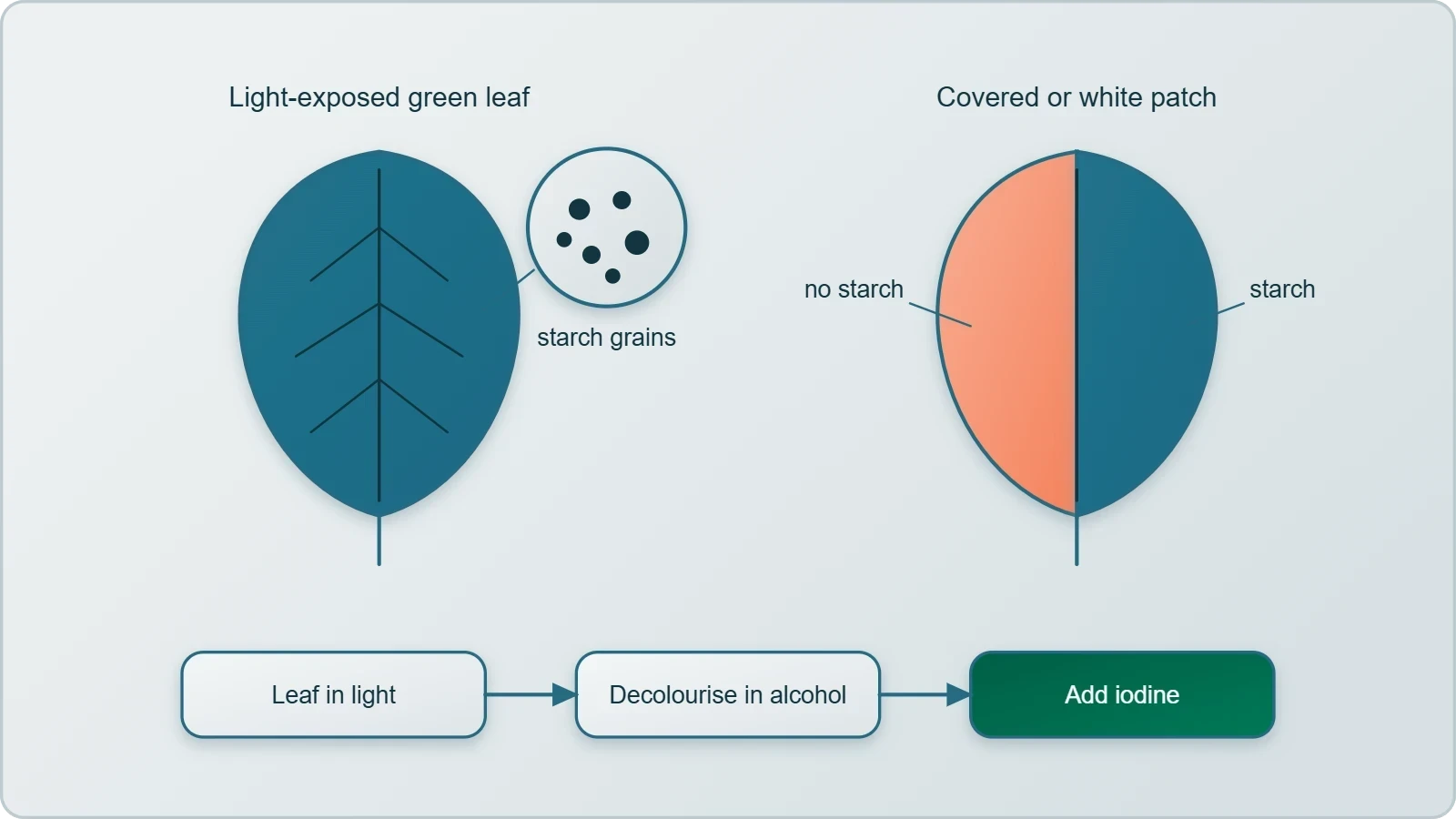
The reasoning also explains why the green parts are singled out. Variegated leaves, with their patches of white and green tissue, provide a natural control: starch appears only in the green regions after illumination. Von Sachs's conclusion that chlorophyll is housed in chloroplasts thus rests on a convergence of anatomical observation and physiological experiment. The pigment is not dissolved freely in the cell sap but sequestered in organelles, and those organelles are concentrated in the mesophyll tissue of the leaf, which is where the bulk of the sugar is manufactured.
What the Evidence Did and Did Not Establish
It is important to be precise about the limits of von Sachs's conclusions. He demonstrated that glucose is produced during growth and stored as starch, and that chlorophyll resides in chloroplasts, but he could not describe the intermediate reactions, the role of light in splitting water, or the involvement of ATP and NADPH. Those discoveries belong to the twentieth century. What he supplied was the localisation: a specific pigment, in a specific organelle, in a specific tissue, associated with the appearance of a specific storage compound.
This localisation was the necessary precondition for everything that followed. Once the chloroplast was identified as the seat of sugar manufacture, later investigators could ask what happens inside it, how light is absorbed, and which chemical species are consumed and released. The next lesson in this sequence takes up precisely that question, examining the membranous system of grana and stroma lamellae and the division of labour between light reactions and carbon reactions. Von Sachs, in effect, drew the map on which later workers plotted the chemistry.
Engelmann, the Prism, and the First Action Spectrum
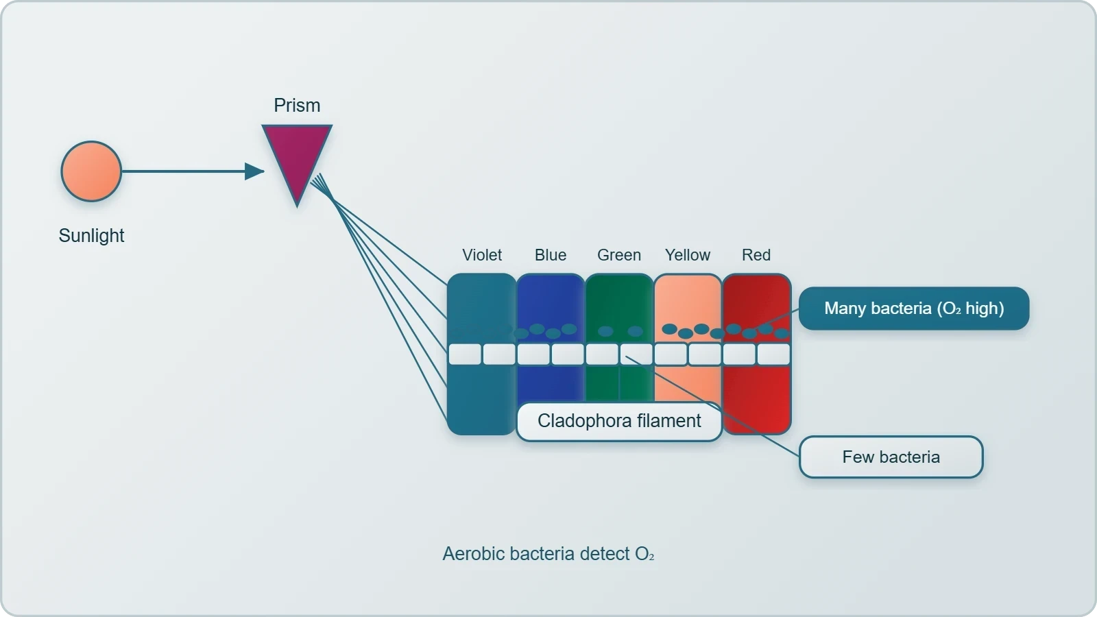
Theodor Wilhelm Engelmann, working between 1843 and 1909, asked a different question: not where sugar is made, but which colours of light drive the process most effectively. His experimental design is one of the most elegant in the history of plant physiology. Using a prism, he split light into its spectral components and then illuminated a green alga, Cladophora, placed in a suspension of aerobic bacteria. The bacteria served as living detectors of oxygen evolution, since aerobic bacteria congregate wherever oxygen is being produced. By observing where the bacteria accumulated along the illuminated filament, Engelmann could read off the alga's photosynthetic activity as a function of wavelength.
He observed that the bacteria accumulated mainly in the region of blue and red light of the split spectrum. A first action spectrum of photosynthesis was thus described, and it resembles roughly the absorption spectra of chlorophyll a and chlorophyll b. The significance of this result cannot be overstated. It established a quantitative correspondence between the wavelengths a pigment absorbs and the wavelengths at which a biological process proceeds, and it did so with equipment no more sophisticated than a prism, a microscope slide, and a culture of bacteria. The action spectrum was, in effect, a biological spectrometer.
The Design Logic of the Bacterial Detector
Why use bacteria rather than a chemical indicator? The answer lies in sensitivity and spatial resolution. Oxygen is a transient, locally variable product, and a chemical assay would average over the whole preparation. Aerobic bacteria, by contrast, migrate toward oxygen and accumulate at its source, producing a visible, position-dependent signal that maps directly onto the illuminated spectrum. The alga provides a linear filament whose segments can be illuminated with different wavelengths simultaneously, so a single preparation yields a complete spectrum rather than a series of separate measurements.
The choice of Cladophora is equally deliberate. It is a filamentous green alga, readily cultured, transparent enough for light to penetrate, and photosynthetic in the same chlorophyll-based manner as higher plants. Engelmann's preparation therefore functions as a model system: the conclusions drawn from it transfer to the leaves of terrestrial plants because the underlying pigment apparatus is conserved. This is a recurring pattern in experimental biology, where a convenient organism stands in for a less tractable one.
Reading the Blue and Red Peaks
The accumulation of bacteria in the blue and red regions of the spectrum corresponds to the absorption maxima of chlorophyll a and b. Chlorophyll absorbs strongly in the red around 660 to 680 nanometres and in the blue-violet around 430 to 450 nanometres, while reflecting green light, which is why leaves appear green. Engelmann's bacteria clustered where the pigment absorbed, confirming that absorption drives photosynthesis. The green region of the spectrum, by contrast, produced little bacterial accumulation, a direct consequence of the pigment's transparency to those wavelengths.
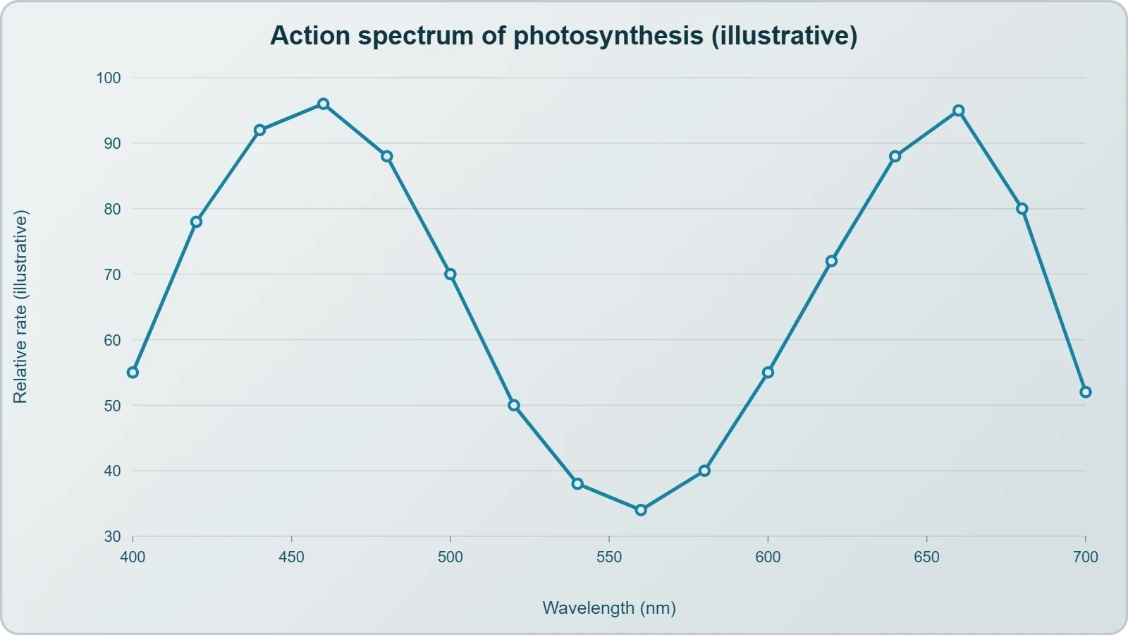
The correspondence is approximate rather than exact, and the word "roughly" in the classical description is well chosen. Action spectra and absorption spectra differ in detail because accessory pigments, energy transfer between pigment molecules, and the geometry of the thylakoid membranes all modulate the response. Nevertheless, the broad agreement was sufficient to establish the principle that the efficiency of photosynthesis varies with wavelength in a manner dictated by the light-absorbing pigments. This principle underpins every subsequent discussion of photosynthetic pigments.
The Empirical Equation and the Mid-Century Consensus
By the middle of the nineteenth century the key features of plant photosynthesis were known, namely, that plants could use light energy to make carbohydrates from carbon dioxide and water. The empirical equation representing the total process of photosynthesis for oxygen-evolving organisms was then understood as a light-driven combination of carbon dioxide and water yielding a carbohydrate and oxygen. In the notation of the period, the carbohydrate was written as [CH₂O], a general formula standing for a six-carbon sugar such as glucose. This compact representation captured the stoichiometry without committing to a mechanism.
The equation is worth writing in its classical form, since it frames everything that follows. The empirical summary reads:
Here [CH₂O] represents a carbohydrate, for example glucose, a six-carbon sugar. The equation is a summary, not a mechanism: it says nothing about which reactant supplies the oxygen, nothing about the intermediate carriers, and nothing about the two-stage character of the process. Those omissions are precisely what van Niel's work would address, and the correction he introduced is the pivot on which the modern understanding of photosynthesis turns.
Why the Empirical Form Was Useful
A summary equation serves a genuine scientific purpose even when its mechanism is unknown. It fixes the stoichiometry, identifies the reactants and products, and provides a target for mechanistic explanation. The [CH₂O] notation is particularly shrewd, because it avoids asserting that glucose itself is the immediate product. Photosynthesis does not simply assemble glucose molecule by molecule; it fixes carbon into a three-carbon intermediate that is later used to build sugars, starch, and other compounds. Writing the product as a generic carbohydrate keeps the equation honest about what is actually known.
The equation also highlights the oxygen-evolving character of plant photosynthesis, which distinguishes it from the bacterial processes van Niel would later study. In oxygen-evolving organisms, water is consumed and oxygen is released, and the balance of atoms must be accounted for. The classical equation balances carbon, hydrogen, and oxygen, but it leaves the provenance of the released oxygen ambiguous. Resolving that ambiguity required an organism in which the products could be compared across different hydrogen donors.
The Unresolved Question of Oxygen's Origin
Before van Niel, it was entirely reasonable to suppose that the oxygen released during photosynthesis came from carbon dioxide. After all, carbon dioxide contains oxygen, and the process consumes it. The alternative—that water is split and its oxygen released—was not obvious, because water was regarded primarily as a solvent and a hydrogen source rather than as the substrate of an oxidation. The empirical equation, with its balanced atoms, is consistent with either interpretation, which is exactly why a decisive experiment was needed.
That experiment came from an unexpected quarter: the study of purple and green sulphur bacteria, organisms that perform a light-dependent carbon fixation but do not release oxygen. By comparing what these bacteria consume and release with what green plants consume and release, van Niel could isolate the variable that determines the identity of the oxidation product. The logic is comparative physiology at its finest, using a non-standard organism to illuminate a standard one.
Van Niel and the Hydrogen Donor Insight
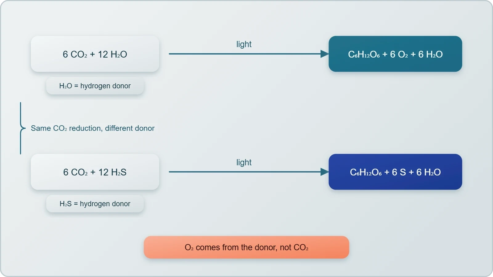
Cornelius van Niel, a microbiologist working between 1897 and 1985, made a milestone contribution to the understanding of photosynthesis. Based on his studies of purple and green bacteria, he demonstrated that photosynthesis is essentially a light-dependent reaction in which hydrogen from a suitable oxidisable compound reduces carbon dioxide to carbohydrates. This formulation shifts the emphasis from carbon dioxide as the central reactant to the hydrogen donor as the variable that determines what else is produced. The general equation he proposed expresses the logic with admirable economy:
In green plants, H₂O is the hydrogen donor and is oxidised to O₂. Some organisms, however, do not release O₂ during photosynthesis. When H₂S is the hydrogen donor for purple and green sulphur bacteria, the oxidation product is sulphur or sulphate, depending on the organism, and not O₂. The parallel is exact: the hydrogen is transferred to carbon dioxide in every case, but the oxidised residue differs according to the donor. Van Niel therefore inferred that the O₂ evolved by the green plant comes from H₂O, not from carbon dioxide.
The Comparative Logic Across Organisms
The strength of van Niel's argument lies in its comparative structure. If photosynthesis were fundamentally a process of reducing carbon dioxide with hydrogen, then the identity of the hydrogen donor should determine the identity of the oxidised by-product. Sulphur bacteria using hydrogen sulphide release sulphur or sulphate; green plants using water release oxygen. The common feature is the reduction of carbon dioxide to carbohydrate, and the variable feature is the donor and its oxidation product. This pattern is far too regular to be coincidental, and it points directly to water as the source of photosynthetic oxygen.
The inference was bold because it could not be tested directly with the techniques then available. Van Niel could not label the oxygen atoms in water and follow them into the gas released. What he could do was construct a general equation that accommodated all the organisms he studied and then read off its implications for plants. The prediction was specific and falsifiable: if water is the donor, then the oxygen released should derive from water. That prediction was later confirmed, vindicating the comparative approach.
Confirmation by Radioisotopic Techniques
The inference was later proved by using radioisotopic techniques. By supplying water labelled with a radioactive or heavy isotope of oxygen and separately labelling carbon dioxide, investigators could determine which reactant contributed the oxygen atoms appearing in the released gas. The result confirmed van Niel's prediction: the oxygen evolved by green plants comes from water. The correct equation representing the overall process of photosynthesis is therefore written with six molecules of carbon dioxide and twelve molecules of water on the reactant side, yielding one molecule of glucose, six molecules of water, and six molecules of oxygen.
Here C₆H₁₂O₆ represents glucose. The equation invites an obvious question: why are twelve molecules of water used as substrate when only six appear as products? The answer lies in the stoichiometry of the light reaction. Twelve water molecules are split to supply the hydrogen needed to reduce six carbon dioxide molecules, and six of the resulting water molecules are reconstituted in the process. The net consumption is six waters, but the substrate requirement is twelve, because the oxygen released comes from the split water and the hydrogen is transferred to carbon dioxide.
The Multistep Nature of the Overall Process
The corrected equation is not a single reaction but a description of a multistep process called photosynthesis. This distinction is essential for understanding everything that follows in the study of the light reaction and the carbon reactions. A single equation conceals the machinery: the absorption of light by pigment molecules, the transfer of excitation energy to a reaction centre, the extraction of electrons from water, the transport of electrons along a chain of carriers, the pumping of protons across a membrane, the synthesis of ATP, the reduction of NADP⁺ to NADPH, and the enzymatic fixation of carbon dioxide in the stroma. Each of these steps is a separate chemical event with its own requirements and controls.
Recognising the multistep character also clarifies why the process is so sensitive to environmental conditions. Temperature affects the enzymatic steps, light intensity affects the photochemical steps, and carbon dioxide concentration affects the carboxylation steps. The overall rate is set by whichever step is slowest under the prevailing conditions, a principle later formalised as Blackman's Law of Limiting Factors. The equation, in other words, is the beginning of the analysis rather than its conclusion.
From Summary Equation to Reaction Scheme
The transition from a summary equation to a reaction scheme is one of the great achievements of twentieth-century plant physiology. The light reactions, occurring in the membranous system of the chloroplast, trap light energy and synthesise ATP and NADPH. The carbon reactions, occurring in the stroma, use these products to synthesise sugar, which in turn forms starch. The two sets of reactions are coupled by the ATP and NADPH generated in the first and consumed in the second, and this coupling is the reason the so-called dark reactions cannot proceed in darkness for long.
The terminology deserves a note of caution. The carbon reactions are called dark reactions by convention, but this should not be construed to mean that they occur in darkness or that they are not light-dependent. They are dependent on the products of the light reactions, and those products are generated only in the light. The label is a convenience, not a claim about mechanism. Keeping this distinction clear prevents a common misconception about how the two stages of photosynthesis relate to one another.
Preparing for the Chloroplast's Division of Labour
The next lesson examines where photosynthesis takes place, and the groundwork has now been laid. The chloroplast contains a membranous system consisting of grana, the stroma lamellae, and the matrix stroma, and there is a clear division of labour within the organelle. The membrane system is responsible for trapping light energy and for the synthesis of ATP and NADPH, while the stroma hosts the enzymatic reactions that synthesise sugar. Von Sachs localised the machinery; van Niel identified the chemistry; the next step is to examine the architecture that makes the chemistry possible.
This progression from experiment to organelle to mechanism is the spine of the subject. Each lesson builds on the last, and each experimental result constrains the interpretations available to the next generation of investigators. The three scientists studied here did not know the details of electron transport or the Calvin cycle, but their conclusions were correct in outline and pointed the way toward those details. That is the mark of enduring experimental work.
Consolidating the Evidence: A Comparative View
The three investigations examined in this lesson can be compared along several dimensions: the question each addressed, the organism used, the signal measured, and the conclusion reached. Setting them side by side clarifies how different experimental strategies converged on a single coherent account of photosynthesis. Von Sachs worked with higher plants and read starch accumulation; Engelmann worked with a filamentous alga and read bacterial accumulation; van Niel worked with purple and green bacteria and read the identity of oxidation products. Each signal was indirect, and each conclusion was an inference from a visible pattern to an invisible process.
The comparison also reveals the complementarity of the approaches. Localisation, spectral response, and chemical stoichiometry are three independent lines of evidence, and their agreement strengthens the overall case. No single experiment could have established the full picture, and no single investigator could have anticipated all the questions the others raised. The cumulative character of the work is what makes it instructive as a model of scientific method.
Key Experimental Parameters at a Glance
The table makes the methodological diversity explicit. Von Sachs relied on histology, Engelmann on microbiology and optics, and van Niel on comparative biochemistry. Yet all three were engaged in the same enterprise: finding a visible proxy for a chemical event. This is a general lesson about experimental design, and it is worth carrying forward into the study of the light reaction, where the proxies become spectroscopic and electrochemical rather than anatomical.
What Each Investigator Contributed to the Modern Picture
Mapping the classical findings onto modern concepts shows that none of the early work was wasted. Von Sachs's starch observation corresponds to the carbon reactions and the storage of fixed carbon. Engelmann's action spectrum corresponds to the absorption properties of chlorophyll a and b. Van Niel's hydrogen-donor argument corresponds to the photolysis of water in the light reaction. The vocabulary has changed, and the mechanistic detail has multiplied, but the conceptual skeleton was already in place by the early twentieth century.
Going Further
Two clarifications help situate this material within the broader study of photosynthesis. The first concerns the distinction between action spectra and absorption spectra. An action spectrum plots the rate of a process against wavelength of light, while an absorption spectrum plots how much light a pigment absorbs at each wavelength. Engelmann measured an action spectrum, since the bacterial accumulation reflected the rate of oxygen evolution at each wavelength, and he compared it with the absorption spectra of chlorophyll a and b. The two are related but not identical, because accessory pigments and energy transfer shape the biological response.
The second clarification concerns the radioisotopic techniques that confirmed van Niel's inference. Radioisotopes are atoms with unstable nuclei that can be detected by their radiation, which is why they can be used as tracers in experiments. By labelling the oxygen in water and in carbon dioxide with distinguishable isotopes, investigators could determine which reactant supplied the oxygen released as gas. The result confirmed that the oxygen comes from water, exactly as van Niel had predicted from his studies of purple and green sulphur bacteria.
Action Versus Absorption Spectra
Keeping the two spectra distinct prevents a common confusion. A pigment may absorb light strongly without driving photosynthesis efficiently, if the absorbed energy is dissipated as heat or fluorescence rather than transferred to a reaction centre. Conversely, the action spectrum integrates the contributions of all pigments in the antenna, so it can differ from the absorption spectrum of any single pigment. The rough resemblance Engelmann observed is therefore a meaningful result, not a trivial identity.
Isotopes as Tracers
The tracer logic is straightforward once the prediction is stated clearly. If the oxygen released during photosynthesis derives from water, then labelling the oxygen atoms in water should label the gas; if it derives from carbon dioxide, labelling the carbon dioxide should do so. The experiment discriminates between two hypotheses that the balanced summary equation cannot distinguish. This is the essence of a decisive experiment: it forces a choice between competing explanations rather than merely confirming that the process occurs.
Summary and Transition to the Chloroplast
The lesson has traced three investigations that converted a suggestive phenomenon into a chemically specific process. Von Sachs provided evidence for the production of glucose when plants grow, showed that glucose is usually stored as starch, and demonstrated that chlorophyll is located in chloroplasts. Engelmann used a prism and aerobic bacteria to describe a first action spectrum of photosynthesis, finding bacterial accumulation mainly in the blue and red regions. Van Niel demonstrated that photosynthesis is essentially a light-dependent reaction in which hydrogen from a suitable oxidisable compound reduces carbon dioxide to carbohydrates, and inferred that the oxygen evolved by green plants comes from water.
The corrected overall equation, with six carbon dioxide and twelve water molecules yielding glucose, six waters, and six oxygens, summarises the process but does not describe a single reaction. It is a multistep process, and the steps are partitioned within the chloroplast between the membrane system and the stroma. The next lesson takes up that partition directly, examining where photosynthesis takes place and how the chloroplast's internal architecture supports the division of labour between light trapping and sugar synthesis.
Key Terms Revisited
These definitions will recur repeatedly as the course proceeds. The action spectrum returns when the absorption properties of chlorophyll a and b are discussed in detail. The hydrogen donor concept returns in the treatment of the light reaction and the photolysis of water. The chloroplast returns in the very next lesson, where its membranous system and stroma are examined as the physical basis of the division of labour. Holding these terms steady makes the later material far easier to assimilate.
Looking Ahead
The next lesson asks where photosynthesis takes place and answers with the chloroplast. Within the chloroplast there is a membranous system consisting of grana, the stroma lamellae, and the matrix stroma, and there is a clear division of labour within the organelle. The membrane system traps light energy and synthesises ATP and NADPH; the stroma hosts the enzymatic reactions that synthesise sugar, which in turn forms starch. The classical experiments studied here established that this organelle is the seat of the process; the next lesson examines how its architecture makes the process possible.
Students should arrive at that lesson with three conclusions firmly in hand. First, the green parts of plants manufacture glucose and store it as starch, and the chlorophyll responsible resides in chloroplasts. Second, the efficiency of photosynthesis varies with wavelength in a manner that roughly matches the absorption spectra of chlorophyll a and b. Third, the oxygen released by green plants comes from water, not from carbon dioxide, as van Niel inferred and radioisotopic techniques later confirmed. These three conclusions are the foundation on which the rest of the course builds.
Also Read
- No links available.
From our network :
- 98% of Global MBA Programs Now Prefer GRE Over GMAT Focus EditionExplore why 98% of global MBA programs now prefer the GRE over GMAT Focus. Learn about the shorter GRE format, verbal logic importance, and strategic versatility for students.
- Mastering DB2 12.1 Instance Design: A Technical Deep Dive into Modern Database ArchitectureComprehensive guide to DB2 12.1 Instance Design. Explore architectural components, HADR setup, buffer pool tuning, and security policies for IBM DB2 12.1. Includes 10 functional SQL and Shell samples.
- Mastering DB2 LUW v12 Tables: A Comprehensive Technical GuideComprehensive guide on DB2 LUW v12 tables. Explore CREATE TABLE syntax, BLU Acceleration, range partitioning, data types, and security features for IBM DB2 LUW v12.
- Analyzing Trump Deportation Numbers Insights Into The 2026 Immigration Crackdown
- Trump Political Strategy How Geopolitical Stunts Serve As Media Diversions
- AI-Powered 'Precision Diagnostic' Replaces Standard GRE Score ReportsDiscover the new GRE Precision Diagnostic. Learn how AI-powered behavioral analytics and time-to-solve tracking are replacing standard GRE score reports to empower students.
- Vite 6/7 'Cold Start' Regression in Massive Module GraphsIn-depth analysis of Vite 6/7 cold start regressions in massive module graphs. Learn about barrel file issues, pre-bundling optimizations, and how to improve Vite dev server performance.
- 10 Physics Numerical Problems with Solutions for IIT JEEPrepare for IIT JEE with 10 physics numerical problems, complete with detailed solutions and conceptual explanations. Ideal for mastering kinematics, thermodynamics, electromagnetism, and optics.
- EV 2.0: The Solid-State Battery Breakthrough and Global Factory ExpansionDiscover the impact of the Solid-State Battery Breakthrough on the EV market. Learn about CATL and BYD stock growth, global factory expansion in Europe, and the future of EV 2.0 technology.
RESOURCES
- Resources will be updated soon.