Eukaryotic gene expression does not end when RNA polymerase releases a primary transcript. Between the moment a precursor RNA emerges from the polymerase and the moment a mature messenger RNA reaches the cytoplasm, the cell performs an elaborate sequence of chemical edits that determine whether a transcript survives, where it localises, how efficiently it is translated, and ultimately which protein isoform the cell produces. RNA processing is therefore not a housekeeping afterthought but a decisive regulatory layer, one that sits at the heart of the molecular basis of inheritance and explains why a genome of roughly twenty thousand protein-coding genes can generate a proteome of vastly greater complexity.

This lesson builds directly on the preceding discussion of advanced DNA replication and repair, shifting attention from the preservation of the genome to the interpretation of its output. We examine the three canonical modifications of eukaryotic transcripts — 5′ capping, splicing, and 3′ cleavage and polyadenylation — and then move into the discovery of catalytic RNA, the combinatorial logic of alternative splicing, and the expansive regulatory universe of non-coding RNAs. Along the way we consider RNA interference and its experimental and therapeutic applications, the mechanisms by which small and long non-coding RNAs silence genes, and the clinical consequences when splicing goes wrong.
The material is pitched at intermediate to advanced learners who already command the central machinery of transcription and translation. Expect mechanistic detail, quantitative reasoning about splice-site selection, and worked examples drawn from human genetics and molecular medicine. By the end of the lesson you should be able to trace a primary transcript through its maturation pathway, predict the consequences of a splice-site mutation, and explain how a twenty-two-nucleotide RNA can silence a gene with remarkable specificity.
On This Page
- The Three Canonical Modifications of Eukaryotic Transcripts
- Ribozymes and the Catalytic Power of RNA
- Alternative Splicing and the Generation of Protein Diversity
- The Expanding World of Non-Coding RNAs
- RNA Interference and Its Applications
- Splicing Defects and Human Disease
- Integrating Processing with Gene Regulation
The Three Canonical Modifications of Eukaryotic Transcripts
RNA polymerase II synthesises precursor mRNA as a bare, chemically fragile molecule that would be unstable and poorly translated if released unchanged into the cytoplasm. The cell therefore couples transcription with a coordinated set of co-transcriptional modifications. The carboxy-terminal domain of the largest polymerase subunit acts as a recruitment platform, binding capping enzymes, splicing factors, and polyadenylation machinery in a defined order. This coupling ensures that processing events occur on the nascent transcript while it is still tethered to the transcription complex, and it explains why processing efficiency is intimately tied to transcriptional elongation rate.
5′ Capping and 3′ End Formation
Capping occurs when the nascent transcript is only twenty to thirty nucleotides long. An RNA triphosphatase removes the terminal γ-phosphate, guanylyltransferase attaches a guanosine monophosphate in a 5′–5′ triphosphate linkage, and methyltransferases add methyl groups to the cap guanine and often to the ribose of the first and second transcribed nucleotides. The resulting 7-methylguanosine cap protects the transcript from exonucleases, is recognised by the cap-binding complex during export, and is bound by eukaryotic initiation factor 4E during translation initiation. Capping therefore links transcript stability directly to translational competence.
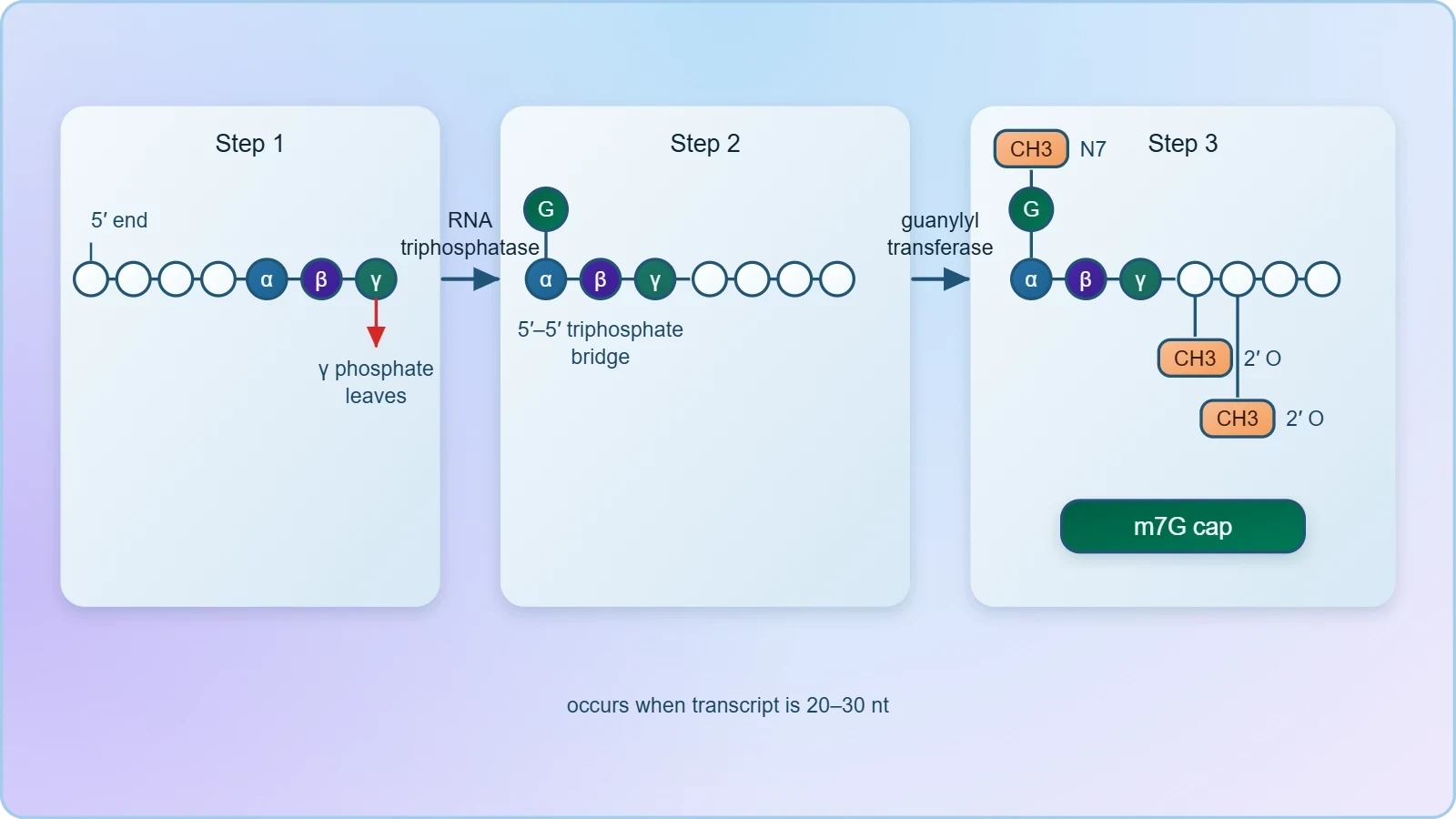
At the other end, the polyadenylation signal AAUAAA directs cleavage of the transcript roughly twenty nucleotides downstream, followed by the addition of a poly(A) tail of approximately two hundred adenosines in mammals. Poly(A) polymerase adds the tail without a template, and poly(A)-binding protein coats it to confer stability and assist nuclear export. The tail length is not fixed; it is progressively shortened in the cytoplasm and serves as a molecular clock that governs transcript half-life. Histone mRNAs are a notable exception, relying instead on a stem-loop structure for their unusual 3′ end formation.
Spliceosome Assembly and the Chemistry of Intron Removal
Splicing removes intervening sequences through two sequential transesterification reactions. In the first, the 2′-hydroxyl of an adenosine within the branch point attacks the 5′ splice site, generating a free 5′ exon and a lariat intermediate held by a 2′–5′ phosphodiester bond. In the second, the liberated 5′ exon attacks the 3′ splice site, joining the exons and releasing the intron lariat, which is subsequently debranched and degraded. No external energy source is required for the chemistry itself; the driving force is the conversion of two phosphodiester bonds into two others.
The reaction is catalysed by the spliceosome, a dynamic ribonucleoprotein machine comprising five small nuclear RNAs (U1, U2, U4, U5, U6) and well over a hundred associated proteins. Spliceosome assembly proceeds through ordered rearrangements in which RNA–RNA and RNA–protein contacts are exchanged, consuming ATP at several proofreading steps. The splice sites themselves are degenerate, so the spliceosome relies on auxiliary elements — exonic and intronic enhancers and silencers — bound by serine-arginine-rich proteins and heterogeneous ribonucleoproteins to distinguish genuine splice sites from the many near-matches scattered throughout a transcript.
Ribozymes and the Catalytic Power of RNA
For decades the division of labour seemed settled: proteins catalyse, nucleic acids store and transmit information. That assumption was overturned in the early 1980s when Thomas Cech and colleagues, studying the self-splicing group I intron of Tetrahymena thermophila ribosomal RNA, found that the intron excised itself in vitro without any protein enzyme. Sidney Altman's laboratory reached a parallel conclusion for the RNA component of bacterial RNase P, which cleaves precursor tRNAs. The term ribozyme was coined for these catalytic RNAs, and the discovery earned Cech and Altman the 1989 Nobel Prize in Chemistry.
Self-Splicing Introns and the Spliceosome as a Ribozyme
Group I and group II introns fold into precise three-dimensional structures that position their own reactive groups for catalysis, using metal ions as cofactors in a manner reminiscent of protein metalloenzymes. Group II introns are especially significant because their mechanism — two transesterifications producing a lariat intermediate — is chemically identical to that of the spliceosome. Structural studies of the spliceosome have since revealed that the catalytic core is formed entirely of RNA, with the U6 snRNA positioning the metal ions that stabilise the leaving groups. The spliceosome is, in effect, a ribozyme that has recruited proteins for regulation and fidelity.
This catalytic versatility has profound evolutionary implications. The RNA world hypothesis proposes that early life relied on RNA both to store genetic information and to catalyse the reactions of metabolism, with protein enzymes and DNA genomes evolving later as more specialised solutions. Ribozymes also retain practical importance: the hammerhead and hairpin ribozymes are small enough to be engineered, and ribozyme-based tools have been explored for targeted RNA cleavage in antiviral and anticancer research. The peptidyl transferase centre of the ribosome, itself an RNA catalyst, is the most abundant ribozyme on Earth.
Ribonuclease P and Catalytic RNA in Modern Cells
RNase P remains a living demonstration that catalytic RNA is not a fossil. In bacteria the enzyme is a ribonucleoprotein in which the RNA subunit performs the cleavage chemistry and the protein subunit mainly provides electrostatic stabilisation. In eukaryotic nuclei, RNase P matures the 5′ ends of all precursor tRNAs, a reaction essential for translation. The persistence of an RNA-based active site across billions of years of evolution is strong evidence that catalysis by RNA is not merely possible but advantageous when substrate recognition depends on base pairing.
Alternative Splicing and the Generation of Protein Diversity
Most human genes contain multiple introns, and the spliceosome must choose among competing splice sites in a context-dependent manner. Alternative splicing exploits this flexibility to produce several distinct mRNAs from a single gene, and it is estimated that more than ninety percent of human multi-exon genes undergo some form of alternative splicing. The resulting isoforms can differ in catalytic activity, subcellular localisation, ligand binding, or stability, allowing a limited genome to encode a far richer proteome. The phenomenon is especially prominent in the nervous system, where it contributes to synaptic complexity.
Patterns of Alternative Splicing
The commonest patterns include exon skipping, mutually exclusive exons, alternative 5′ and 3′ splice sites, and intron retention. Exon skipping accounts for the majority of alternative events in mammals. Mutually exclusive exons allow a gene to swap functional modules, as in the Dscam gene of Drosophila, which can generate tens of thousands of distinct isoforms through combinatorial exon choice. Intron retention, once dismissed as noise, is now recognised as a regulated mechanism that can produce unstable transcripts destined for nonsense-mediated decay, thereby fine-tuning expression levels.
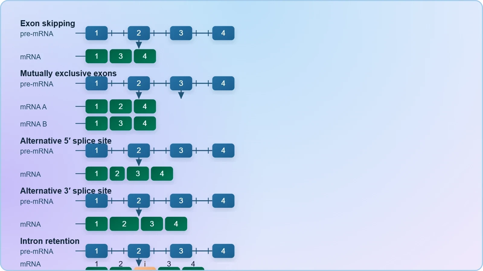
Splice-site selection is governed by the relative strength of competing sites, the concentration and phosphorylation state of splicing regulators, and the kinetics of transcription. The serine-arginine-rich protein family generally promotes exon inclusion, while heterogeneous ribonucleoproteins tend to antagonise them. Because these regulators differ between tissues and developmental stages, the same transcript can be spliced differently in neurons and muscle. This regulatory logic explains why alternative splicing is particularly sensitive to cellular stress and why splicing profiles shift markedly during differentiation and tumorigenesis.
Worked Example: Calculating Isoform Diversity
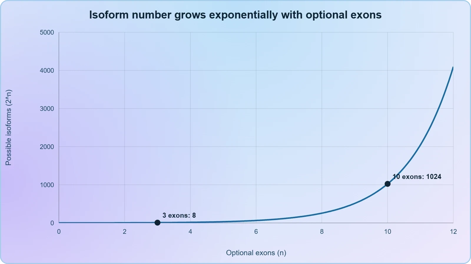
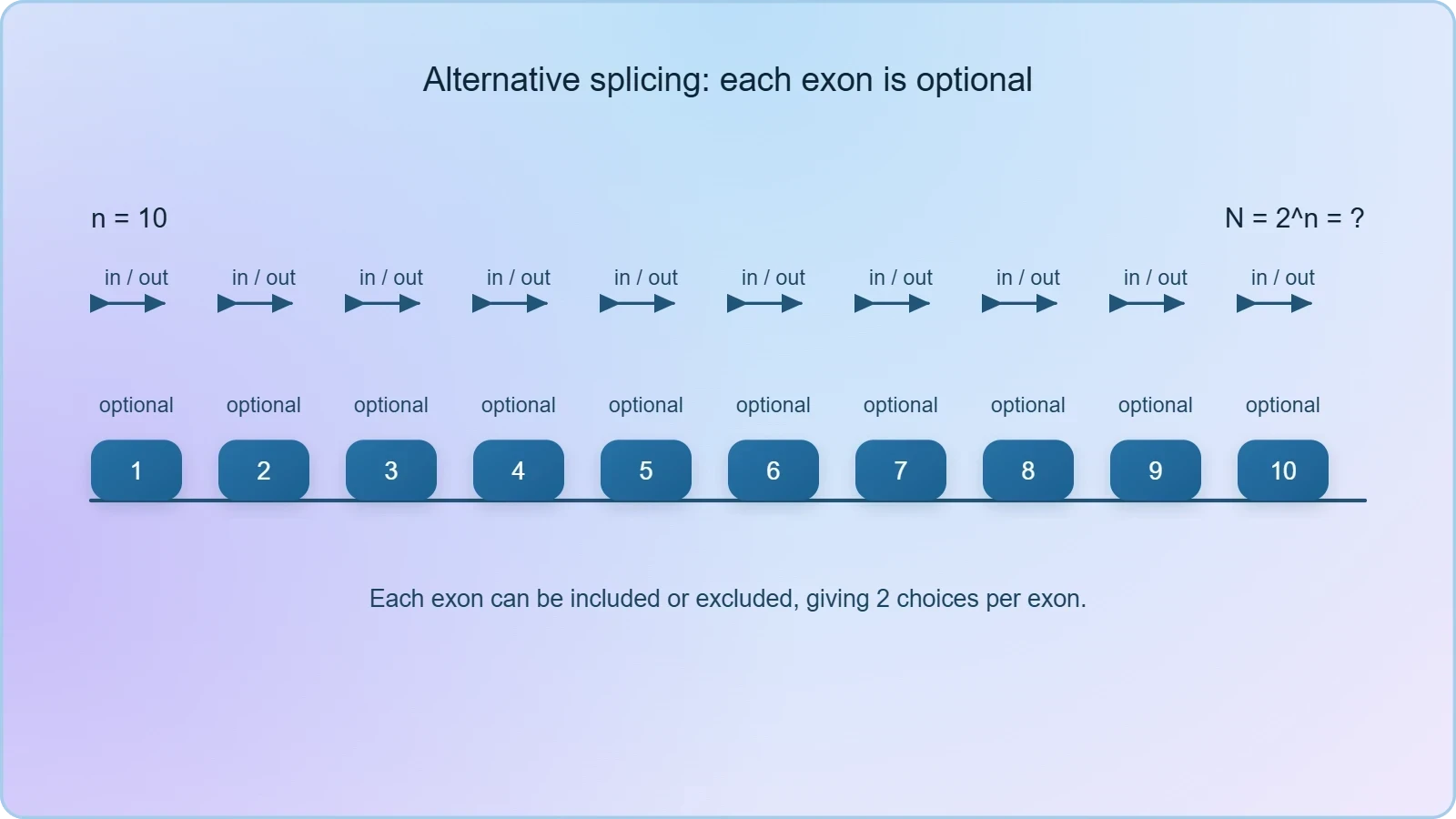
Consider a hypothetical gene with three alternatively spliced exons, each of which may be included or excluded independently. The number of possible mature mRNAs is the product of the independent binary choices, giving ##2^{3} = 8## distinct transcripts. If each exon can additionally be retained in one of two mutually exclusive variants, the combinatorial space expands further. The general relationship for ##n## independently optional exons is:
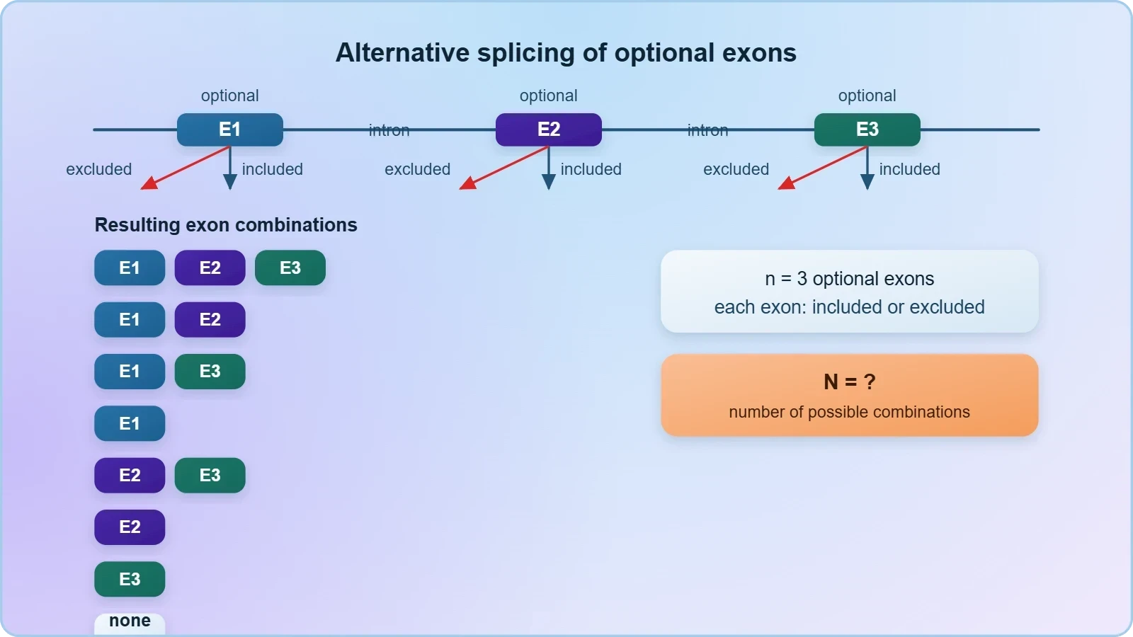
For a gene with ten optional exons, ##N = 1024## potential isoforms, which illustrates how a modest number of exons can encode enormous diversity. Real genes rarely realise the full theoretical space because splice-site strength and regulator availability constrain the outcomes, but the calculation captures the essential combinatorial principle. It also explains why mutations that alter a single splice site can have consequences disproportionate to the size of the affected exon.
The Expanding World of Non-Coding RNAs
Only a small fraction of the human genome encodes protein, yet the majority of it is transcribed. The resulting non-coding RNAs were long regarded as transcriptional noise, but they are now recognised as essential regulators of gene expression, chromatin architecture, and genome stability. Non-coding RNAs are classified by size, biogenesis pathway, and mode of action. Small RNAs of roughly twenty to thirty nucleotides act through base pairing with complementary targets, while long non-coding RNAs exceeding two hundred nucleotides often function as scaffolds, decoys, or guides that recruit chromatin-modifying complexes to specific loci.
Small Regulatory RNAs: miRNA, siRNA, and piRNA
MicroRNAs are encoded in the genome and processed from hairpin precursors by the RNase III enzymes Drosha and Dicer. The mature microRNA is loaded into the RNA-induced silencing complex, where it guides Argonaute proteins to complementary sites usually located in the 3′ untranslated region of target mRNAs. Extensive pairing triggers cleavage, whereas partial pairing represses translation and accelerates deadenylation. Because a single microRNA can regulate hundreds of targets, these molecules function as network-level rheostats rather than simple switches, and their misregulation is implicated in cancer, metabolic disease, and developmental disorders.
Small interfering RNAs arise from long double-stranded RNA and typically direct cleavage of perfectly complementary targets. In plants and invertebrates they also amplify through RNA-dependent RNA polymerases, producing systemic silencing. Piwi-interacting RNAs are a distinct class expressed mainly in the germ line, where they silence transposable elements and safeguard genome integrity across generations. Together these pathways illustrate a recurring theme: short RNAs bound to Argonaute-family proteins provide sequence-specific recognition that proteins alone cannot achieve.
Long Non-Coding RNAs and Gene Silencing
Long non-coding RNAs operate through diverse mechanisms. Xist, the archetypal example, coats the inactive X chromosome in female mammals and recruits repressive complexes that establish and maintain dosage compensation. Other long non-coding RNAs act as molecular decoys that sequester transcription factors, as scaffolds that assemble chromatin modifiers, or as guides that deliver silencing complexes to particular genomic addresses. Their expression is frequently tissue-specific, and genome-wide association studies have mapped numerous disease-associated variants to non-coding RNA loci, underscoring their regulatory importance.
Gene silencing by non-coding RNAs extends to the transcriptional level. RNA-directed DNA methylation in plants and heterochromatin formation in fission yeast demonstrate that small RNAs can feed back on chromatin to enforce stable repression. In mammals, Argonaute proteins can associate with chromatin and promote repressive histone modifications at certain promoters. These observations blur the traditional boundary between transcriptional and post-transcriptional control and suggest that RNA is a general-purpose regulator of genome function.
RNA Interference and Its Applications
RNA interference is the sequence-specific silencing of gene expression triggered by double-stranded RNA. Andrew Fire and Craig Mello demonstrated in Caenorhabditis elegans that injection of double-stranded RNA produced potent and heritable silencing, a discovery recognised with the 2006 Nobel Prize in Physiology or Medicine. Mechanistically, Dicer cleaves the trigger into short duplexes, one strand of which is loaded into Argonaute within the RNA-induced silencing complex. The guide strand then directs cleavage or repression of complementary transcripts, and in some organisms the signal spreads systemically.
Experimental and Therapeutic Uses of RNAi
RNAi has become a standard laboratory tool for loss-of-function studies, permitting researchers to deplete a chosen transcript without altering the genome. Genome-wide short hairpin RNA and small interfering RNA libraries enable systematic screens for genes affecting proliferation, drug sensitivity, or viral replication. In agriculture, engineered small RNAs confer resistance to pests and viruses. The pathway also underpins crop traits and has been explored for vector control, illustrating how a basic discovery in nematode biology translated into practical biotechnology within two decades.
Therapeutically, small interfering RNAs have matured into approved medicines. Patisiran, formulated in lipid nanoparticles, silences transthyretin to treat hereditary transthyretin amyloidosis, and inclisiran targets PCSK9 to lower LDL cholesterol with infrequent dosing. Chemical modifications such as 2′-O-methyl and phosphorothioate linkages improve stability and reduce immunogenicity, while GalNAc conjugation directs oligonucleotides to hepatocytes. Delivery beyond the liver remains the central obstacle, and the balance between on-target potency and off-target silencing demands careful guide-strand design.
Off-Target Effects and Design Considerations
Specificity is never absolute. Partial complementarity between a guide strand and unintended transcripts can produce measurable repression, and saturation of the endogenous microRNA pathway may perturb normal regulation. Computational design therefore seeks guides with minimal seed-region matches elsewhere in the transcriptome, and experimental validation with multiple independent reagents remains essential. Dose control matters as well, because high concentrations of exogenous small RNA can overwhelm Argonaute availability and trigger innate immune responses through double-stranded RNA sensors.
Splicing Defects and Human Disease
Because splicing must precisely remove introns while preserving the reading frame, mutations that alter splice-site recognition are frequently pathogenic. A conservative estimate attributes fifteen to thirty percent of disease-causing point mutations to aberrant splicing, and the proportion rises in genes with many introns. The consequences range from exon skipping and intron retention to activation of cryptic splice sites, each of which can shift the reading frame, introduce premature termination codons, or delete a functional domain. Many such transcripts are degraded by nonsense-mediated decay, producing haploinsufficiency rather than a toxic protein.
Representative Splicing Diseases
Beta-thalassaemia provides a classic illustration. Intronic mutations that create or destroy splice sites reduce correct processing of the beta-globin transcript, lowering functional haemoglobin and causing anaemia. Spinal muscular atrophy results from loss of SMN1; the paralogue SMN2 produces mostly an exon-7-skipped transcript because a single nucleotide difference weakens a splicing enhancer, and the small amount of full-length protein it generates determines disease severity. This observation directly motivated the development of antisense oligonucleotides that redirect SMN2 splicing.
Myotonic dystrophy type 1 arises when expanded CTG repeats in DMPK produce transcripts that sequester muscleblind-like splicing regulators in nuclear foci. The resulting misregulation of alternative splicing across many downstream genes explains the multisystemic phenotype, including myotonia and cardiac conduction defects. Familial dysautonomia, by contrast, is caused by a point mutation that disrupts a splice site in IKBKAP, leading to skipping of exon 20 in neuronal tissue. Each example demonstrates that splicing fidelity is a therapeutic target in its own right.
Antisense Oligonucleotides as Splicing Modifiers
Splice-switching antisense oligonucleotides are short chemically modified nucleic acids that base-pair with a pre-mRNA and sterically block the spliceosome from recognising a chosen site. Nusinersen, approved for spinal muscular atrophy, masks a silencer in SMN2 intron 7 and restores exon 7 inclusion, increasing full-length protein. Eteplirsen and related compounds induce skipping of a mutated dystrophin exon to restore the reading frame in Duchenne muscular dystrophy. These agents must reach the nucleus, resist nucleases, and avoid immune activation, which is why phosphorothioate backbones and sugar modifications are standard.
The broader lesson is that RNA processing is not merely a maturation step but a point of therapeutic leverage. Small molecules that modulate splicing, including those targeting the SF3B1 component of the spliceosome, are under investigation in oncology, where spliceosome mutations drive specific myeloid malignancies. Understanding the chemistry and regulation of splicing therefore connects fundamental molecular biology to precision medicine, and it prepares the ground for the next lesson, which examines the central dogma and the exceptions that challenge its simplicity.
Integrating Processing with Gene Regulation
RNA processing sits at the intersection of transcription, chromatin state, and RNA surveillance. The phosphorylation pattern of the polymerase CTD determines which processing factors are recruited and when, so changes in elongation rate can alter splice-site choice. Chromatin marks influence exon recognition, and splicing in turn feeds back on transcription. Meanwhile, quality-control systems such as nonsense-mediated decay, the nuclear exosome, and the RNA-induced silencing complex inspect the finished product and eliminate transcripts that fail processing. The mature mRNA that reaches the ribosome is therefore the survivor of a rigorous selection process.
Quality Control and Surveillance Pathways
Nonsense-mediated decay recognises premature termination codons, typically those more than fifty to fifty-five nucleotides upstream of the final exon–exon junction, and targets the transcript for destruction. This mechanism protects the cell from truncated proteins and doubles as a regulatory device, since alternative splicing that introduces a premature stop codon can deliberately route a transcript to decay. The nuclear exosome degrades improperly processed or retained transcripts, and the exon junction complex deposited during splicing marks the transcript for export and subsequent surveillance.
These pathways explain why a splicing mutation often produces a simple loss of function rather than a dominant toxic protein. They also create therapeutic opportunities: drugs that suppress premature termination codon recognition, such as ataluren, aim to read through nonsense mutations in selected genetic diseases. Understanding surveillance is therefore inseparable from understanding processing, and together they define the functional output of a gene.
Key Takeaways for the Molecular Biologist
Three principles deserve emphasis. First, processing is co-transcriptional and physically coupled to the polymerase, so transcriptional and post-transcriptional control cannot be cleanly separated. Second, RNA is a catalyst and a regulator, not merely an intermediary, as ribozymes and non-coding RNAs demonstrate. Third, the combinatorial logic of alternative splicing and the sequence specificity of small RNAs together generate regulatory complexity far exceeding the gene count. These principles recur throughout genome science and prepare the ground for the exceptions to the central dogma examined next.
Also Read
- No links available.
From our network :
- Do You Need a New Router or Is Your Internet Plan the Problem?A practical comparison of the signs that point to router limits versus signs your broadband speed or provider is the bottleneck, so you can decide what to replace before spending money.
- Trump Political Strategy How Geopolitical Stunts Serve As Media Diversions
- Where to Put Your Wireless Router for Better CoverageA practical, physics-grounded guide to router placement: why the centre of the home beats the corner, how walls and metal sap signal, and how to run a simple survey that proves the improvement.
- How to Print Cheaply: Cutting the Cost per PageA practical, measured guide to lowering what each printed page costs — duplex and draft modes, third-party ink, yield mathematics, and knowing when a new printer beats refills.
- 10 Physics Numerical Problems with Solutions for IIT JEEPrepare for IIT JEE with 10 physics numerical problems, complete with detailed solutions and conceptual explanations. Ideal for mastering kinematics, thermodynamics, electromagnetism, and optics.
- 98% of Global MBA Programs Now Prefer GRE Over GMAT Focus EditionExplore why 98% of global MBA programs now prefer the GRE over GMAT Focus. Learn about the shorter GRE format, verbal logic importance, and strategic versatility for students.
- Analyzing Trump Deportation Numbers Insights Into The 2026 Immigration Crackdown
- EV 2.0: The Solid-State Battery Breakthrough and Global Factory ExpansionDiscover the impact of the Solid-State Battery Breakthrough on the EV market. Learn about CATL and BYD stock growth, global factory expansion in Europe, and the future of EV 2.0 technology.
- AI-Powered 'Precision Diagnostic' Replaces Standard GRE Score ReportsDiscover the new GRE Precision Diagnostic. Learn how AI-powered behavioral analytics and time-to-solve tracking are replacing standard GRE score reports to empower students.
RESOURCES
- Resources will be updated soon.