Control and coordination in the frog represent one of the most instructive examples of how a vertebrate body governs itself through two interlocking systems: the fast, electrically wired nervous system and the slower, chemically broadcast endocrine glands. This closing lesson of the course examines how Rana tigrina integrates these two networks, how its sense organs translate the outside world into nerve signals, and how its reproductive machinery ensures the continuation of the species through external fertilisation and a dramatic larval transformation. The material builds directly on the circulatory and excretory systems studied previously, since hormones travel in blood and the reproductive ducts share the cloaca with the urinary tract.
The lesson follows the source material in its own order: first the endocrine glands and the architecture of the nervous system, then the brain and spinal cord, then the five categories of sense organs, then the male and female reproductive systems, and finally the frog's value to mankind and to the ecosystem. Every definition, count and anatomical relationship is preserved exactly as given, including the ten pairs of cranial nerves, the 10–12 vasa efferentia, and the 2500 to 3000 ova a mature female can lay at one time. A short "Going further" block at the end adds well-established background on amphibian life cycles without altering the core content.
Because this is the final lesson, it also serves as a synthesis point. The neural and endocrine systems described here regulate the very organs — heart, kidneys, gut and gonads — that earlier lessons dissected one by one. Understanding how the frog senses its pond, coordinates its response, and reproduces within it completes the picture of structural organisation in animals that this course set out to build, moving from cells and tissues upward to whole organ systems and, ultimately, to the whole organism behaving in its environment.
On This Page
- The Endocrine Glands and the Dual System of Coordination
- Organisation of the Nervous System and the Structure of the Brain
- Sense Organs: Touch, Taste, Smell, Vision and Hearing
- The Male Reproductive System
- The Female Reproductive System, Fertilisation and Metamorphosis
- Benefits to Mankind and Ecological Balance
The Endocrine Glands and the Dual System of Coordination
The system for control and coordination is highly evolved in the frog, and it includes both the neural system and the endocrine glands. Chemical coordination of the various organs of the body is achieved by hormones, which are secreted by the endocrine glands and carried through the bloodstream to their targets. This arrangement means that the circulatory system, examined in the previous lesson, is not merely a transport route for respiratory gases and nitrogenous waste but also the delivery network for chemical messengers that adjust the activity of distant organs over seconds to hours rather than milliseconds.
The Prominent Endocrine Glands of the Frog
The prominent endocrine glands found in the frog are the pituitary, thyroid, parathyroid, thymus, pineal body, pancreatic islets, adrenals and gonads. Each gland contributes a distinct regulatory voice: the pituitary acts as a master gland influencing other glands, the thyroid governs metabolic rate, the parathyroids manage calcium balance, the thymus supports immune maturation, the pineal body is associated with biological rhythms, the pancreatic islets regulate blood sugar, the adrenals prepare the body for stress, and the gonads produce the sex hormones that drive reproduction. Together they form a diffuse but coordinated chemical network.
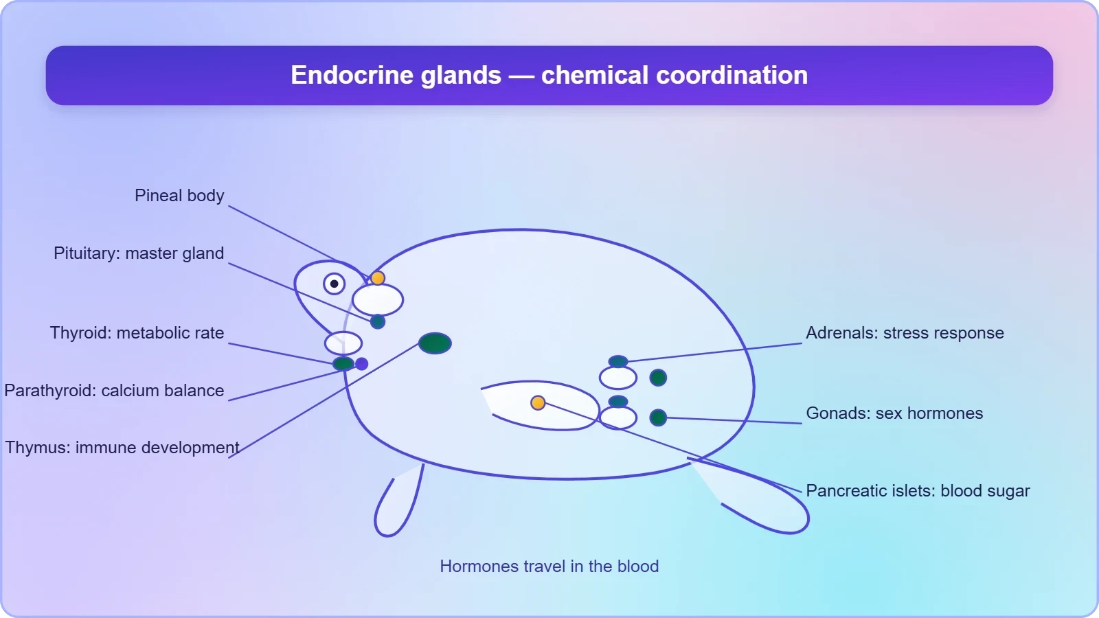
What makes this list pedagogically valuable is that it mirrors, in simplified form, the endocrine map of vertebrates generally. A learner who can name these eight gland groups in the frog and state their broad functions has acquired a transferable framework. The gonads deserve particular attention in this lesson because they sit at the junction of the endocrine and reproductive systems: they secrete hormones while simultaneously producing the gametes whose release is described later in this lesson.
How Neural and Chemical Coordination Divide the Work
Neural coordination is rapid, targeted and short-lived; chemical coordination through hormones is slower, broadcast through the blood and longer-lasting. The frog relies on both because different problems demand different timescales. Escaping a predator requires split-second nerve impulses to skeletal muscle, while seasonal breeding, metabolic adjustment and growth require hormonal signals that persist for days or weeks. The two systems are not rivals but partners, and the hypothalamus–pituitary axis is the classic bridge between them, translating neural information into hormonal commands.
This division of labour echoes the broader theme of the course: cells, tissues, organs and organ systems split up the work in a way that ensures the survival of the body as a whole. Just as epithelial, connective, muscular and neural tissues specialise, so do the two coordinating systems specialise by speed and reach. Recognising this parallel helps the learner see structural organisation as a single principle operating at every level of the frog's body.
Organisation of the Nervous System and the Structure of the Brain
The nervous system of the frog is organised into three divisions: a central nervous system comprising the brain and spinal cord, a peripheral nervous system comprising the cranial and spinal nerves, and an autonomic nervous system comprising the sympathetic and parasympathetic components. This tripartite scheme is the standard vertebrate plan, and the frog illustrates it cleanly. The central division integrates information, the peripheral division carries sensory input inward and motor commands outward, and the autonomic division governs the involuntary machinery of the internal organs, including the gut, heart and glands discussed elsewhere in the course.
Cranial Nerves, the Cranium and the Spinal Cord
There are ten pairs of cranial nerves arising from the brain. The brain itself is enclosed in a bony structure called the brain box, or cranium, which protects the delicate neural tissue. The medulla oblongata, the posterior part of the hind-brain, passes out through the foramen magnum and continues into the spinal cord, which is enclosed in the vertebral column. This continuity between brain and cord, and the bony protection at both ends, is a structural feature worth committing to memory because it recurs in every vertebrate dissection a learner will later encounter.
The figure of ten cranial nerve pairs is a specific, examinable detail from the material and should be retained exactly. It is worth noting how the peripheral nerves connect the central nervous system to the sense organs described in the next section and to the muscles that execute movement, completing the reflex arc from stimulus to response. The autonomic nerves, meanwhile, reach the smooth muscle and glands of the viscera, providing the involuntary background control that keeps the frog's internal environment stable.
Fore-brain, Mid-brain and Hind-brain
The brain is divided into fore-brain, mid-brain and hind-brain. The fore-brain includes the olfactory lobes, paired cerebral hemispheres and the unpaired diencephalon. The mid-brain is characterised by a pair of optic lobes. The hind-brain consists of the cerebellum and the medulla oblongata. Each subdivision carries a functional signature: olfactory lobes serve smell, cerebral hemispheres serve higher integration, the diencephalon links neural and endocrine control, the optic lobes serve vision, the cerebellum coordinates balance and muscular activity, and the medulla governs vital involuntary functions.
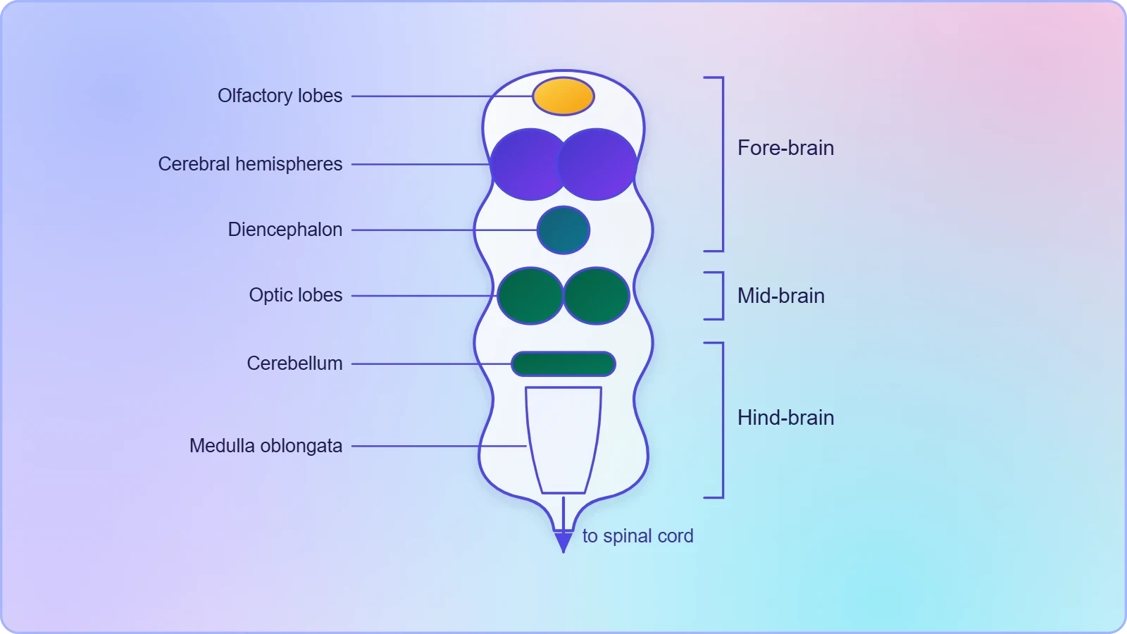
Mapping structure to function in this way transforms a list of names into a working model of the brain. The olfactory lobes and optic lobes, in particular, foreshadow the sense organs examined next, since the frog's sensory world is dominated by smell and sight. The cerebellum's role in balance also connects naturally to the ear, which the material describes as an organ of hearing as well as balancing, or equilibrium.
Sense Organs: Touch, Taste, Smell, Vision and Hearing
The frog has different types of sense organs, namely organs of touch (sensory papillae), taste (taste buds), smell (nasal epithelium), vision (eyes) and hearing (tympanum with internal ears). Out of these, the eyes and internal ears are well-organised structures, while the rest are cellular aggregations around nerve endings. This distinction between organised organs and simpler receptor clusters is an important conceptual point: not every sense requires a complex organ, and the frog's skin, mouth and nasal lining carry out chemosensation and mechanosensation through dispersed cells rather than discrete structures.
The Eyes and the Tympanum
The eyes in a frog are a pair of spherical structures situated in the orbit in the skull. These are simple eyes, possessing only one unit. The external ear is absent in frogs, and only the tympanum can be seen externally. The ear is an organ of hearing as well as balancing, or equilibrium. The absence of an external ear is a distinctive feature of the frog's morphology and connects back to the head-and-trunk body plan described in the earlier morphology lesson, where the tympanum appears as a visible disc on the side of the head.
The description of the eye as "simple" deserves emphasis: it possesses only one unit, in contrast to the compound eyes of many arthropods. This is a useful comparative point for learners. The dual role of the ear in hearing and balance also links neatly to the cerebellum, whose coordinating function depends on the equilibrium information the ear supplies, closing a loop between sensory input and motor control.
Chemical and Tactile Senses
Touch is served by sensory papillae, taste by taste buds, and smell by the nasal epithelium. These are cellular aggregations around nerve endings rather than encapsulated organs, yet they provide the frog with essential information about its immediate environment: the texture of a surface, the chemistry of potential food, and the scent of prey or predator. For a semi-aquatic animal that hunts by snapping at moving prey, these distributed senses complement the more conspicuous eyes and tympanum, giving a complete picture of how the frog perceives its world.
It is worth pausing on the logic of the list. The material groups five senses and then divides them by structural complexity, not by importance. This teaches a valuable habit of mind: classify by organisation as well as by function. The same principle of division of labour that organises tissues and organ systems also organises the sensory apparatus, with some senses delegated to specialised organs and others to dispersed receptor cells.
The Male Reproductive System
Frogs have well-organised male and female reproductive systems. The male reproductive organs consist of a pair of yellowish ovoid testes, which are found adhered to the upper part of the kidneys by a double fold of peritoneum called the mesorchium. This peritoneal attachment is a structural detail worth noting because it explains how the testes are held in position near the kidneys, a proximity that becomes functionally significant when the sperm pathway is traced through the kidney tissue itself.
The Sperm Pathway: Vasa Efferentia, Bidder's Canal and the Urinogenital Duct
Vasa efferentia are 10–12 in number and arise from the testes. They enter the kidneys on their side and open into Bidder's canal. Finally the pathway communicates with the urinogenital duct that comes out of the kidneys and opens into the cloaca. The cloaca is a small, median chamber that is used to pass faecal matter, urine and sperms to the exterior. This shared exit is the anatomical reason the system is called urinogenital: three distinct streams converge on a single chamber.
The number 10–12 for the vasa efferentia is a specific figure to retain. The route is unusual among vertebrates in that sperm must pass through kidney tissue via Bidder's canal before reaching the urinogenital duct, and this is precisely why the excretory and reproductive systems of the male frog cannot be studied in isolation. The previous lesson on the excretory system therefore provides essential groundwork for understanding this pathway.
The Cloaca as a Common Chamber
The cloaca receives the products of digestion, excretion and reproduction, and it opens to the exterior through the cloacal aperture. In the male, the urinogenital duct delivers both urine and sperms into this chamber. Understanding the cloaca as a convergence point helps the learner see the frog's body not as a set of sealed compartments but as an integrated system in which several organ systems share a final common pathway. This is a recurring theme in vertebrate anatomy and a useful anchor for later study.
The material also notes the thin-walled urinary bladder lying ventral to the rectum, which opens into the cloaca, and that the frog excretes urea and is thus ureotelic. These details belong to the excretory lesson but are worth recalling here because they complete the picture of what the cloaca handles. The male system, in short, is a study in shared plumbing, where reproductive and excretory functions are structurally intertwined.
The Female Reproductive System, Fertilisation and Metamorphosis
The female reproductive organs include a pair of ovaries. The ovaries are situated near the kidneys, and there is no functional connection with the kidneys. A pair of oviducts arising from the ovaries opens into the cloaca separately. This absence of a functional connection with the kidneys is a striking contrast with the male system, where sperm must traverse renal tissue. The female duct system is therefore simpler and more direct, a difference that learners should be able to state and explain.
Ova, External Fertilisation and the Tadpole
A mature female can lay 2500 to 3000 ova at a time. Fertilisation is external and takes place in water. Development involves a larval stage called the tadpole, and the tadpole undergoes metamorphosis to form the adult. The enormous egg output is a reproductive strategy suited to external fertilisation in water, where the chances of any single egg being fertilised and surviving are low. The numbers and the sequence — ova, external fertilisation, tadpole, metamorphosis, adult — should be retained exactly as given.
The contrast between the sexes is instructive. The male delivers sperms through a shared urinogenital route, while the female releases ova through oviducts that open separately into the cloaca. Both systems converge on the cloaca, and both depend on water for the union of gametes. This dependence on water is the ecological thread that ties the reproductive system to the frog's amphibious way of life and to the habitats in which it must breed.
Metamorphosis and the Amphibian Life Cycle
Metamorphosis is a change of body form during development, and in frogs the aquatic tadpole transforms into the adult frog. This transformation is the most visible expression of the frog's dual existence. The tadpole is adapted to life in water, while the adult is adapted to life both in water and on land, breathing through skin in water and through lungs on land as described in the respiratory lesson. Reproduction and development thus complete the structural story of the frog by connecting the adult form to its next generation.
Frogs are amphibians, a group whose name refers to their two modes of life, in water and on land. The reproductive sequence — external fertilisation in water followed by an aquatic larval stage — is the mechanism that preserves this dual life cycle across generations. Seen in this light, the reproductive system is not an isolated topic but the means by which the entire structural organisation of the frog is transmitted and renewed.
Benefits to Mankind and Ecological Balance
Frogs are beneficial for mankind because they eat insects and protect the crop. This simple statement carries real economic weight: insectivorous feeding makes the frog a natural pest controller in agricultural landscapes. Frogs also maintain ecological balance because they serve as an important link of food chain and food web in the ecosystem. In some countries the muscular legs of frog are used as food by man. These three points — crop protection, ecological linkage and direct food use — summarise the material's account of the frog's value.
Frogs in Food Chains and Food Webs
As an important link in food chains and food webs, the frog occupies a middle position: it consumes insects and other small invertebrates and is itself consumed by snakes, birds and larger predators. Removing frogs from an ecosystem therefore ripples in both directions, reducing pest control below and starving predators above. This ecological role connects the structural organisation studied throughout the course to the functioning of whole communities, showing that anatomy and physiology have consequences beyond the individual organism.
The material's emphasis on ecological balance is a fitting close to the course. The frog's body — its sensory organs for detecting prey, its nervous and endocrine systems for coordinating the strike, its reproductive system for sustaining populations — is adapted to the role it plays in the food web. Structure and ecological function are two sides of the same coin, and the frog illustrates this integration as well as any organism a beginner could study.
Consolidating the Course
This final lesson consolidates the course by showing how control, coordination, sensation and reproduction depend on the systems studied earlier. Hormones travel in the blood described in the circulatory lesson; the reproductive ducts share the cloaca with the excretory tract; the sense organs feed the nervous system that the brain orchestrates. The frog emerges not as a list of parts but as an integrated whole, and the principle of division of labour that began with cells and tissues reaches its fullest expression in the behaving, reproducing organism.
Learners preparing for examinations should be able to draw the male and female reproductive systems, trace the sperm pathway through the vasa efferentia and Bidder's canal, name the eight endocrine glands, list the five sense organs with their structural grades, and state the sequence from external fertilisation to metamorphosis. These are the concrete, examinable outcomes of the lesson, and each is anchored in the material presented above rather than in outside sources.
Going further
Metamorphosis is a change of body form during development; in frogs the aquatic tadpole transforms into the adult frog. Frogs are amphibians, a group whose name refers to their two modes of life, in water and on land. These two background notes help place the lesson's reproductive sequence in a wider biological context without altering any of the material's definitions, numbers or order of ideas.
Also Read
- No links available.
From our network :
- Vite 6/7 'Cold Start' Regression in Massive Module GraphsIn-depth analysis of Vite 6/7 cold start regressions in massive module graphs. Learn about barrel file issues, pre-bundling optimizations, and how to improve Vite dev server performance.
- Mastering DB2 12.1 Instance Design: A Technical Deep Dive into Modern Database ArchitectureComprehensive guide to DB2 12.1 Instance Design. Explore architectural components, HADR setup, buffer pool tuning, and security policies for IBM DB2 12.1. Includes 10 functional SQL and Shell samples.
- EV 2.0: The Solid-State Battery Breakthrough and Global Factory ExpansionDiscover the impact of the Solid-State Battery Breakthrough on the EV market. Learn about CATL and BYD stock growth, global factory expansion in Europe, and the future of EV 2.0 technology.
- 10 Physics Numerical Problems with Solutions for IIT JEEPrepare for IIT JEE with 10 physics numerical problems, complete with detailed solutions and conceptual explanations. Ideal for mastering kinematics, thermodynamics, electromagnetism, and optics.
- 98% of Global MBA Programs Now Prefer GRE Over GMAT Focus EditionExplore why 98% of global MBA programs now prefer the GRE over GMAT Focus. Learn about the shorter GRE format, verbal logic importance, and strategic versatility for students.
- AI-Powered 'Precision Diagnostic' Replaces Standard GRE Score ReportsDiscover the new GRE Precision Diagnostic. Learn how AI-powered behavioral analytics and time-to-solve tracking are replacing standard GRE score reports to empower students.
- Analyzing Trump Deportation Numbers Insights Into The 2026 Immigration Crackdown
- Mastering DB2 LUW v12 Tables: A Comprehensive Technical GuideComprehensive guide on DB2 LUW v12 tables. Explore CREATE TABLE syntax, BLU Acceleration, range partitioning, data types, and security features for IBM DB2 LUW v12.
- Trump Political Strategy How Geopolitical Stunts Serve As Media Diversions
RESOURCES
- Resources will be updated soon.