Neural control and coordination represents one of the most elegant organising principles in all of biology: a system in which excitable cells, wired into precise circuits, allow an organism to sense its environment, integrate that information, and respond with speed and accuracy. This closing lesson draws together the threads woven through the preceding nine sessions, revisiting the neural system as an integrating apparatus, the neuron as its functional unit, the electrical and chemical events that carry a nerve impulse, and the anatomical divisions of the human neural system from the spinal cord to the deepest structures of the brain. The aim is consolidation rather than novelty, so that each concept settles into a coherent whole.
The material at the heart of this lesson establishes a clear chain of reasoning. Neurons are excitable because of a differential concentration gradient of ions across their membranes; the resulting electrical potential difference at rest is the resting potential; the impulse travels along the axon as a wave of depolarisation and repolarisation; and transmission between neurons occurs at synapses through chemicals called neurotransmitters. Upon that foundation the human neural system is built, divided into the central neural system and the peripheral neural system, with the brain further partitioned into forebrain, midbrain and hindbrain. Each of these ideas is revisited here in the order the chapter presents them.
Because this is the final lesson of the course, it also serves as a revision map. Learners preparing for examinations will find the comparisons and structured summaries below particularly useful: central versus peripheral organisation, resting versus action potential, and the division of labour among the major brain regions. A short block headed "Going further" appears near the end, offering a few well-established observations about why rapid communication and sensory integration matter to the whole organism. Nothing in that block alters the core material; it simply places the chapter's facts in a wider biological context.
On This Page
The Neural System as an Integrating and Coordinating Apparatus
The neural system coordinates and integrates functions as well as metabolic and homeostatic activities of all the organs. That single sentence from the chapter summary carries considerable weight, because it defines the system by what it accomplishes rather than by what it is made of. Coordination here is not a vague metaphor; it is the practical outcome of neurons monitoring conditions, comparing them against requirements, and issuing corrective signals. Metabolic activity in the liver, homeostatic regulation of body temperature, and the rhythmic work of the heart all fall within the reach of neural influence, even where endocrine mechanisms share the burden.
Integration, the companion term, refers to the assembly of many separate inputs into a single coherent output. A neuron may receive thousands of synaptic contacts, some excitatory and some inhibitory, and the impulse it ultimately generates reflects the summed balance of those influences. This is why the chapter pairs coordination with integration: coordination without integration would produce scattered, contradictory commands, while integration without coordination would produce decisions that never reach the organs they concern. Together they allow the body to behave as one regulated entity rather than a collection of independent tissues.
Excitable Cells and the Ionic Basis of Excitability
Neurons, the functional units of the neural system, are excitable cells due to a differential concentration gradient of ions across the membrane. Excitability is therefore not a mysterious property but a physical consequence of unequal ion distribution. Because ions carry charge and cannot freely cross the lipid bilayer, their separation creates an electrical potential difference, and any change in membrane permeability immediately alters that potential. The neuron exploits this arrangement, converting chemical gradients into electrical signals and back again.
This ionic logic explains why the chapter treats the resting potential and the nerve impulse as two states of the same system rather than as unrelated phenomena. The resting condition is a charged membrane waiting for a trigger; the impulse is a transient, self-propagating reversal of that charge. Understanding one illuminates the other, and examination questions that ask students to compare resting potential with action potential are really testing whether this continuity has been grasped. The table below sets out the essential contrast in a form suited to rapid revision.

From Resting Potential to Nerve Impulse
The electrical potential difference across the resting neural membrane is called the resting potential. This definition deserves to be memorised precisely, because it anchors several later explanations. The membrane at rest is polarised, meaning its two faces differ in charge, and this polarisation is maintained by the ionic gradients established across the membrane. When a stimulus of sufficient strength arrives, the permeability of the membrane changes locally, the polarised state is lost at that point, and the region becomes depolarised. This local depolarisation is the beginning of the impulse.
The nerve impulse is conducted along the axon membrane in the form of a wave of depolarisation and repolarisation. The phrase "wave" is instructive: the disturbance does not travel as a physical object moving down the axon but as a moving zone of changed permeability, with depolarisation ahead and repolarisation behind restoring the resting condition. Polarisation of the membrane therefore describes the charged resting state, depolarisation describes its loss during excitation, and repolarisation describes the restoration that follows. These three terms, used consistently, answer the chapter's exercise on explaining membrane polarisation and depolarisation.
Synapses, Neurotransmitters and the Transmission of Impulses
A synapse is formed by the membranes of a pre-synaptic neuron and a post-synaptic neuron, which may or may not be separated by a gap called the synaptic cleft. This definition contains two important points. First, the synapse is defined by the junction of two membranes, not by the gap alone; a synapse can exist where the membranes are closely apposed. Second, the synaptic cleft is a possibility rather than a necessity in the definition, which prepares the learner for the distinction between electrical and chemical synapses without overstating either.
Chemicals involved in the transmission of impulses at chemical synapses are called neurotransmitters. At such a synapse the electrical signal of the arriving impulse is converted into a chemical signal, which crosses the cleft and acts upon the post-synaptic membrane, regenerating an electrical response on the far side. This conversion is the reason chemical synapses introduce a brief delay while also permitting integration, since the quantity of neurotransmitter released and the responsiveness of the post-synaptic membrane both influence whether a new impulse is generated. The comparison below summarises the two organisational divisions of the human system alongside the synaptic distinction.
The Synaptic Cleft and Chemical Transmission
Transmission across a chemical synapse proceeds in a defined sequence that the chapter expects learners to narrate. The impulse arrives at the pre-synaptic membrane; a neurotransmitter is released into the synaptic cleft; the chemical acts on the post-synaptic membrane; and a new electrical response is initiated there. Each step depends on the one before it, so an examination answer that omits the release step, or that places the neurotransmitter on the wrong side of the cleft, loses marks even if the general direction of transmission is correct.
The presence of the synaptic cleft is what makes chemical transmission necessary. Where two membranes face one another across a gap, an electrical current cannot simply continue; a chemical intermediary must bridge the separation. This is why the chapter defines neurotransmitters specifically as the chemicals involved in transmission at chemical synapses. The distinction also explains why synapses are described as the sites where impulses are either passed onward or modulated, a point that connects naturally to the integrative role of the neural system discussed earlier.
Why the Synaptic Arrangement Matters
Synapses are not merely junctions; they are decision points. Because transmission depends on the release of neurotransmitter and on the response of the post-synaptic membrane, the same arriving impulse may be relayed faithfully, weakened, or blocked. This gives the neural system a flexibility that a continuous wire could not provide. The chapter's emphasis on the pre-synaptic and post-synaptic membranes, and on the cleft between them, is therefore an emphasis on control rather than on mere connectivity.
For revision purposes, the synapse is best remembered as a three-part structure with a chemical step at its centre. The pre-synaptic membrane sends, the cleft separates, and the post-synaptic membrane receives, with neurotransmitters carrying the message across. Keeping this sequence in mind makes the exercise on transmission across a chemical synapse straightforward, and it also prepares the learner for the brain's circuitry, where enormous numbers of such junctions allow sensory input to be weighed and responses to be shaped before any command is issued.
The Architecture of the Human Neural System
The human neural system consists of two parts: the central neural system (CNS) and the peripheral neural system. The CNS consists of the brain and spinal cord, and it is here that the chapter locates the integrative work of the system. The peripheral neural system, by contrast, comprises the neural tissue that lies outside these central structures and connects them with the rest of the body. This two-part division is the framework within which every subsequent anatomical detail is placed, and it is the first comparison the chapter's exercises ask learners to make.
Keeping the division clear prevents a common confusion in examination answers, where peripheral structures are sometimes listed as though they belonged to the brain. The rule is simple: if a structure is part of the brain or the spinal cord, it belongs to the CNS; if it lies outside them, it belongs to the peripheral neural system. The table below organises the major brain regions and their functions, providing a single reference point for the anatomical portion of the chapter.
The Forebrain: Cerebrum, Thalamus and Hypothalamus
The forebrain consists of the cerebrum, thalamus and hypothalamus. The cerebrum is longitudinally divided into two halves that are connected by the corpus callosum, a detail that answers the structural question in the exercises. A very important part of the forebrain called the hypothalamus controls body temperature, eating and drinking. This short list of functions is worth committing to memory, because it captures the hypothalamus as a regulator of internal conditions, linking the neural system directly to the homeostatic coordination described at the opening of the chapter.
Inner parts of the cerebral hemispheres and a group of associated deep structures form a complex structure called the limbic system. The chapter assigns the limbic system a distinctive set of concerns: olfaction, autonomic responses, regulation of sexual behaviour, expression of emotional reactions, and motivation. Notice how this list spans sensory, visceral and behavioural domains at once, which is precisely why the limbic system is described as a complex rather than as a single organ. For revision, the five limbic functions should be recalled as a group, since they are frequently examined together.
The Midbrain and Hindbrain: Sensory Integration and Vital Centres
The midbrain receives and integrates visual, tactile and auditory inputs. Its role is therefore integrative in the most literal sense: it brings together information arriving through different senses. The hindbrain comprises pons, cerebellum and medulla, and each of these three structures has a distinct responsibility in the chapter's account. The cerebellum integrates information received from the semicircular canals of the ear and the auditory system, which places it at the junction of balance-related and sound-related input. The medulla contains centres which control respiration, cardiovascular reflexes and gastric secretions.
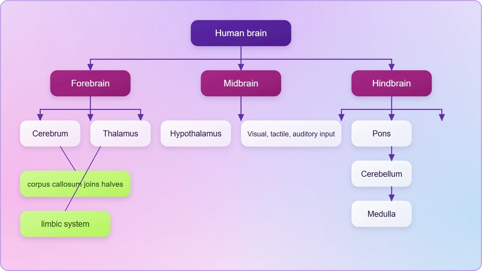
The pons consists of fibre tracts that interconnect different regions of the brain. Although this description is brief, it identifies the pons as a connector rather than a processing centre in the chapter's scheme, and that is the characterisation learners should reproduce. Taken together, the hindbrain emerges as the region where vital involuntary functions and cross-regional communication are housed, while the midbrain handles sensory integration and the forebrain carries the weight of regulation, emotion and higher coordination. The table below consolidates these functional assignments for quick reference.
Consolidating the Chapter: Definitions and Comparisons
Revision succeeds when definitions are exact and comparisons are structured. The chapter supplies a compact set of terms whose wording should be reproduced faithfully: resting potential, nerve impulse, synapse, synaptic cleft, neurotransmitter, central neural system, peripheral neural system, corpus callosum, limbic system. Each term carries a precise meaning, and paraphrasing too loosely is the most common source of lost marks. The exercise asking for a brief description of the structure of the brain, for instance, is best answered by moving systematically from the three major divisions to the structures within each.
The comparison questions deserve particular attention because they test relationships rather than isolated facts. Comparing the CNS with the peripheral neural system requires the learner to state what each contains and where each is situated. Comparing resting potential with action potential requires the learner to distinguish a static charged state from a propagating wave of depolarisation and repolarisation. The table below gathers the chapter's core definitions into a single revision card, which can be used for self-testing before the examination.
Working Through the Exercise Questions
The first exercise asks for a brief description of the structure of the brain. A sound answer proceeds in the chapter's own order: the brain is divided into forebrain, midbrain and hindbrain; the forebrain consists of cerebrum, thalamus and hypothalamus; the cerebrum is longitudinally divided into two halves connected by the corpus callosum; the midbrain receives and integrates visual, tactile and auditory inputs; and the hindbrain comprises pons, cerebellum and medulla, each with its stated role. Adding the limbic system and the functions of the hypothalamus completes the description without straying beyond the material.
The second exercise asks for two comparisons, and the third asks for explanations of polarisation, depolarisation and transmission across a chemical synapse. These are best answered with the chapter's own vocabulary. Polarisation refers to the charged state of the membrane at rest; depolarisation refers to the loss of that state when the membrane is excited; and transmission across a chemical synapse requires the release of neurotransmitter from the pre-synaptic side and its action on the post-synaptic membrane. The table below maps each exercise to the section of the chapter that answers it, so that revision can be targeted efficiently.
Going further
The nervous system enables rapid communication between different parts of the body. Where chemical messengers carried in the blood may take minutes to act, a nerve impulse travels along an axon in a fraction of a second, allowing an organism to react to a change in its surroundings almost immediately. This speed is the practical advantage of a wired system, and it explains why neural mechanisms dominate wherever rapid responses are required, from withdrawing a hand from a hot surface to adjusting the heartbeat during exertion.
The brain integrates sensory information and coordinates responses. Visual, tactile and auditory inputs arriving through the midbrain, balance-related signals reaching the cerebellum, and visceral information routed through the limbic system are all weighed against one another before an appropriate output is produced. Seen in this light, the anatomical divisions studied in this chapter are not separate organs performing unrelated tasks but parts of a single integrating apparatus, which is precisely the theme with which the chapter opens and with which this course now closes.
Closing the Course: What to Carry Forward
Ten lessons have moved from the general principle of neural coordination to the specific anatomy of the human brain, and the final summary restores the whole picture. The neural system coordinates and integrates functions as well as metabolic and homeostatic activities of all the organs. Neurons are excitable cells due to a differential concentration gradient of ions across the membrane. The resting potential is the electrical potential difference across the resting neural membrane, and the nerve impulse is conducted along the axon membrane as a wave of depolarisation and repolarisation. These statements form the conceptual spine of the chapter.
Upon that spine hang the synaptic and anatomical details. A synapse is formed by the membranes of a pre-synaptic and a post-synaptic neuron, which may or may not be separated by a synaptic cleft, and neurotransmitters are the chemicals involved in transmission at chemical synapses. The human neural system consists of the central neural system and the peripheral neural system; the CNS consists of the brain and spinal cord; and the brain divides into forebrain, midbrain and hindbrain. Each of these statements has been examined in its own lesson and is now gathered into a single, coherent account.
For learners moving on to further study, the most valuable habit to carry forward is the one this chapter rewards: precise use of defined terms, and a willingness to connect structure to function. The hypothalamus controls body temperature, eating and drinking because it sits at the junction of neural and homeostatic regulation. The medulla controls respiration, cardiovascular reflexes and gastric secretions because vital involuntary functions require a dedicated centre. The cerebellum integrates information from the semicircular canals and the auditory system because balance and hearing must be reconciled. Structure and function, held together, make the material memorable.
Finally, the exercises remain the best test of understanding. Describing the structure of the brain, comparing the CNS with the peripheral neural system, contrasting resting and action potentials, and explaining polarisation, depolarisation and synaptic transmission together cover the chapter's essential content. A learner who can answer all of these without consulting the text has mastered the material in the sense that matters: not the recitation of isolated facts, but the ability to move fluently between the electrical behaviour of a single membrane and the coordinated activity of an entire nervous system.
Also Read
- No links available.
From our network :
- Analyzing Trump Deportation Numbers Insights Into The 2026 Immigration Crackdown
- Mastering DB2 LUW v12 Tables: A Comprehensive Technical GuideComprehensive guide on DB2 LUW v12 tables. Explore CREATE TABLE syntax, BLU Acceleration, range partitioning, data types, and security features for IBM DB2 LUW v12.
- AI-Powered 'Precision Diagnostic' Replaces Standard GRE Score ReportsDiscover the new GRE Precision Diagnostic. Learn how AI-powered behavioral analytics and time-to-solve tracking are replacing standard GRE score reports to empower students.
- 98% of Global MBA Programs Now Prefer GRE Over GMAT Focus EditionExplore why 98% of global MBA programs now prefer the GRE over GMAT Focus. Learn about the shorter GRE format, verbal logic importance, and strategic versatility for students.
- 10 Physics Numerical Problems with Solutions for IIT JEEPrepare for IIT JEE with 10 physics numerical problems, complete with detailed solutions and conceptual explanations. Ideal for mastering kinematics, thermodynamics, electromagnetism, and optics.
- Vite 6/7 'Cold Start' Regression in Massive Module GraphsIn-depth analysis of Vite 6/7 cold start regressions in massive module graphs. Learn about barrel file issues, pre-bundling optimizations, and how to improve Vite dev server performance.
- Mastering DB2 12.1 Instance Design: A Technical Deep Dive into Modern Database ArchitectureComprehensive guide to DB2 12.1 Instance Design. Explore architectural components, HADR setup, buffer pool tuning, and security policies for IBM DB2 12.1. Includes 10 functional SQL and Shell samples.
- Trump Political Strategy How Geopolitical Stunts Serve As Media Diversions
- EV 2.0: The Solid-State Battery Breakthrough and Global Factory ExpansionDiscover the impact of the Solid-State Battery Breakthrough on the EV market. Learn about CATL and BYD stock growth, global factory expansion in Europe, and the future of EV 2.0 technology.
RESOURCES
- neural control and coordination chapter 18 - NCERTncert.nic.inBrain stem forms the connections between the brain and spinal cord. Reprint 2026-27. Page 8. NEURAL CONTROL AND COORDINATION. 237. SUMMARY. The neural system ...
- NEET Neural Control Summary by Vipin | PDF - Scribdscribd.comNeural Control and Coordination Summary Lecture Classnotes - Free download as PDF File (.pdf), Text File (.txt) or read online for free.
- Chapter 21: Neural Control and Coordination - LabXchangelabxchange.orgChapter 21: Neural Control and Coordination. Core Concept. All ... Summary of The Nervous System. OpenStax. Text. Favorite. Case studies. Real-world ...
- The neural control of interlimb coordination during mammalian ...pmc.ncbi.nlm.nih.govIn summary, experimental studies in mammals have shown that locomotor movements of the four limbs are generated by distinct spinal neuronal networks, one for ...
- Class 11 Neural Control Overview | PDF | Chemical Synapse - Scribdscribd.comThe CNS includes the brain and spinal cord for information processing and control. The PNS comprises all nerves of the body associated with the…
- Neural control of the female urethral and anal rhabdosphincters and ...pubmed.ncbi.nlm.nih.govFinally, a detailed summary of the neurochemical anatomy of Onuf's nucleus and the pharmacological control of the rhabdosphincter are provided. Publication ...
- Neurobiology: Reconstructing the Neural Control of Leg Coordinationsciencedirect.comSummary. Walking is adaptable because the timing of movements of individual legs can be varied while maintaining leg coordination. Recent work in stick ...
- In brief: How does the nervous system work? - InformedHealth.orgncbi.nlm.nih.govMay 4, 2023 ... Metabolic processes are also controlled by the nervous system. Go to: What makes up our nervous system? There are many…
- Neural and autonomic regulation during brief mindfulness ... - Frontiersfrontiersin.org... neural signatures and their coordination with autonomic rhythms during stress, regulation, and control contexts. In addition, the study incorporates ...
- Activity, Summary and Case Study - Neural Control and Coordinationbrainkart.comMay 12, 2018 ... Neural system coordinates and integrates the functions of all organs and responds to changes in the internal and external environments.
- neural control and coordination chapter 18 - Smart Achieverssmartachievers.onlineIn this chapter, you will learn about the neural system of human, mechanisms of neural coordination like transmission of nerve impulse, impulse conduction ...
- High-level summary of the AI Act | EU Artificial Intelligence Actartificialintelligenceact.euCritical infrastructure: AI systems used as safety components in the management and operation of critical digital infrastructure, road traffic and the supply of ...
- Parkinson's Disease | National Institute of Neurological Disorders ...ninds.nih.govJul 23, 2026 ... This brain area helps control how smooth and controlled a person's movements are. ... For research articles and summaries on…
- Neural Control of Wing Coordination in Flies: Current Biologycell.comDec 11, 2014 ... Summary. At the onset of each flight bout in flies, neural circuits in the CNS must rapidly integrate multimodal sensory…
- Proton signaling links epithelial sensing to neural control of host ...nature.comMar 27, 2026 ... ... coordination between epithelial sensing and systemic responses. Here ... In summary, our study identifies a proton-mediated gut-to ...