The neuron stands as the structural and functional unit of the neural system, a microscopic entity whose architecture encodes the entire logic of nervous communication. Every thought, reflex, and sensation depends on the coordinated behaviour of these cells, which receive, integrate, and transmit information across astonishingly precise pathways. Understanding the neuron is therefore not a peripheral detail but the foundation upon which the study of neural control and coordination rests. This lesson examines the neuron's component parts, its classification by structure, and the sheathing arrangements that influence how signals travel along its fibres.
Building directly on the previous discussion of the human neural system, where the central and peripheral divisions were mapped, we now descend to the cellular level to see what actually performs the work those divisions organise. The cell body, dendrites, and axon each carry distinct responsibilities, and the presence of Nissl's granules, synaptic knobs, and neurotransmitters reveals how structure serves function at every turn. Classification into multipolar, bipolar, and unipolar types, together with the distinction between myelinated and non-myelinated axons, completes a picture that prepares the ground for the next lesson on impulse generation and conduction.
This treatment follows the material closely, preserving its terminology, definitions, and order of ideas, while a short "Going further" block adds well-established background where it aids comprehension. Learners at the intermediate level will find the content sequenced so that each structural feature is tied to a functional consequence, making the eventual study of membrane polarisation and ion movement far more intuitive. The neuron, once its anatomy is secure, becomes a readable instrument rather than an abstract diagram.
On This Page
The Neuron as the Structural and Functional Unit
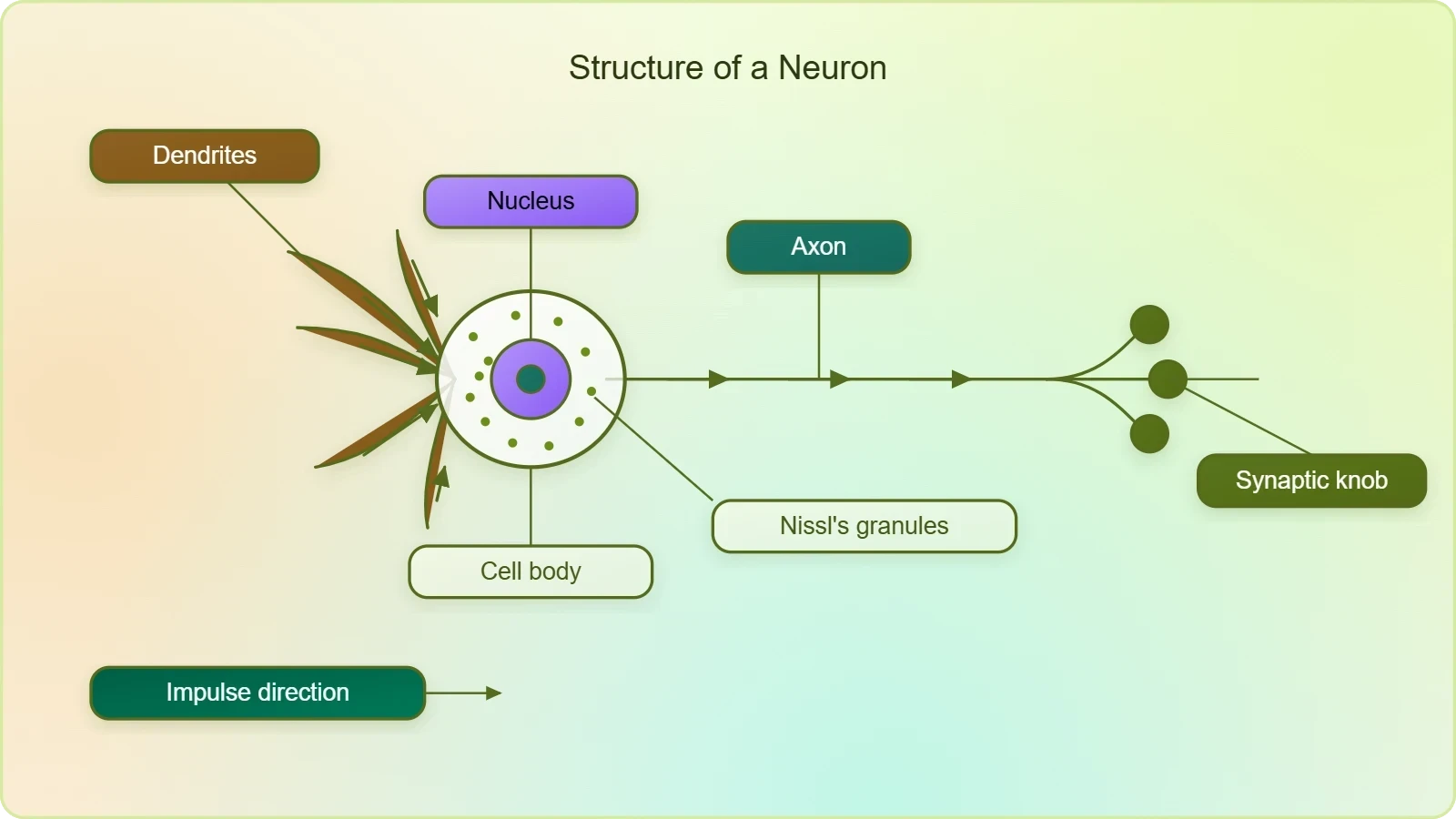
The neuron is described as a microscopic structure composed of three principal parts: the cell body, the dendrites, and the axon. This tripartite organisation is not arbitrary; each component contributes a specific capability that the others cannot supply. The cell body houses the metabolic machinery, the dendrites gather incoming signals, and the axon dispatches outgoing ones. Together they form a cell specialised for the reception, integration, and transmission of information, which is precisely why the neuron is regarded as both the structural and the functional unit of the neural system.
The Cell Body and Its Contents
The cell body, or cyton, contains the nucleus and cytoplasm from which the other processes extend. Within its cytoplasm are found Nissl's granules, granular bodies that give the cell body its characteristic appearance and that are associated with the synthesis of proteins needed for neural function. These granules are conspicuously present in the cell body and in the dendrites but are absent from the axon, a distribution that reflects the division of labour within the cell. The cell body thus functions as the administrative and manufacturing centre of the neuron.
Short fibres termed dendrites extend from the cell body and contain Nissl's granules as well. Their role is to transmit impulses towards the cell body, collecting signals from neighbouring neurons or from sensory receptors and funnelling them inward. The branching of dendrites increases the surface area available for receiving input, allowing a single neuron to integrate information from many sources simultaneously. This inward direction of flow is a defining feature of dendritic function and stands in deliberate contrast to the outward flow along the axon.
The Axon, Synaptic Knobs, and Neurotransmitters
The axon is a long fibre whose distal end is branched, and each branch terminates as a bulb-like structure called a synaptic knob. These knobs possess synaptic vesicles containing chemicals called neurotransmitters, the molecular messengers that carry the signal across the junction to the next cell. The axon transmits nerve impulses away from the cell body to a synapse or to a neuro-muscular junction, ensuring that information moves in a single, directed path. The presence of neurotransmitter-filled vesicles at the axon terminals links the electrical event within the neuron to the chemical event between neurons.
The arrangement of synaptic knobs at the ends of axonal branches means that a single axon can influence several target cells at once, distributing its signal across a network rather than to one destination. At the neuro-muscular junction, the same mechanism allows a neural impulse to command the contraction of muscle tissue, bridging the nervous and muscular systems. The neurotransmitter released from the synaptic vesicles thus serves as the final translator of an electrical instruction into a chemical one, a conversion essential to every coordinated movement and response.
Classification of Neurons by Number of Axons and Dendrites
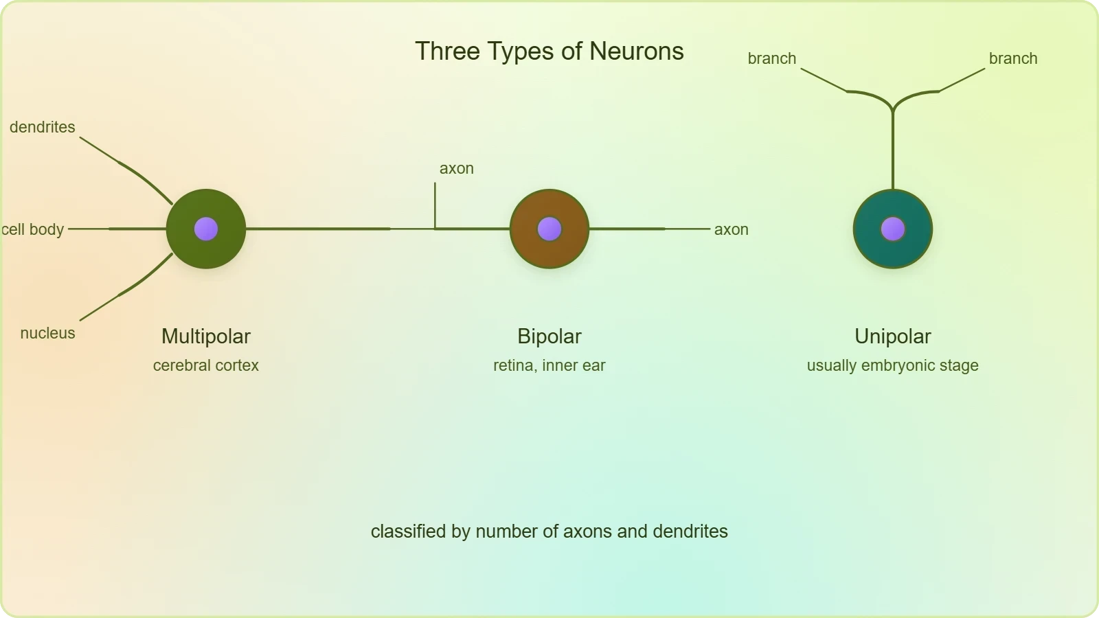
Based on the number of axons and dendrites, neurons are divided into three types: multipolar, bipolar, and unipolar. This classification is a direct reading of cellular geometry, and each type is associated with a characteristic location in the body. The scheme is simple to state but powerful in practice, because the shape of a neuron hints at how it collects and distributes signals. Recognising these three forms allows a learner to interpret histological descriptions and to anticipate the functional style of a neuron before any physiological detail is supplied.
Multipolar and Bipolar Neurons
Multipolar neurons possess one axon and two or more dendrites and are found in the cerebral cortex. Their multiple dendritic processes make them adept at receiving convergent input from many sources, which suits the dense integrative work of cortical tissue. Bipolar neurons possess one axon and one dendrite and are found in the retina of the eye. This simpler geometry, with a single receiving process and a single transmitting process, matches the retina's role in conveying visual information along a relatively direct pathway from photoreceptors onward.
The contrast between these two forms illustrates a general principle: the more dendritic branches a neuron carries, the greater its capacity to gather and combine signals. Multipolar neurons, with their extensive dendritic trees, are therefore common where complex integration is required, while bipolar neurons serve pathways where a more linear transfer of information is sufficient. Both types retain the same fundamental division of labour, with dendrites conducting inward and the axon conducting outward, but the balance of branching differs according to the demands of the circuit.
Unipolar Neurons and the Embryonic Stage
Unipolar neurons have a cell body with one axon only and are found usually in the embryonic stage. Their simplicity marks them as an early or transitional form rather than the dominant architecture of the mature nervous system. The classification thus carries developmental information as well as structural information, reminding the learner that neuronal form is not fixed across all contexts. In the mature nervous system the multipolar and bipolar types predominate in the locations described, while the unipolar type is chiefly noted for its association with embryonic development.
Presenting the three types together reinforces the logic of classification by number of processes. A learner who can count axons and dendrites can assign any described neuron to its category and, from that assignment, infer its typical location. This habit of reasoning from structure to setting is precisely the skill the lesson aims to build, and it prepares the student for the physiological discussion that follows, where the geometry of the axon and its sheath will determine how an impulse is conducted.
Myelinated and Non-Myelinated Axons
There are two types of axons, namely, myelinated and non-myelinated. The distinction concerns the sheath surrounding the fibre rather than the fibre's internal structure, and it has direct consequences for where each type is found and how it behaves. Myelinated nerve fibres are enveloped with Schwann cells, which form a myelin sheath around the axon. Non-myelinated fibres are also enclosed by a Schwann cell, but one that does not form a myelin sheath around the axon. The presence or absence of this sheath is the single criterion that separates the two categories.
Schwann Cells, the Myelin Sheath, and Nodes of Ranvier
In myelinated fibres, the Schwann cells wrap the axon to produce the myelin sheath, a layered covering that runs along the length of the fibre. The gaps between two adjacent myelin sheaths are called nodes of Ranvier, and these interruptions punctuate the sheath at intervals. Myelinated nerve fibres are found in spinal and cranial nerves, where their sheathed construction is characteristic. The nodes of Ranvier are not defects in the covering but a regular feature of its organisation, and they will assume physiological importance when impulse conduction is examined in the next lesson.
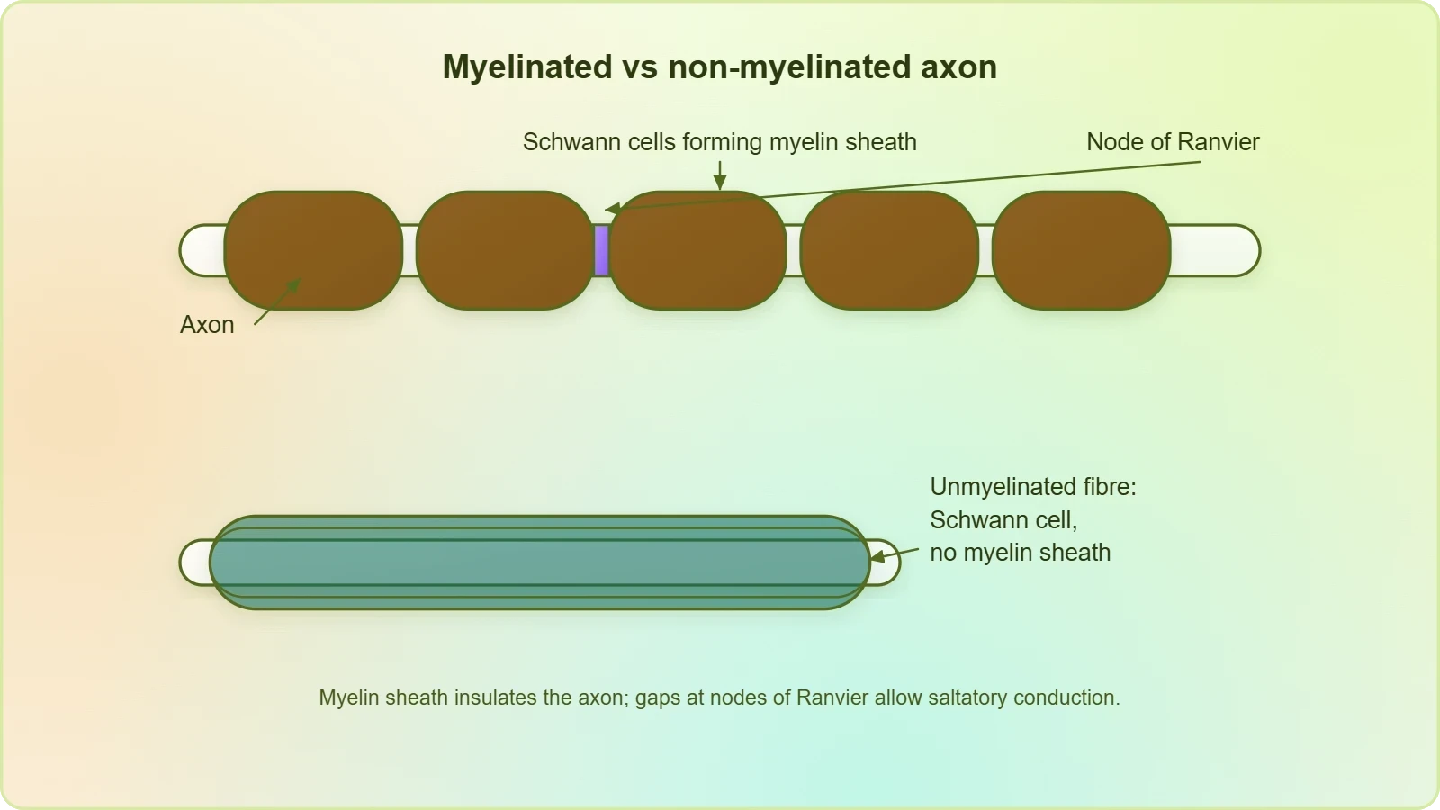
The relationship between Schwann cells and the axon is therefore one of enclosure and segmentation. Each Schwann cell contributes a segment of sheath, and the boundaries between segments define the nodes. This arrangement gives the myelinated fibre a distinctive beaded appearance under the microscope, with the sheath interrupted at regular points. Recognising this pattern is essential for interpreting histological images of spinal and cranial nerves, where myelinated fibres are the norm and the nodes appear as predictable gaps in an otherwise continuous covering.
Non-Myelinated Fibres and Their Distribution
Unmyelinated nerve fibre is enclosed by a Schwann cell that does not form a myelin sheath around the axon, and is commonly found in autonomous and the somatic neural systems. The Schwann cell is present in both categories, which is a point worth emphasising: the difference lies in whether a myelin sheath is formed, not in whether a Schwann cell is associated with the fibre. This subtlety prevents a common misreading in which non-myelinated fibres are imagined to lack supporting cells altogether.
The distribution of the two fibre types across the nervous system reflects the different demands placed on each division. Myelinated fibres dominate the spinal and cranial nerves, while non-myelinated fibres are commonly found in the autonomous and somatic neural systems. A learner should be able to state, for any fibre described, whether it is myelinated or non-myelinated, which cells enclose it, and where it is typically located. These three facts form a compact but complete account of axonal sheathing as the material presents it.
Direction of Impulse Flow and the Synaptic Junction
The neuron's internal geography enforces a direction of flow that is central to its function. Dendrites transmit impulses towards the cell body, while the axon transmits nerve impulses away from the cell body to a synapse or to a neuro-muscular junction. This polarity of structure mirrors the polarity of signalling and ensures that information moves through the cell in an orderly sequence rather than dispersing in all directions. The synapse, where the axonal signal meets the next cell, is the point at which the neuron's electrical activity is converted into a chemical message.
From Axon Terminal to Synapse
At the distal end, the axon branches, and each branch terminates as a synaptic knob possessing synaptic vesicles that contain neurotransmitters. When the impulse reaches this terminal, the chemicals held in the vesicles are available to carry the signal across the junction. The synapse is thus a specialised site of transfer, and the neuro-muscular junction is a particular instance of it where the receiving cell is a muscle fibre. The material presents this as the destination of the axonal impulse, completing the outward journey that began at the cell body.
This arrangement means that the neuron never simply discharges into empty space; its output is always addressed to a defined target, whether another neuron at a synapse or a muscle at the neuro-muscular junction. The synaptic knobs and their vesicles are the physical instruments of that address. A learner should appreciate that the chemical step at the synapse is not an alternative to electrical transmission but its necessary continuation, allowing a signal to cross the gap between cells that an electrical current alone could not bridge.
Why Structure Predicts Function
Every structural feature described in this lesson carries a functional implication. Nissl's granules in the cell body and dendrites support the synthetic work of the receiving region; the branching of dendrites widens the catchment of incoming signals; the long axon with its terminal knobs delivers output to distant targets; and the myelin sheath with its nodes organises the fibre for the conduction events to come. Reading the neuron in this way turns anatomy into a predictive account of behaviour, which is the habit the lesson seeks to instil.
This structural literacy also prepares the learner for the next lesson, where the polarised state of the neural membrane and the selective permeability of ion channels will be examined. The axon, already understood as a sheathed fibre with nodes and terminals, becomes the stage on which those electrical events unfold. Having established the parts and their arrangement, the student can approach impulse generation and conduction not as a list of unfamiliar facts but as the natural activity of a structure already known.
Going Further
Neurons are specialised cells that transmit electrical impulses in the nervous system, and this specialisation is reflected in the features described above. The excitable nature of the neural membrane, mentioned in the material as the basis of impulse generation, is a property of these cells that distinguishes them from most others in the body. Recognising the neuron as an electrically active cell helps the learner connect its anatomy to its behaviour, since the structures examined here exist to support the rapid transmission of impulses across sometimes considerable distances within the organism.
The Myelin Sheath as an Insulator
The myelin sheath acts as an insulator around some nerve fibres, a property that follows from its layered, fatty composition. Insulation confines the electrical events of the axon to the fibre itself and prevents the signal from dissipating into surrounding tissue. This background point complements the material's description of the sheath without altering it: the material establishes that Schwann cells form the myelin sheath and that gaps between adjacent sheaths are the nodes of Ranvier, while the insulating role explains why such a covering is advantageous to a conducting fibre.
Appreciating the sheath as an insulator also clarifies why the nodes of Ranvier matter. If the covering insulates the fibre, the interruptions in that covering become the points at which the membrane is exposed, and the physiological significance of this arrangement will be developed when conduction is studied. For the present lesson, the essential point is that the myelin sheath is a structural feature with a clear functional rationale, and that its presence or absence defines the two categories of axon described in the material.
Consolidating the Neuron's Architecture
The lesson has moved from the whole neuron to its parts and back again, and the picture that emerges is of a cell whose every feature is purposeful. The cell body with its Nissl's granules, the dendrites that conduct inward, the axon that conducts outward, and the synaptic knobs with their neurotransmitter-filled vesicles together constitute the structural and functional unit of the neural system. Classification by number of processes and by the presence of a myelin sheath adds two further layers of order, allowing any neuron to be placed within a coherent scheme.
Checking Understanding
A learner who can define the three parts of the neuron, state the direction of impulse flow along dendrites and axon, name the three structural types with their locations, and distinguish myelinated from non-myelinated fibres has secured the core of the lesson. These are not isolated facts but connected propositions: the direction of flow explains the division between dendrites and axon, the number of processes defines the type, and the presence of a myelin sheath defines the fibre category. Testing oneself on these connections is more valuable than memorising the list alone.
It is equally important to keep the boundaries of the lesson in view. The generation and conduction of the nerve impulse, including the polarised state of the membrane and the selective permeability of ion channels, belongs to the next lesson and is not developed here. The present treatment stops at the structure of the neuron and the sheathing of its axon, leaving the electrical behaviour of the membrane for the discussion that follows. This deliberate separation keeps each lesson focused and allows the material to be mastered in sequence.
Preparing for Impulse Generation and Conduction
The structures described here are the very structures that will carry the impulse in the next lesson. The axon, with its myelin sheath and nodes of Ranvier, is the fibre along which conduction occurs; the synaptic knobs are the terminals at which the signal is handed on; and the polarised membrane of the neuron is the medium in which the impulse arises. Holding this anatomy in mind makes the physiology easier to follow, because each new concept can be attached to a known part rather than to an abstract diagram.
With the neuron established as the unit of the neural system, the learner is ready to ask how an impulse is generated and conducted along the fibre. That question will be answered by examining the ion channels of the neural membrane, the unequal distribution of ions across it, and the active transport that maintains the gradients. The groundwork laid in this lesson, from Nissl's granules to nodes of Ranvier, ensures that the answer will find a ready structure in which to take its place.
Also Read
- No links available.
From our network :
- AI-Powered 'Precision Diagnostic' Replaces Standard GRE Score ReportsDiscover the new GRE Precision Diagnostic. Learn how AI-powered behavioral analytics and time-to-solve tracking are replacing standard GRE score reports to empower students.
- Vite 6/7 'Cold Start' Regression in Massive Module GraphsIn-depth analysis of Vite 6/7 cold start regressions in massive module graphs. Learn about barrel file issues, pre-bundling optimizations, and how to improve Vite dev server performance.
- 98% of Global MBA Programs Now Prefer GRE Over GMAT Focus EditionExplore why 98% of global MBA programs now prefer the GRE over GMAT Focus. Learn about the shorter GRE format, verbal logic importance, and strategic versatility for students.
- Trump Political Strategy How Geopolitical Stunts Serve As Media Diversions
- EV 2.0: The Solid-State Battery Breakthrough and Global Factory ExpansionDiscover the impact of the Solid-State Battery Breakthrough on the EV market. Learn about CATL and BYD stock growth, global factory expansion in Europe, and the future of EV 2.0 technology.
- Analyzing Trump Deportation Numbers Insights Into The 2026 Immigration Crackdown
- Mastering DB2 12.1 Instance Design: A Technical Deep Dive into Modern Database ArchitectureComprehensive guide to DB2 12.1 Instance Design. Explore architectural components, HADR setup, buffer pool tuning, and security policies for IBM DB2 12.1. Includes 10 functional SQL and Shell samples.
- 10 Physics Numerical Problems with Solutions for IIT JEEPrepare for IIT JEE with 10 physics numerical problems, complete with detailed solutions and conceptual explanations. Ideal for mastering kinematics, thermodynamics, electromagnetism, and optics.
- Mastering DB2 LUW v12 Tables: A Comprehensive Technical GuideComprehensive guide on DB2 LUW v12 tables. Explore CREATE TABLE syntax, BLU Acceleration, range partitioning, data types, and security features for IBM DB2 LUW v12.
RESOURCES
- Overview of neuron structure and function (article) - Khan Academykhanacademy.orgThese include nerve cells (or neurons) and glial cells (or glia). Neurons are the basic functional units of the nervous system, and they generate…
- Neuroanatomy, Neurons - StatPearls - NCBI Bookshelfncbi.nlm.nih.govStructure and Function. Neurons are unique in their ability to receive and ... neuron depending on its function and location. In addition to afferent ...
- Wiring optimization can relate neuronal structure and function - PNASpnas.orgIn this paper, we solve for the neuronal layout of an entire nervous system of the nematode C. elegans using the updated wiring diagram…
- An Easy Guide to Neuron Diagrams and Types - Healthlinehealthline.comBut they can also be found in parts of the nervous system that help the nose and ear function. Pyramidal neurons: These neurons have…
- Topographic structure and function of locus coeruleus noradrenaline ...pubmed.ncbi.nlm.nih.govSep 16, 2026 ... Noradrenaline (also known as norepinephrine) is released throughout most of the central nervous system by neurons in the locus coeruleus…
- Nervous System: What It Is, Parts, Function & Disordersmy.clevelandclinic.orgNov 16, 2023 ... Your nervous system plays a role in everything you do. The three main parts of your nervous system are your…
- Muscle and neuronal nicotinic acetylcholine receptors ... - PubMedpubmed.ncbi.nlm.nih.govStructure, function and pathogenicity. FEBS J. 2007 Aug;274(15):3799-845. doi ... They are widely expressed in the central nervous system, while, in the ...
- Stress signalling pathways that impair prefrontal cortex structure and ...pmc.ncbi.nlm.nih.gov... role of helping to depolarize the neuron when the cell has become hyperpolarized. ... In: Handbook of Physiology, The Nervous System, Higher Functions…
- Neuron - Simply Psychologysimplypsychology.orgAug 28, 2026 ... A neuron is a nerve cell that processes and transmits information through electrical and chemical signals in the nervous system.
- Neuronal Nicotinic Acetylcholine Receptor Structure and Function ...pmc.ncbi.nlm.nih.govFor example, α3β4* nAChRs, which are commonly found in the peripheral nervous system, are expressed at high levels only in the medial habenula, interpeduncular ...
- What is a neuron? - Queensland Brain Instituteqbi.uq.edu.auNeurons (also called neurones or nerve cells) are the fundamental units of the brain and nervous system, the cells responsible for receiving sensory input ...
- Brain Basics: The Life and Death of a Neuronninds.nih.govFeb 25, 2025 ... Architecture of a Neuron: its parts include the cell body, nucleus, dendrite, ... neurons found in the brain and the…
- Topographic structure and function of locus coeruleus noradrenaline ...nature.comSep 16, 2026 ... Noradrenaline (also known as norepinephrine) is released throughout most of the central nervous system by neurons in the locus coeruleus1–4.
- Brain Anatomy and How the Brain Works | Johns Hopkins Medicinehopkinsmedicine.orgTo do this, the central nervous system relies on billions of neurons (nerve cells). Main Parts of the Brain and Their Functions. At a…
- Spiking dynamics of individual neurons reflect changes in ... - Naturenature.comJul 30, 2025 ... ... structural features of neural circuits based on single-neuron activity. ... neural systems, spiking neural networks (SNNs) utilize discrete ...