The midbrain occupies a small volume of the human brain, yet its position and internal architecture make it one of the most consequential relay stations in the entire nervous system. Situated between the thalamus and hypothalamus of the forebrain above and the pons of the hindbrain below, it forms a short but vital bridge through which ascending sensory information and descending motor commands must pass. Learners who have already studied the cerebrum, thalamus and hypothalamus in the previous lesson will recognise that the midbrain does not duplicate their functions; instead, it serves as a compact corridor and integration point that links the higher centres with the lower brain regions and the spinal cord.
This lesson examines the midbrain exactly as the source material presents it: its location between the thalamus/hypothalamus of the forebrain and the pons of the hindbrain, the canal called the cerebral aqueduct that passes through it, and the dorsal portion consisting mainly of four round swellings known as the corpora quadrigemina. These three facts form the backbone of the discussion, and every explanation here builds outward from them rather than replacing them. The terminology, definitions and order of ideas are preserved so that the lesson remains faithful to the material while remaining readable for an intermediate learner preparing for the hindbrain and brain stem that follow.
Because the midbrain is often treated as a brief footnote between the forebrain and hindbrain, it is easy to underestimate its role. A careful reading shows that its very position explains much of its importance: anything travelling between the cerebral hemispheres and the medulla must traverse this narrow region. By the end of this lesson, you should be able to state the midbrain's boundaries, describe the cerebral aqueduct and the corpora quadrigemina, and explain why the midbrain is counted among the three major regions of the brain stem. A short "Going further" block adds well-established background without disturbing the core material.
On This Page
- Location of the Midbrain Between Forebrain and Hindbrain
- The Cerebral Aqueduct Passing Through the Midbrain
- The Corpora Quadrigemina on the Dorsal Portion
- The Midbrain as Part of the Brain Stem
- Consolidating the Midbrain: Definitions and Distinctions
- Going Further: Background Beyond the Core Material
- Preparing for the Hindbrain and Brain Stem
Location of the Midbrain Between Forebrain and Hindbrain
The defining feature of the midbrain is its position. It lies between the thalamus and hypothalamus of the forebrain and the pons of the hindbrain, occupying the narrow interval where the two larger divisions meet. This location is not incidental; it places the midbrain directly in the path of the major fibre systems that connect the cerebrum with the lower brain and spinal cord. When you visualise the brain from the side, the midbrain appears as a short stalk-like segment above the pons, and this simple picture is the most reliable starting point for understanding its relationships.
Anatomical Boundaries and Neighbouring Structures
Above the midbrain sit the thalamus and hypothalamus, the diencephalic structures studied in the previous lesson. The thalamus acts as a major coordinating centre for sensory and motor signalling, while the hypothalamus contains centres controlling body temperature and the urges for eating and drinking. Below the midbrain lies the pons, a mass of fibre tracts that interconnect different regions of the brain. The midbrain therefore sits precisely at the junction of the two great divisions, and its upper and lower boundaries are defined by these neighbouring structures rather than by any capsule of its own.
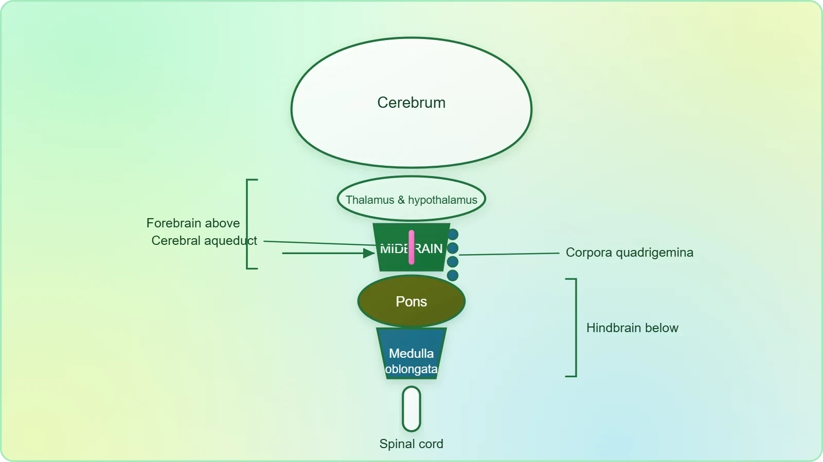
This arrangement means that the midbrain is continuous with both the forebrain above and the hindbrain below. There is no sharp membrane separating them; the transition is one of structure and function rather than of enclosure. For the learner, the practical consequence is that the midbrain cannot be studied in isolation. Its boundaries are the thalamus and hypothalamus superiorly and the pons inferiorly, and any description of its role must acknowledge that it receives and forwards traffic generated in those adjacent regions.
Why Position Determines Function
Position determines function in the nervous system because nerve fibres must physically travel from one region to another. Any structure sitting between two major divisions will inevitably carry the fibres that connect them, and the midbrain is no exception. Its location between the forebrain and the pons means that ascending pathways carrying sensory information toward the cerebral cortex and descending pathways carrying motor commands toward the spinal cord both pass through its territory. The midbrain is thus a corridor as much as a centre, and its strategic placement explains why even a small lesion in this region can produce effects far beyond its size.
Understanding this principle prepares the learner for the next lesson, where the pons and medulla are described as containing fibre tracts and vital centres. The midbrain, the pons and the medulla together form the brain stem, and the brain stem forms the connections between the brain and spinal cord. The midbrain is therefore the uppermost of the three major regions of the brain stem, and its position at the top of that column is the first fact to fix in memory.
The Cerebral Aqueduct Passing Through the Midbrain
A canal called the cerebral aqueduct passes through the midbrain. This single sentence from the material carries considerable functional weight, because a canal running through a structure implies a continuous channel connecting the spaces above and below it. The cerebral aqueduct is the narrow passage that traverses the midbrain from its upper to its lower end, and its presence explains why the midbrain is described as a conduit in more than one sense. It is a corridor for nerve fibres and, at the same time, a channel for the fluid that fills the internal cavities of the brain.
Structure and Course of the Canal
The cerebral aqueduct runs through the midbrain as a slender, midline channel. It is not a wide cavity but a narrow tube, and this narrowness is itself significant: a small blockage in such a confined passage can have disproportionate consequences for the fluid system of the brain. The canal passes through the midbrain in a roughly vertical course, entering at the upper end near the thalamus and hypothalamus and emerging at the lower end toward the pons. Its walls are formed by the surrounding midbrain tissue, and it is therefore best imagined as a tunnel bored through the substance of the midbrain rather than as a separate organ.
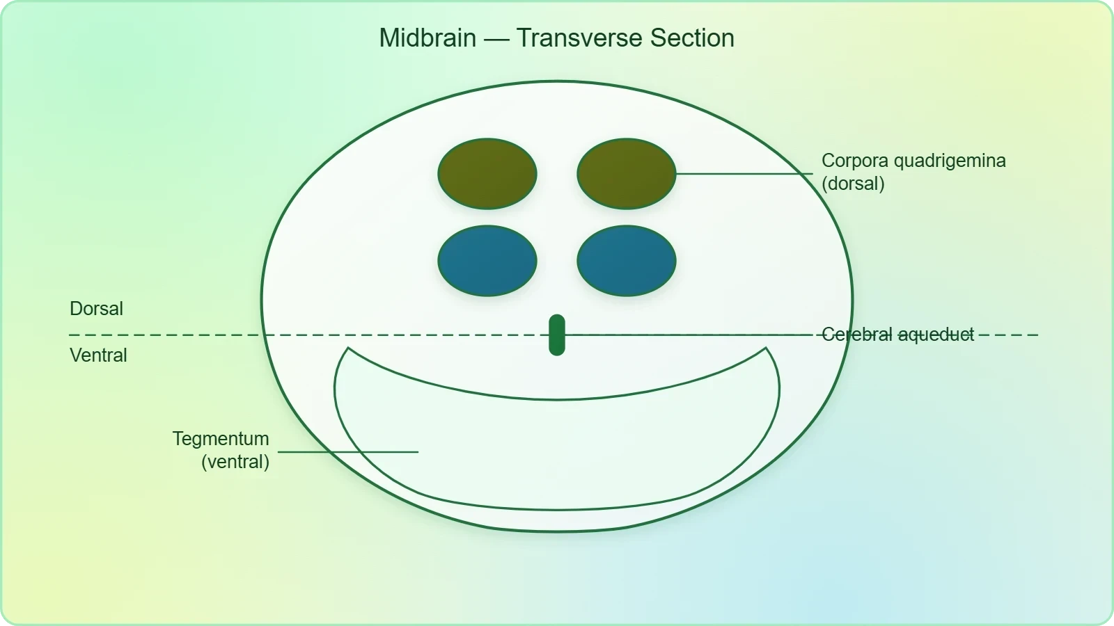
Because the aqueduct is embedded within the midbrain, its course defines the internal geography of the region. Structures lying dorsal to the canal differ from those lying ventral to it, and the dorsal portion is where the corpora quadrigemina are found. This dorsal-ventral distinction is a useful organising principle for the learner: the canal acts as a reference line, and the swellings described in the material sit on the dorsal side of that line.
Functional Significance of the Passage
The functional significance of the cerebral aqueduct lies in what it connects. It provides a continuous channel between the fluid-filled spaces of the forebrain and those of the hindbrain, allowing the cerebrospinal fluid to circulate through the ventricular system. This circulation is essential for the chemical environment of the nervous tissue, and the aqueduct is the narrowest point in that route. When the passage is narrowed or obstructed, fluid accumulates above the blockage, and the pressure effects can be serious. The midbrain therefore participates in the fluid economy of the brain simply by housing this canal.
For the learner, the key point is that the cerebral aqueduct is a defining feature of the midbrain, mentioned in the material alongside its location and its dorsal swellings. It is not a structure of the forebrain or the hindbrain but of the midbrain specifically, and it passes through the midbrain to link the regions on either side. Remembering this single fact anchors the midbrain in the broader architecture of the central neural system.
The Corpora Quadrigemina on the Dorsal Portion
The dorsal portion of the midbrain consists mainly of four round swellings, or lobes, called the corpora quadrigemina. This is the most distinctive surface feature of the region and the one most likely to appear in an examination question. The name itself is descriptive: "corpora" refers to bodies, "quadrigemina" to a set of four, and the term therefore names the four rounded elevations that together form the dorsal roof of the midbrain. They are arranged as two pairs, one pair on each side of the midline, and they sit above the cerebral aqueduct.
Arrangement and Appearance of the Four Swellings
The four swellings are grouped into two pairs, an upper pair and a lower pair, positioned symmetrically about the midline. Each swelling is rounded and lobe-like, which is why the material describes them as round swellings or lobes. Their dorsal position means they are visible on the upper surface of the midbrain when it is viewed from behind, and their paired arrangement gives the region a characteristic four-part appearance. The term corpora quadrigemina captures this arrangement precisely and should be associated in the learner's mind with the number four.
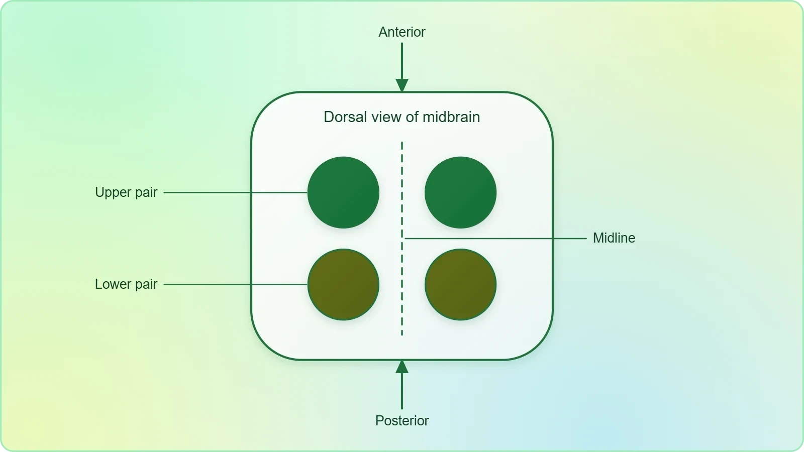
Because the swellings are described as forming the main content of the dorsal portion, they dominate the external anatomy of the midbrain's upper surface. The ventral portion, by contrast, is not described in the material in the same detail, and the lesson should not invent structures there. The disciplined approach is to state what the material states: the dorsal portion consists mainly of four round swellings called the corpora quadrigemina, and these are the defining surface feature of the region.
Role as a Dorsal Landmark
The corpora quadrigemina serve as a dorsal landmark that helps the learner orient the midbrain in three dimensions. If the cerebral aqueduct is the internal reference line, the corpora quadrigemina are the external reference point on the dorsal side. Together they allow a mental model in which the canal runs through the centre and the four swellings cap the roof. This model is simple, accurate to the material, and sufficient for the intermediate level at which this course is pitched.
The swellings also illustrate a broader principle of neuroanatomy: surface features often reflect underlying functional groupings. The material does not elaborate on the functions of the individual lobes, and this lesson will not go beyond it. What matters at this stage is recognition and naming, so that when the hindbrain and brain stem are studied in the next lesson, the midbrain can be placed correctly at the top of the brain stem with its dorsal swellings intact.
The Midbrain as Part of the Brain Stem
Three major regions make up the brain stem: the midbrain, the pons and the medulla oblongata. This statement from the material places the midbrain within a larger functional unit and explains why its study matters beyond its own boundaries. The brain stem forms the connections between the brain and spinal cord, and the midbrain is its uppermost component. Recognising this membership is essential, because the next lesson will describe the pons and medulla in detail, and the learner must be able to assemble all three regions into a single coherent column.
Membership of the Brain Stem Column
The brain stem is a column of tissue that links the higher brain centres with the spinal cord, and its three regions are arranged in sequence from top to bottom. The midbrain sits at the top, the pons in the middle, and the medulla oblongata at the bottom where it joins the spinal cord. This vertical arrangement mirrors the flow of information: ascending fibres travel upward through the column, and descending fibres travel downward. The midbrain's membership in this column is what gives its position between the thalamus and the pons its full meaning.
It is worth noting that the brain stem is defined by its three regions, not by the addition of any other structure. The material is explicit on this point, and the lesson should preserve it. The midbrain is therefore not an appendage to the brain stem but one of its three constituent parts, and any account of the brain stem that omits it is incomplete. This is the conceptual bridge to the next lesson, where the pons and medulla will be examined as the remaining two members.
Connecting the Brain and Spinal Cord
The brain stem forms the connections between the brain and spinal cord, and the midbrain contributes to this connecting function by carrying fibres that pass between the forebrain and the lower regions. Every ascending signal destined for the cerebral cortex and every descending command destined for the spinal cord must traverse the brain stem, and the midbrain is the first segment of that journey from above. Its role is therefore both structural and functional: structurally it is a segment of the column, and functionally it is a conduit for the traffic that the column carries.
This dual role explains why the midbrain is studied between the forebrain and the hindbrain rather than in isolation. The forebrain lesson established the higher centres; this lesson establishes the connecting segment; the next lesson will establish the lower centres and the full brain stem. The sequence is deliberate, and each lesson builds on the one before it without repeating material that belongs elsewhere.
Consolidating the Midbrain: Definitions and Distinctions
Consolidation requires that the learner can state the midbrain's definition without hesitation and distinguish it from its neighbours. The material gives three defining facts: its location between the thalamus/hypothalamus of the forebrain and the pons of the hindbrain, the cerebral aqueduct that passes through it, and the corpora quadrigemina on its dorsal portion. These three facts, taken together, constitute a complete working definition at the intermediate level. Anything beyond them belongs to more advanced study and should not be imported into this lesson.
Key Definitions to Retain
The first definition to retain is positional: the midbrain is located between the thalamus and hypothalamus of the forebrain and the pons of the hindbrain. The second is internal: a canal called the cerebral aqueduct passes through the midbrain. The third is external: the dorsal portion of the midbrain consists mainly of four round swellings called the corpora quadrigemina. Each definition is short, each is drawn directly from the material, and each can be recalled independently. Together they allow a learner to describe the midbrain accurately in a few sentences.
It is useful to pair each definition with a common error. The positional definition is sometimes confused with the pons, which lies below rather than within the midbrain. The internal definition is sometimes attributed to the forebrain, although the aqueduct is a feature of the midbrain. The external definition is sometimes reduced to two swellings rather than four, losing the "quadrigemina" of the name. Naming these errors explicitly helps the learner avoid them in examination answers and in later study of the brain stem.
Distinguishing the Midbrain from Adjacent Regions
The midbrain is distinguished from the forebrain by the absence of the cerebrum, thalamus and hypothalamus within its own boundaries, and from the hindbrain by the absence of the pons, cerebellum and medulla. Its neighbours are named in the material precisely so that the boundaries can be drawn. The thalamus and hypothalamus lie above it, the pons lies below it, and the midbrain occupies the interval between them. This simple spatial scheme is more reliable than any list of functions, because the functions of the midbrain are not elaborated in the material.
Distinguishing the midbrain from adjacent regions also clarifies the order of ideas in the course. The forebrain lesson covered the cerebrum, thalamus and hypothalamus; this lesson covers the midbrain; the next lesson covers the hindbrain and brain stem. Each region is treated in turn, and the boundaries between them are respected. The learner who keeps this order in mind will find that the brain stem, when it is assembled in the next lesson, fits together as a coherent whole rather than as a set of disconnected facts.
Going Further: Background Beyond the Core Material
The following block adds well-established background that supports understanding without altering the core material. It is kept separate so that the learner can distinguish what the lesson requires from what is offered as context. Nothing here contradicts the material; the additional points simply place the midbrain in a slightly wider anatomical setting. Read it after the core sections have been mastered, and treat it as enrichment rather than as examination content.
The Midbrain as Part of the Brain Stem
The midbrain is part of the brain stem, a point already stated in the material and worth reinforcing here. The brain stem is the stalk-like column that connects the brain with the spinal cord, and the midbrain is its uppermost segment. Because the brain stem carries the great ascending and descending pathways, its regions are often studied together as a functional unit. The midbrain's membership in this unit explains why its lesions can affect functions that seem remote from its small size, and why the next lesson on the hindbrain and brain stem completes the picture.
Recognising the midbrain as part of the brain stem also clarifies the terminology used in clinical and anatomical descriptions. When the brain stem is described as a whole, the midbrain is named first, followed by the pons and the medulla. This order reflects the vertical arrangement of the column and matches the order of the lessons in this course. The learner who internalises this sequence will find that the three regions are easier to remember together than separately.
The Cerebral Aqueduct and the Ventricular System
The cerebral aqueduct connects the third and fourth ventricles of the brain. This is well-established background that extends the material's statement that a canal called the cerebral aqueduct passes through the midbrain. The third ventricle lies above the midbrain in the region of the thalamus and hypothalamus, and the fourth ventricle lies below it in the region of the hindbrain. The aqueduct is the narrow channel between them, and its course through the midbrain is what makes the midbrain a participant in the circulation of cerebrospinal fluid.
This background helps explain why the aqueduct is clinically important despite its small calibre. Because it is the narrowest part of the ventricular pathway, it is a natural point of vulnerability, and obstruction at this level produces a characteristic pattern of fluid accumulation above the block. The material does not discuss these consequences, and this lesson does not require them, but the learner who understands the aqueduct's connections will find the anatomy more coherent and the clinical references in later study easier to follow.
Preparing for the Hindbrain and Brain Stem
This lesson has established the midbrain as the segment between the thalamus and hypothalamus of the forebrain and the pons of the hindbrain, pierced by the cerebral aqueduct and capped dorsally by the corpora quadrigemina. It has also placed the midbrain within the brain stem, the three-part column that forms the connections between the brain and spinal cord. The next lesson will complete the column by describing the pons, the cerebellum and the medulla oblongata, and will assemble the full brain stem from its three major regions.
Before moving on, verify that you can state the three defining facts without reference to the text, that you can name the structures immediately above and below the midbrain, and that you can explain why the midbrain is counted among the three regions of the brain stem. If any of these points is uncertain, return to the relevant section and re-read it. The hindbrain lesson assumes this foundation, and a secure grasp of the midbrain will make the transition to the pons and medulla straightforward.
Also Read
- No links available.
From our network :
- Vite 6/7 'Cold Start' Regression in Massive Module GraphsIn-depth analysis of Vite 6/7 cold start regressions in massive module graphs. Learn about barrel file issues, pre-bundling optimizations, and how to improve Vite dev server performance.
- Mastering DB2 LUW v12 Tables: A Comprehensive Technical GuideComprehensive guide on DB2 LUW v12 tables. Explore CREATE TABLE syntax, BLU Acceleration, range partitioning, data types, and security features for IBM DB2 LUW v12.
- Mastering DB2 12.1 Instance Design: A Technical Deep Dive into Modern Database ArchitectureComprehensive guide to DB2 12.1 Instance Design. Explore architectural components, HADR setup, buffer pool tuning, and security policies for IBM DB2 12.1. Includes 10 functional SQL and Shell samples.
- Analyzing Trump Deportation Numbers Insights Into The 2026 Immigration Crackdown
- Trump Political Strategy How Geopolitical Stunts Serve As Media Diversions
- EV 2.0: The Solid-State Battery Breakthrough and Global Factory ExpansionDiscover the impact of the Solid-State Battery Breakthrough on the EV market. Learn about CATL and BYD stock growth, global factory expansion in Europe, and the future of EV 2.0 technology.
- 10 Physics Numerical Problems with Solutions for IIT JEEPrepare for IIT JEE with 10 physics numerical problems, complete with detailed solutions and conceptual explanations. Ideal for mastering kinematics, thermodynamics, electromagnetism, and optics.
- AI-Powered 'Precision Diagnostic' Replaces Standard GRE Score ReportsDiscover the new GRE Precision Diagnostic. Learn how AI-powered behavioral analytics and time-to-solve tracking are replacing standard GRE score reports to empower students.
- 98% of Global MBA Programs Now Prefer GRE Over GMAT Focus EditionExplore why 98% of global MBA programs now prefer the GRE over GMAT Focus. Learn about the shorter GRE format, verbal logic importance, and strategic versatility for students.
RESOURCES
- Corpora quadrigemina - an overview | ScienceDirect Topicssciencedirect.com... midbrain and resulting occlusion of the cerebral aqueduct. The subarachnoid ... The tectum, or roof, of the mesencephalon was partially exposed during dissection ...
- Neuroanatomy, Mesencephalon Midbrain - StatPearls - NCBI - NIHncbi.nlm.nih.govOn the posterior surface of the midbrain, there is a structure called the corpora quadrigemina. ... cerebral aqueduct, cerebral peduncle, and crus cerebri.
- Cerebral aqueduct - Wikipediaen.wikipedia.orgThe midbrain tegmentum is situated anteriorly to the cerebral aqueduct. ... The superior and inferior colliculi that make up the corpora quadrigemina are situated ...
- The Midbrain - Colliculi - Peduncles - TeachMeAnatomyteachmeanatomy.infoExternal Anatomy of the Midbrain · Tectum – located posterior to the cerebral aqueduct · Paired cerebral peduncles – located anteriorly and laterally. Internally, ...
- Comparative anatomy of dissected optic lobes, optic ventricles ...pmc.ncbi.nlm.nih.govNov 23, 2021 ... ... aqueduct for the midbrain ventricle in all vertebrates with a cerebral ventricular system. The lateral radiograph of the entire…
- Midbrain: Anatomy, location, parts, definition - Kenhubkenhub.com... corpora quadrigemina. ... The dorsal part of the tegmentum is traversed by the cerebral aqueduct, which connects the third and fourth ventricles of…
- Human Anatomy & Physiology Laboratory OER: AP1 Lab Module 15 ...georgiasouthern.libguides.comCorpora Quadrigemina - in the midbrain; formed of four “bumps” called ... Cerebral Aqueduct - in the midbrain; part of the ventricular system of…
- midbrain development Gene Ontology Term (GO:0030901)informatics.jax.org... cerebral peduncles and a dorsal tectum containing the corpora quadrigemina and that ... corpora quadrigemina development; midbrain dopaminergic neuron ...
- Corpora quadrigemina | Radiology Reference Article - Radiopaediaradiopaedia.orgOct 20, 2024 ... Midbrain anatomy · Supracerebellar arachnoid cyst. Related articles: Anatomy: Brain ... aqueduct of Sylvius · fourth ventricle · superior ...
- Brainstem: Location, anatomy, parts, function | Kenhubkenhub.com... corpora quadrigemina (posteriorly). It continues along a ... the tectum is dorsal to the cerebral aqueduct and fourth ventricle, primarily in the midbrain.
- Mesencephalon - wikidocwikidoc.orgAug 9, 2012 ... In biological anatomy, the mesencephalon (or midbrain) is the ... cerebral aqueduct. Corpora Quadrigemina. It consists of four solid ...
- Gross Anatomy of the Brain and Cranial Nervespearsonhighered.comThe cerebral peduncles and the rounded corpora quadrigemina make up the midbrain tissue anterior and posterior (respectively) to the cerebral aqueduct. 2 ...
- Anatomical Presentation Of The Inferior Colliculus Of The Brainstemsciepro.comJun 11, 2026 ... ... cerebral aqueduct region, with its surface contour forming the lower component of the corpora quadrigemina. The brachium of the…
- Solved Cerebral aqueduct Corpora quadrigemina Pituitary - Cheggchegg.comApr 3, 2021 ... Corpora quadrigemina: A part of the midbrain, it includes the superior and inferior colliculi (involved in visual and auditory reflexes)…
- The Midbrain – Human Neuroanatomyuen.pressbooks.pubFeb 25, 2026 ... It consist of four symmetrical bumps collectively called the corpora quadrigemina (Latin, “four twinned bodies”). ... Anatomy of the Brain ...