Breathing is not a single uniform act performed identically by every living creature; it is a varied and adaptable process shaped by where an organism lives and how complex its body plan has become. Across the animal kingdom, the exchange of oxygen from the atmosphere with carbon dioxide produced by cells takes many anatomical forms, each calibrated to the demands of a particular habitat and level of organisation. Understanding this diversity is essential before examining the human respiratory system, because it reveals the underlying logic that governs how gases move from the environment into living tissue.
This lesson traces respiratory organs across animal groups, beginning with the simplest invertebrates that rely on diffusion over their entire body surface and moving through the moist cuticle of earthworms, the tracheal tubes of insects, the gills of aquatic arthropods, molluscs and fishes, and the lungs of terrestrial vertebrates. Each structure represents a solution to the same fundamental problem: how to bring oxygen close enough to cells while expelling carbon dioxide efficiently. The progression from surface diffusion to specialised, vascularised organs mirrors the increasing metabolic demands and body complexity of the animals concerned.
By the end of this lesson, learners should be able to identify which respiratory mechanism belongs to which group of animals, explain why habitat and organisation determine that mechanism, and distinguish branchial from pulmonary and cutaneous respiration. This foundation prepares the ground for the detailed study of the human respiratory system that follows, where the same principles of thin, moist, vascularised exchange surfaces reappear in a far more elaborate form.
On This Page
- The Governing Principle: Habitat and Level of Organisation
- Simple Diffusion and the Moist Cuticle
- Tracheal Tubes: Bringing Air Directly to Tissues
- Branchial Respiration: Gills in Water
- Pulmonary and Cutaneous Respiration on Land
- Comparing Respiratory Strategies Across Animal Groups
- Matching Organs to Habitats and Organisation
- What Every Respiratory Surface Must Provide
- Respiratory Organs by Animal Group
- Branchial, Pulmonary and Cutaneous Respiration
- Exchange Surface and Diffusion Distance
- Aquatic Versus Terrestrial Exchange
- Level of Organisation and Respiratory Demand
- Lesson Roadmap: Where This Fits
- Going Further
The Governing Principle: Habitat and Level of Organisation
Mechanisms of breathing vary among different groups of animals depending mainly on their habitats and levels of organisation. This single sentence carries the organising logic of the entire lesson. An animal's habitat determines whether the surrounding medium is water or air, and that medium dictates the physical constraints under which gas exchange must occur. Meanwhile, the level of organisation determines whether the body is a simple sheet of cells in direct contact with the environment or a complex, multi-layered structure requiring dedicated transport networks to reach interior tissues.
Lower invertebrates such as sponges, coelenterates and flatworms sit at the simplest end of this spectrum. Their bodies are thin enough, and their metabolic rates low enough, that every cell lies close to the external environment. No specialised organ is needed because no cell is far from the source of oxygen. As bodies become thicker and more metabolically active, however, diffusion alone becomes insufficient, and specialised respiratory structures emerge to bridge the growing distance between the environment and the tissues.
Why Simple Bodies Need No Respiratory Organs
In sponges, coelenterates and flatworms, oxygen is exchanged with carbon dioxide by simple diffusion over the entire body surface. Every part of the body acts as a respiratory surface, and the gases simply move down their concentration gradients across the outer membrane. This arrangement works because the diffusion distance from the exterior to the innermost cells remains very short, and because these organisms generally possess low metabolic demands that a modest flux of gases can satisfy.
The elegance of this solution lies in its economy: no gills, no lungs, no circulatory plumbing dedicated to respiration. Yet the same simplicity imposes a hard ceiling on body size and thickness. Once an animal's tissues are stacked several cell layers deep, the outermost cells consume the incoming oxygen before it can reach the interior, and diffusion over the body surface ceases to be adequate. This constraint explains why more complex animals must develop dedicated exchange organs.
The Transition Toward Specialised Exchange Surfaces
As organisation increases, the respiratory surface is progressively internalised and elaborated. Earthworms retain a body-surface strategy but refine it with a moist cuticle, while insects internalise the exchange surface into a network of tubes that pipes atmospheric air directly to the tissues. Aquatic arthropods and molluscs concentrate the exchange surface into vascularised gills, and terrestrial forms concentrate it into vascularised lungs. Each step represents an increasing investment in structure to serve a larger, more demanding body.
This progression is not a ladder of superiority but a series of adaptations matched to circumstance. An insect's tracheal system is exquisitely suited to its small, active body, just as a fish's gills are matched to the oxygen-poor medium of water. The organising principle remains constant: the respiratory organ must present a large, thin, moist surface to the environment and connect it, directly or through the blood, to the cells that need oxygen.
Simple Diffusion and the Moist Cuticle
The simplest respiratory strategy in the animal kingdom is direct exchange across the body surface, and it persists in a refined form in earthworms through the moist cuticle. These two examples, taken together, illustrate how the same basic principle of surface exchange can be maintained as body organisation becomes slightly more complex. They also introduce the crucial requirement that the exchange surface remain moist, a condition that will recur in every subsequent respiratory organ discussed in this lesson.
Understanding these surface-based mechanisms is important because they establish the baseline against which all specialised organs are measured. When a gill or a lung is described as an elaboration of the body surface, the comparison is literal: these organs are folded, vascularised extensions of the same thin, moist membrane that covers a sponge or an earthworm. The difference lies in the scale of folding and the intimacy of the connection to the circulatory system.
Diffusion Over the Entire Body Surface
Lower invertebrates like sponges, coelenterates and flatworms exchange oxygen with carbon dioxide by simple diffusion over their entire body surface. There is no localisation of respiratory function; the whole exterior of the animal participates. For these organisms, the body surface is simultaneously the respiratory organ, and the effectiveness of exchange depends on the surface remaining thin and permeable while the animal's tissues remain close to it.
This whole-body approach has a quiet efficiency. Because gases need only cross a single membrane and diffuse a short distance, the process requires no energy expenditure for ventilation and no circulatory system to distribute the gases. The animal's own movements and the ambient currents of its environment suffice to renew the medium at the surface. The limitation, as noted, is that the strategy cannot scale to thick, bulky bodies with high metabolic rates.
The Earthworm's Moist Cuticle
Earthworms use their moist cuticle as the respiratory surface. The cuticle must remain moist for gas exchange to proceed, which is why earthworms are found in damp soil and why their skin appears glistening. Oxygen from the surrounding air dissolves in the moisture of the cuticle and diffuses inward, while carbon dioxide diffuses outward along the same pathway. The moist cuticle thus functions as a thin, hydrated exchange membrane covering the entire body.
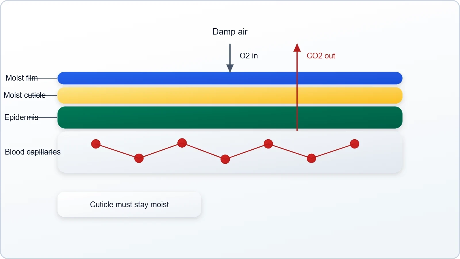
This arrangement represents a modest advance over simple diffusion because the earthworm possesses a circulatory system that helps distribute gases once they have entered through the cuticle. The exchange surface itself, however, remains the body surface. The earthworm therefore occupies an intermediate position: it has not evolved a dedicated respiratory organ, yet it has coupled its cutaneous exchange to an internal transport system, anticipating the vascularised organs of more complex animals.
Tracheal Tubes: Bringing Air Directly to Tissues
Insects solve the problem of gas exchange in a manner strikingly different from the aquatic and cutaneous strategies already described. Rather than dissolving gases in a moist surface and relying on the blood to carry them, insects have a network of tubes, the tracheal tubes, that transport atmospheric air within the body. This system delivers air close to the tissues themselves, bypassing the need for a blood-borne respiratory pigment in many cases and representing one of the most direct respiratory solutions in the animal kingdom.
The tracheal system is a beautiful illustration of how a respiratory organ can be internalised without becoming a lung. The tubes open to the exterior through small pores and branch repeatedly, penetrating deep into the body until their finest endings lie adjacent to individual cells. Air moves through this network, and gases exchange directly with the tissues at the tube endings, so that the entire body becomes a distributed respiratory organ served by an internal piping system.
Anatomy of the Tracheal Network
The tracheal tubes form a branching network that begins at the body surface and ramifies throughout the insect's interior. Atmospheric air enters the system and travels along the tubes, which grow progressively finer as they penetrate the tissues. Because the finest branches reach close to the cells, the diffusion distance for oxygen and carbon dioxide remains short despite the insect's relatively complex body plan. The tubes thus extend the principle of short diffusion distance into the interior of the animal.
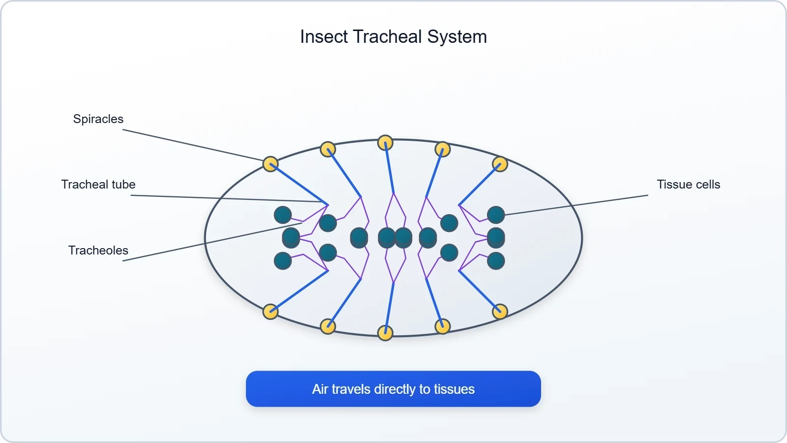
This design confers a notable advantage: the air within the tracheal tubes is renewed directly from the atmosphere, so the insect does not depend on a circulatory fluid to ferry oxygen from a remote exchange surface. The respiratory medium is delivered to the doorstep of the cells. The system's efficiency is reflected in the high metabolic rates and vigorous activity of many insects, though the reliance on diffusion within the finest tubes does impose limits on body size.
How the Tracheal Strategy Differs from Gills and Lungs
The tracheal strategy differs fundamentally from branchial and pulmonary respiration in that it does not concentrate the exchange surface into a single localised organ. Gills and lungs are vascularised structures in which gases cross into the blood, which then carries them to the tissues. In the tracheal system, by contrast, air is conducted directly to the tissues, and the exchange occurs at the ends of the tubes rather than at a centralised organ surface.
This contrast sharpens our understanding of what a respiratory organ actually does. A gill or a lung is a device for bringing the medium and the blood into intimate contact across a thin, moist barrier. The tracheal system achieves the same end by bringing the medium to the tissue directly. Both are solutions to the same problem of short diffusion distance, and both demonstrate that the form of the respiratory organ is dictated by the animal's habitat and organisation.
Branchial Respiration: Gills in Water
Special vascularised structures called gills are used by most of the aquatic arthropods and molluscs for branchial respiration, and among vertebrates, fishes use gills as well. The gill is the characteristic respiratory organ of aquatic life, and its prevalence across such distantly related groups reflects the demanding physics of extracting oxygen from water. Water holds far less oxygen than air, so aquatic animals must pass large volumes of it across an efficient exchange surface to meet their metabolic needs.
The term branchial respiration refers specifically to gas exchange across gills, and the defining feature of these structures is their vascularisation. Gills are richly supplied with blood vessels that lie close to the surface, so that oxygen dissolved in the water can diffuse across the thin gill membrane into the blood, while carbon dioxide diffuses from the blood into the water. This intimate association of exchange surface and circulatory system is what distinguishes a true respiratory organ from a simple body surface.
Gills in Aquatic Arthropods and Molluscs
Most aquatic arthropods and molluscs rely on gills for branchial respiration. These animals inhabit water and must extract dissolved oxygen from a medium that contains only a fraction of the oxygen available in air. Their gills present a large, thin, vascularised surface to the surrounding water, maximising the opportunity for oxygen to diffuse into the blood. The gills are typically positioned and shaped so that water flows across them, continually renewing the oxygen supply at the exchange surface.
The diversity of gill forms across arthropods and molluscs is considerable, yet the underlying principle is uniform. Each gill is an outgrowth or fold of the body surface, thinned and packed with blood vessels, over which water is passed. The animal's movements or the currents of its habitat keep fresh water in contact with the gill surface. In this way, the gill solves the twin problems of a dilute oxygen medium and a body too large for surface diffusion alone.
Gills in Fishes
Among vertebrates, fishes use gills, and this is the defining respiratory feature that separates them from the air-breathing vertebrates. Fish gills are vascularised structures through which water is passed, and the dissolved oxygen crosses the gill membrane into the blood while carbon dioxide passes out. The gills are the site of branchial respiration in fishes, and their efficiency allows fishes to thrive in aquatic habitats where oxygen is relatively scarce.
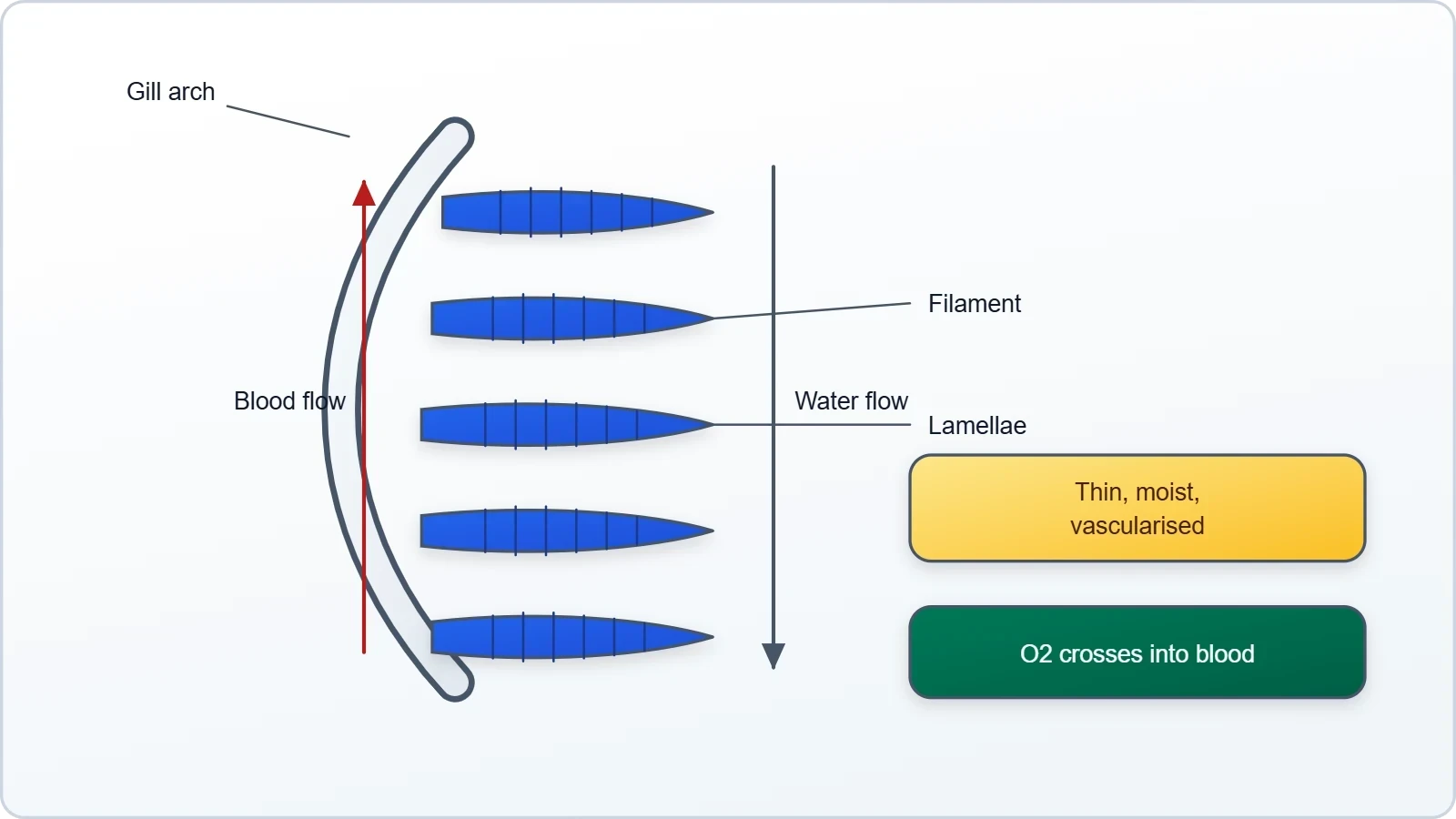
The fish gill exemplifies the general principle that a respiratory surface must be thin, moist and vascularised. Because the gill is bathed in water, it remains naturally moist, and its extensive vascularisation ensures that blood is brought into close proximity with the external medium. The continuous flow of water across the gills, maintained by the fish's ventilatory movements, keeps the oxygen gradient favourable so that diffusion can proceed without interruption.
Pulmonary and Cutaneous Respiration on Land
Vascularised bags called lungs are used by the terrestrial forms for the exchange of gases, a mode of exchange known as pulmonary respiration. Among vertebrates, amphibians, reptiles, birds and mammals respire through lungs, while amphibians like frogs can respire through their moist skin as well, a mode termed cutaneous respiration. The move onto land brought a new respiratory medium, air, and with it new opportunities and new hazards for gas exchange.
Air is far richer in oxygen than water and far less viscous, so terrestrial animals can meet their oxygen needs with smaller ventilatory volumes. Yet air is also drying, and a respiratory surface exposed to it risks desiccation. This is why lungs are internalised, protected within the body, and why their surfaces are kept moist by secretions. The internalised lung represents the terrestrial answer to the same requirement that governs every respiratory organ: a thin, moist, vascularised surface in contact with the medium.
Lungs Across Terrestrial Vertebrates
Amphibians, reptiles, birds and mammals all respire through lungs, though the structure and efficiency of these lungs vary considerably across the groups. The lung is a vascularised bag, an internalised chamber whose walls are richly supplied with blood vessels and whose interior surface is kept moist. Air drawn into the lung contacts this surface, and oxygen diffuses into the blood while carbon dioxide diffuses out, to be expelled with the next breath.
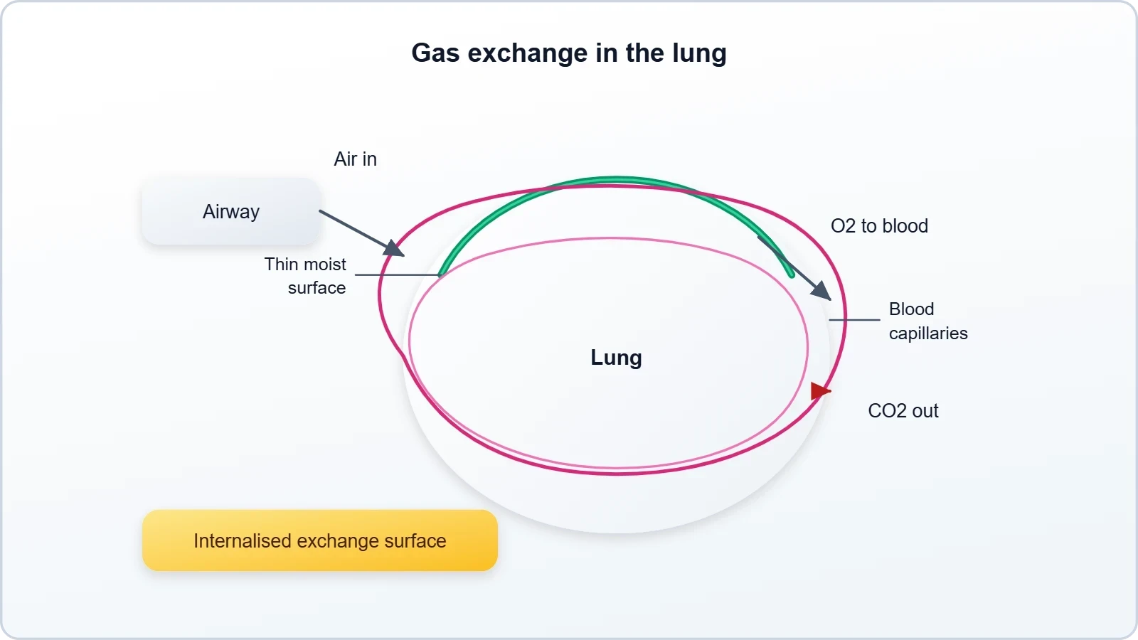
The adoption of pulmonary respiration by all terrestrial vertebrate groups marks a decisive shift in respiratory strategy. The exchange surface is no longer the exterior of the body but an internal chamber connected to the atmosphere by an airway. This internalisation protects the delicate exchange membrane from mechanical damage and from drying, while the vascularised lung wall ensures that the gases are carried efficiently between the exchange surface and the tissues.
Cutaneous Respiration in Frogs
Amphibians like frogs can respire through their moist skin, a mode of exchange called cutaneous respiration. This supplementary route complements pulmonary respiration and is possible because the frog's skin is thin, moist and well supplied with blood vessels. Oxygen from the air or from water dissolves in the skin's moisture and diffuses into the blood, while carbon dioxide diffuses outward, allowing the frog to exchange gases even when its lungs are not actively ventilated.
Cutaneous respiration is a striking reminder that respiratory strategies can coexist within a single animal. The frog possesses lungs for pulmonary respiration yet retains the ancestral capacity for exchange across the body surface. This dual arrangement reflects the amphibian's position at the boundary between aquatic and terrestrial life, and it demonstrates that the respiratory organ an animal uses is not an either-or choice but a reflection of its habitat and level of organisation.
Comparing Respiratory Strategies Across Animal Groups
The respiratory strategies surveyed in this lesson form a coherent set of solutions to a single problem, and comparing them side by side clarifies the logic that connects them. Each strategy reflects the animal's habitat, whether water or air, and its level of organisation, whether a thin-bodied invertebrate or a complex vertebrate. The comparison also reveals the recurring requirements that any respiratory surface must satisfy, regardless of the animal that possesses it.
Reading across the groups, a clear pattern emerges. Simple diffusion and the moist cuticle serve animals whose bodies are thin and whose exchange surface is the body itself. The tracheal system internalises the medium and delivers it directly to the tissues. Gills and lungs concentrate the exchange surface into vascularised organs, one suited to water and the other to air. Cutaneous respiration persists alongside lungs in amphibians as a supplementary route. Together these strategies span the full range of animal organisation.
Matching Organs to Habitats and Organisation
The match between respiratory organ and habitat is direct and instructive. Aquatic animals, whether arthropods, molluscs or fishes, use gills because water is their medium and gills are adapted to extract oxygen from it. Terrestrial animals use lungs because air is their medium and lungs are adapted to exchange gases with it while protecting the surface from drying. The tracheal system of insects is a terrestrial solution of a different kind, delivering air directly to the tissues.
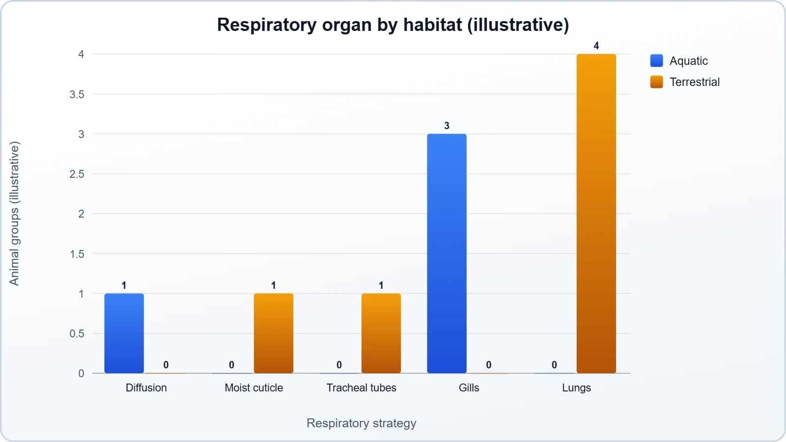
The match between respiratory organ and level of organisation is equally clear. Lower invertebrates with thin bodies need no specialised organ, while larger and more complex animals require dedicated structures. The earthworm's moist cuticle and the frog's cutaneous respiration show that surface exchange can persist alongside more advanced arrangements, but the dominant trend is toward specialised, vascularised organs as organisation increases.
What Every Respiratory Surface Must Provide
Beneath the diversity of gills, lungs, tracheal tubes and moist cuticles lies a common set of requirements. Every respiratory surface must be thin, so that gases can diffuse across it over a short distance. It must be moist, so that gases can dissolve and cross the membrane. And in animals with a circulatory system, it must be vascularised, so that blood can carry gases between the exchange surface and the tissues. These requirements recur in every group examined.
Recognising these shared requirements prepares the learner for the study of the human respiratory system, where the same principles are embodied in the airways and lungs. The human lung is a vascularised bag, its surface thin and moist, its interior connected to the atmosphere by a conducting airway. The lessons learned from sponges, earthworms, insects, fishes and frogs thus illuminate the human case, showing that our own respiratory organ is one expression of a universal biological strategy.
Going Further
Beyond the material itself, two background points help consolidate understanding. First, a respiratory surface must be thin and moist so that gases can diffuse across it. Thinness keeps the diffusion distance short, and moisture allows the gases to dissolve and cross the membrane. These two properties explain why gills remain wet in water, why lungs are internalised and lined with moisture, and why the earthworm's cuticle must stay damp for exchange to continue.
Second, gills are vascularised structures, meaning they contain many blood vessels close to the surface. This vascularisation is what allows a gill to function as a true respiratory organ rather than a mere flap of tissue: oxygen that crosses the thin gill membrane enters the blood immediately and is carried away, while carbon dioxide arrives in the blood and diffuses out. The same principle of close association between exchange surface and blood vessels underlies the vascularised lungs of terrestrial vertebrates.
These background points do not alter the lesson's core content but illuminate why the structures described take the forms they do. They also anticipate the detailed treatment of gas exchange and transport that follows in later lessons, where the movement of oxygen and carbon dioxide across respiratory surfaces and through the blood is examined in quantitative terms. For now, the essential takeaway is that every respiratory organ, from moist cuticle to lung, is a variation on the theme of a thin, moist, vascularised surface.
Also Read
- No links available.
From our network :
- Mastering DB2 12.1 Instance Design: A Technical Deep Dive into Modern Database ArchitectureComprehensive guide to DB2 12.1 Instance Design. Explore architectural components, HADR setup, buffer pool tuning, and security policies for IBM DB2 12.1. Includes 10 functional SQL and Shell samples.
- EV 2.0: The Solid-State Battery Breakthrough and Global Factory ExpansionDiscover the impact of the Solid-State Battery Breakthrough on the EV market. Learn about CATL and BYD stock growth, global factory expansion in Europe, and the future of EV 2.0 technology.
- Vite 6/7 'Cold Start' Regression in Massive Module GraphsIn-depth analysis of Vite 6/7 cold start regressions in massive module graphs. Learn about barrel file issues, pre-bundling optimizations, and how to improve Vite dev server performance.
- Analyzing Trump Deportation Numbers Insights Into The 2026 Immigration Crackdown
- Trump Political Strategy How Geopolitical Stunts Serve As Media Diversions
- 10 Physics Numerical Problems with Solutions for IIT JEEPrepare for IIT JEE with 10 physics numerical problems, complete with detailed solutions and conceptual explanations. Ideal for mastering kinematics, thermodynamics, electromagnetism, and optics.
- AI-Powered 'Precision Diagnostic' Replaces Standard GRE Score ReportsDiscover the new GRE Precision Diagnostic. Learn how AI-powered behavioral analytics and time-to-solve tracking are replacing standard GRE score reports to empower students.
- Mastering DB2 LUW v12 Tables: A Comprehensive Technical GuideComprehensive guide on DB2 LUW v12 tables. Explore CREATE TABLE syntax, BLU Acceleration, range partitioning, data types, and security features for IBM DB2 LUW v12.
- 98% of Global MBA Programs Now Prefer GRE Over GMAT Focus EditionExplore why 98% of global MBA programs now prefer the GRE over GMAT Focus. Learn about the shorter GRE format, verbal logic importance, and strategic versatility for students.
RESOURCES
- Antimicrobial use Guidelines for Treatment of Respiratory Tract ...pmc.ncbi.nlm.nih.govAccordingly, the International Society for Companion Animal Infectious Diseases convened a Working Group of clinical microbiologists, pharmacologists, and ...
- Respiratory Tract Cancer Incidences across Industry Groups - PubMedpubmed.ncbi.nlm.nih.govOct 25, 2022 ... The number of cases and incidence rates of laryngeal and lung cancers have been increasing globally. Therefore, identifying the occupational ...
- Respiratory Tract Cancer Incidences across Industry Groups - PMCpmc.ncbi.nlm.nih.govMoreover, workers employed in fishing, mining, transportation, construction, animal production, and healthcare industries have a high risk of lung cancer. As an ...
- Monitoring of antimicrobial susceptibility of respiratory ... - PubMedpubmed.ncbi.nlm.nih.govOct 15, 2016 ... Monitoring of antimicrobial susceptibility of respiratory tract pathogens isolated from diseased cattle and pigs across Europe, 2009-2012: ...
- Unlocking the energetic secrets of collective animal movementoeb.harvard.eduFeb 20, 2024 ... ... group movements by animals moving through a fluid could reduce the energy cost of locomotion ... respiratory organs from…
- Prophylactic intranasal administration of a TLR2/6 agonist reduces ...sciencedirect.com... throughout the lower respiratory tract [11]. A topical treatment ... Animals progressively gained weight through the course of the study across all groups ...
- SARS-CoV-2 Omicron variant causes mild pathology in the upper ...nature.comJun 20, 2022 ... ... throughout the respiratory tract. Results. VOC Omicron infection ... respiratory tract compared to those of the other investigated groups.
- Lungs, gills, and tracheas | Zoology | Research Starters - EBSCOebsco.comLungs, gills, and tracheas are specialized respiratory organs that enable gas exchange in various animal groups ... animals through the process of natural ...
- Particle Pollution Exposure | US EPAepa.govApr 8, 2026 ... Phagocytes (i.e., macrophages, monocytes, and polymorphonuclear leukocytes) are present throughout the respiratory tract. Alveolar ...
- Middle East respiratory syndrome coronavirus (MERS-CoV)who.intDec 11, 2025 ... Around 70% of new or emerging diseases are of animal origin, including MERS-CoV which is transmitted between dromedary camels and…
- Hazard Classification Guidance for Manufacturers, Importers, and ...osha.govinclude the hazard statement “corrosive to the respiratory tract” along with the already required ... classification for respiratory tract irritation;. (d) ...
- Highly Pathogenic Avian Influenza A(H5N1) Virus - CDCcdc.gov... organs) from infected birds or other animals, OR ... High virus titers were detected in respiratory organs, with medium titers in other organs…
- Respiratory System organs | CK-12 Foundationflexbooks.ck12.orgNov 1, 2026 ... Understanding the diversity of respiratory organs across animal groups is crucial for grasping how various species survive in markedly different ...
- NORA Sector and Cross-Sector Approach - CDCcdc.govMay 19, 2025 ... The 10 sector groups follow the North American Industry Classification System (NAICS) from the U.S. Census Bureau.
- Appendix A to § 1910.1200 - Health Hazard Criteria (Mandatory)osha.gov... classification for respiratory tract irritation;. (d) There are currently no scientifically validated animal tests that deal specifically with RTI; however ...