The forebrain is the largest and most functionally elaborate division of the human brain, and it is here that sensation is interpreted, movement is initiated, memory is stored, and emotion acquires its distinctive human colour. This seventh lesson in the neural control and coordination series examines the three structures that together constitute the forebrain: the cerebrum, the thalamus and the hypothalamus. Each performs a distinct office, yet each depends on the others, and the coordination between them is what allows a person to recognise a familiar face, recall a name, and feel the warmth of recognition in the same instant.
Building directly on the previous lesson, which established the brain as a whole and described the protective arrangements of meninges, cerebrospinal fluid and the skull, this lesson moves inward to the forebrain itself. The cerebrum is treated first, with its longitudinal cleft, its two hemispheres, the corpus callosum that bridges them, and the folded grey cortex that carries motor, sensory and association areas. The thalamus and hypothalamus follow, and the lesson closes with the limbic system, the deep complex of structures that links the inner cerebral hemispheres with the hypothalamus in the service of emotion, motivation and sexual behaviour.
Learners should read this lesson with an eye to structure and function together. The grey matter of the cortex is grey because neuron cell bodies are concentrated there; the white matter is white because myelinated fibres run beneath it. The thalamus is a coordinating centre for sensory and motor signalling, while the hypothalamus holds centres for temperature, hunger and thirst and secretes hypothalamic hormones. These are not isolated facts but a connected account of how the forebrain organises the body's most complex behaviours, and the material that follows preserves that order of ideas faithfully.
On This Page
- The Cerebrum: Hemispheres, Corpus Callosum and the Folded Cortex
- Functional Areas of the Cerebral Cortex
- The Thalamus as a Coordinating Centre
- The Hypothalamus: Centres, Hormones and Homeostasis
- The Limbic System: Emotion, Motivation and Sexual Behaviour
- Integrating the Forebrain and Preparing for the Midbrain
The Cerebrum: Hemispheres, Corpus Callosum and the Folded Cortex
The cerebrum forms the major part of the human brain, and its sheer bulk is the first fact any learner should register. A deep cleft divides it longitudinally into two halves, termed the left and right cerebral hemispheres. These hemispheres are not independent organs but partners, connected by a tract of nerve fibres called the corpus callosum. The surface layer of cells covering each hemisphere is the cerebral cortex, and it is thrown into prominent folds, a design that greatly increases the surface area available for neurons within the confined volume of the skull.
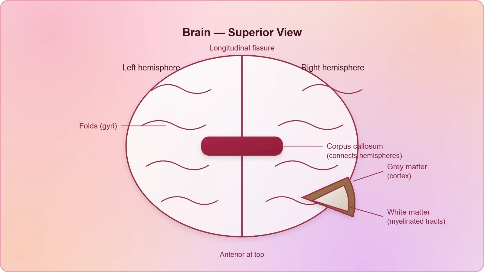
The Cerebral Cortex as Grey Matter
The cerebral cortex is referred to as grey matter because of its greyish appearance, and the reason for that colour is precise: neuron cell bodies are concentrated here. This concentration of cell bodies at the surface, rather than deep within the brain, is a defining architectural feature of the cerebral hemispheres. The folds of the cortex are not decorative; they permit a far greater number of neurons to occupy the cortical sheet than a smooth surface would allow, and the functional richness of human cognition is bound up with this arrangement.
Within the cortex, three broad categories of territory are recognised. There are motor areas, concerned with the initiation of movement; sensory areas, concerned with the reception and interpretation of incoming signals; and large regions that are neither clearly sensory nor motor in function. These last regions, the association areas, are responsible for complex functions such as intersensory associations, memory and communication. The association areas are, in a real sense, where the human mind does its most characteristic work, joining sight to sound, and both to meaning.
White Matter Beneath the Cortex
Beneath the grey cortical layer lie the fibres of the tracts, and these fibres are covered with the myelin sheath. This myelinated inner part of the cerebral hemisphere gives an opaque white appearance to the layer and is therefore called the white matter. The distinction between grey and white matter is thus a distinction between cell bodies gathered at the surface and myelinated conducting fibres running beneath them, and the two together constitute the substance of each cerebral hemisphere.
The functional logic of this arrangement deserves emphasis. Grey matter at the cortex is where signals are integrated and decisions are made; white matter beneath is where signals travel, insulated by myelin so that conduction is rapid and reliable. The corpus callosum, itself a tract of nerve fibres, is the great commissure through which the two hemispheres exchange information, ensuring that the left and right halves of the cerebrum act as a single coordinated organ rather than two separate ones.
Functional Areas of the Cerebral Cortex
The cerebral cortex is not a uniform sheet but a mosaic of functionally distinct territories. The material identifies three broad categories: motor areas, sensory areas, and the association areas that are neither clearly sensory nor motor in function. This tripartite division is the essential framework for understanding cortical function, and it explains how the cerebrum can simultaneously command movement, receive sensation, and perform the higher integrative work of memory and communication. Each category occupies its own regions of the cortical surface, and their coordinated activity underlies behaviour.
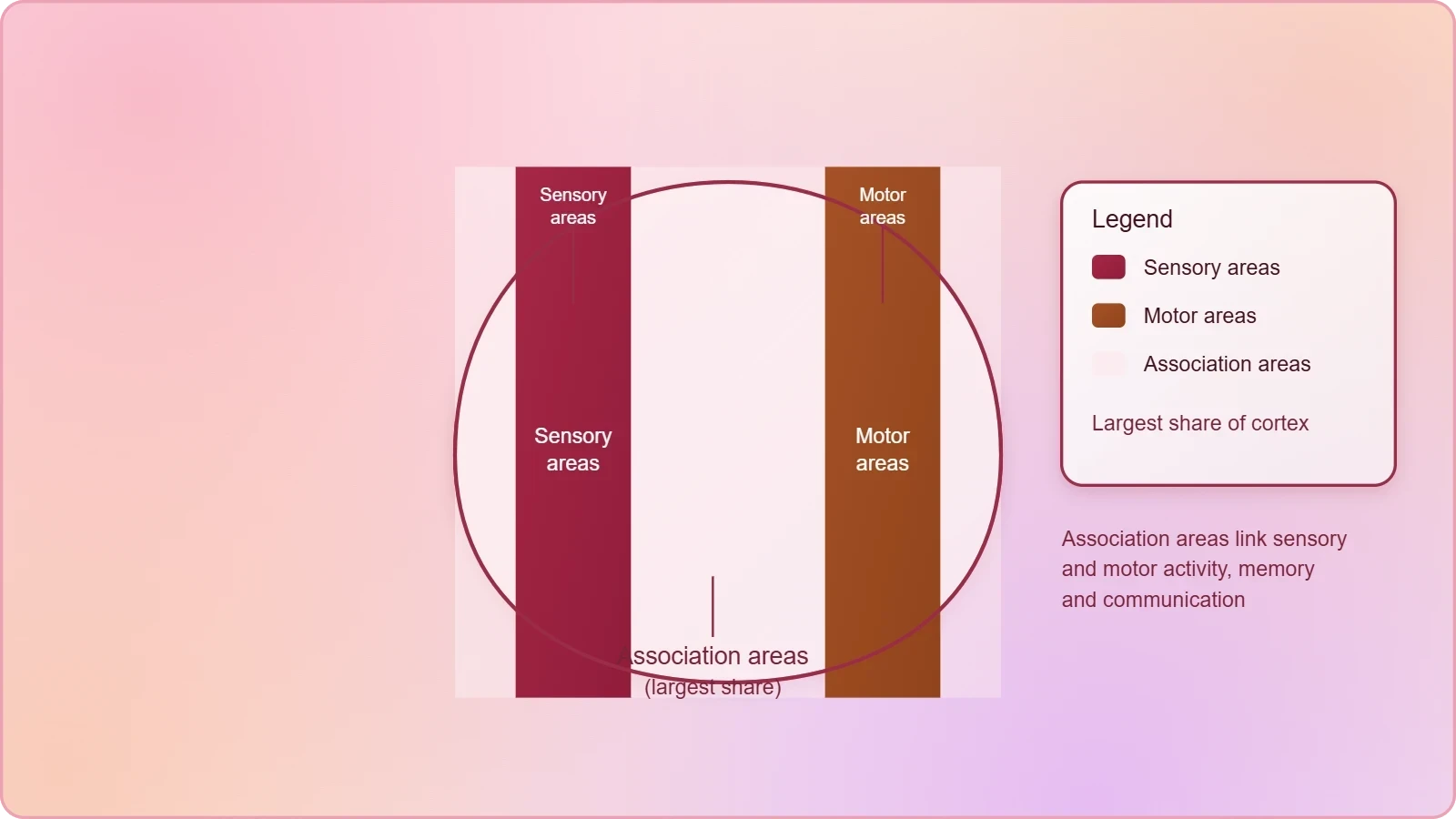
Motor and Sensory Areas
Motor areas are the cortical territories concerned with the initiation and control of movement, issuing the commands that ultimately reach the muscles through descending pathways. Sensory areas receive and interpret the signals arriving from the sense organs and from the body surface, translating raw neural traffic into the perceived qualities of touch, sight, sound and the rest. Together these two categories form the input and output faces of the cortex, the points at which the brain meets the world and acts upon it.
The separation of motor from sensory territory is a fundamental principle of cortical organisation, and it reflects the division of labour within the nervous system as a whole. Sensory signals ascend to the cortex, are interpreted there, and motor commands descend from it. Between the arrival of sensation and the departure of movement lies the vast intermediate territory of the association areas, where the raw material of experience is processed into perception, memory and considered response.
Association Areas and Complex Function
The association areas are large regions of the cortex that are neither clearly sensory nor motor in function, and they are responsible for complex functions such as intersensory associations, memory and communication. Intersensory association means the linking of information from different senses into a unified experience; memory means the retention and recall of that experience; communication means its expression to others. These are precisely the functions that distinguish human mental life, and they are localised not in the primary sensory or motor zones but in the association cortex that surrounds them.
It is worth pausing on the significance of this arrangement. A brain equipped only with sensory and motor areas could receive stimuli and produce movements, but it could not bind sight to sound, hold an image in memory, or convey an idea to another person. The association areas supply exactly these capacities. Their large extent in the human cortex is one of the anatomical facts most often linked to the richness of human cognition, and the material rightly gives them prominence alongside the primary areas.
The Thalamus as a Coordinating Centre
The cerebrum wraps around a structure called the thalamus, which is a major coordinating centre for sensory and motor signalling. This single sentence from the material carries a great deal of functional weight. The thalamus is not a sensory organ itself, nor a motor organ, but a coordinating centre: a place where sensory and motor signals are brought together and organised. Its position, enveloped by the cerebral hemispheres, is anatomically apt for a structure whose business is the integration of traffic passing between the body and the cortex.
Position and Relationship to the Cerebrum
The thalamus lies deep within the brain, wrapped around by the cerebrum, and this relationship is central to understanding the forebrain as an integrated whole. The cerebral hemispheres do not sit above the thalamus as a separate lid; they enclose it. The thalamus therefore occupies a strategic central position from which it can coordinate the sensory and motor signalling that passes between the lower parts of the nervous system and the cerebral cortex, acting as a hub through which information is routed and organised.
Learners should note that the material describes the thalamus as a coordinating centre rather than merely a relay. Coordination implies organisation and regulation, not passive transmission. The thalamus is thus a genuine participant in the processing of sensory and motor information, and its intimate anatomical association with the cerebrum reflects the closeness of their functional partnership in the forebrain.
Coordinating Sensory and Motor Signalling
As a major coordinating centre for sensory and motor signalling, the thalamus stands at the junction of the ascending and descending pathways of the forebrain. Sensory information on its way to the cortex and motor information on its way from the cortex both pass through or are organised at this level. The thalamus thus contributes to the smooth integration of sensation and action, ensuring that the cortex receives information in an ordered form and that motor commands are dispatched in coordination with sensory context.
This coordinating role explains why the thalamus is grouped with the cerebrum and hypothalamus as a component of the forebrain rather than being treated as a mere way-station of the brainstem. Its function is integrative, and its position is central. For the learner, the essential point is that the thalamus is a major coordinating centre for sensory and motor signalling, a definition that should be retained precisely as the material states it.
The Hypothalamus: Centres, Hormones and Homeostasis
Another very important part of the brain, the hypothalamus, lies at the base of the thalamus. Despite its modest size, it contains a number of centres that control body temperature and the urge for eating and drinking. It also contains several groups of neurosecretory cells, which secrete hormones called hypothalamic hormones. The hypothalamus is therefore both a neural control centre and an endocrine organ, a dual identity that makes it one of the most consequential small structures in the entire nervous system.
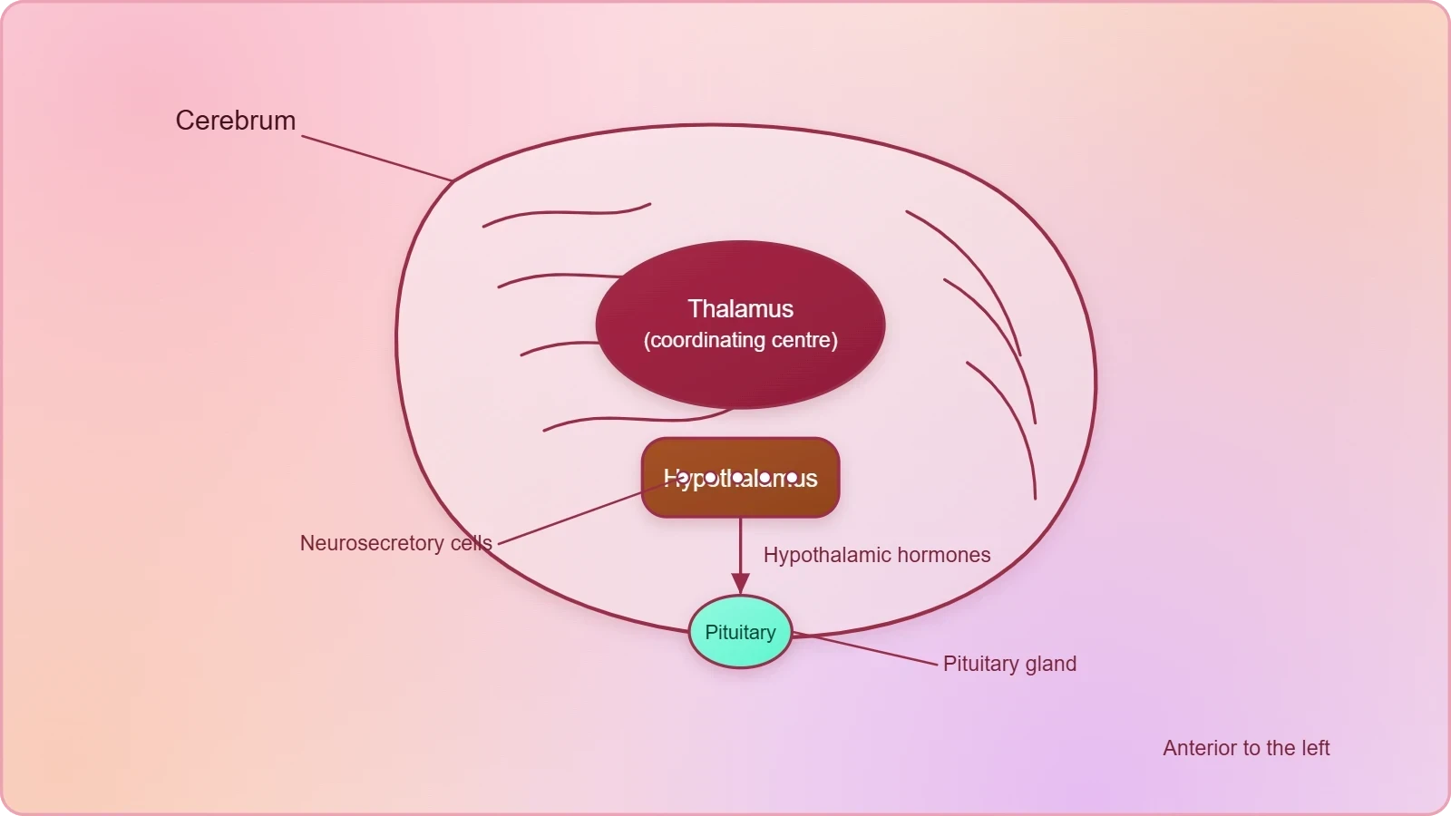
Centres for Temperature, Hunger and Thirst
The centres within the hypothalamus that control body temperature, the urge for eating and the urge for drinking are the neural machinery of homeostasis. Temperature regulation requires the continuous comparison of actual body temperature with an optimal set point and the initiation of corrective responses; hunger and thirst require the monitoring of the body's energy and water status and the generation of the drives that prompt intake. These are not simple reflexes but sustained regulatory activities, and the hypothalamus is their seat.
Because these centres operate continuously and largely below the level of conscious awareness, their importance is easily underestimated. A person does not decide to maintain a constant body temperature or to feel thirsty after water loss; the hypothalamus manages these matters on the body's behalf. The material's phrase, that the hypothalamus contains a number of centres controlling these functions, captures the essential point: the hypothalamus is a collection of specialised regulatory centres, each devoted to a particular aspect of internal balance.
Neurosecretory Cells and Hypothalamic Hormones
The hypothalamus also contains several groups of neurosecretory cells, which secrete hormones called hypothalamic hormones. These cells are remarkable because they combine the properties of neurons and endocrine cells: they receive and conduct neural signals, and they release chemical messengers into the blood. The hypothalamic hormones they secrete are the chemical link between the nervous and endocrine systems, and their existence is one of the clearest demonstrations that the two systems are not separate domains but a single integrated control network.
For the learner, the term neurosecretory cell should be understood literally. It is a cell that secretes, and it is a cell of neural character. The hypothalamus thus participates in chemical signalling as well as neural regulation, and its hormones act as the hypothalamic contribution to the body's endocrine coordination. This dual role, neural and hormonal, is the defining feature of the hypothalamus and the reason it is described as a very important part of the brain.
The Limbic System: Emotion, Motivation and Sexual Behaviour
The inner parts of the cerebral hemispheres, together with a group of associated deep structures such as the amygdala and hippocampus, form a complex structure called the limbic lobe or limbic system. Along with the hypothalamus, it is involved in the regulation of sexual behaviour, the expression of emotional reactions such as excitement, pleasure, rage and fear, and motivation. The limbic system thus supplies the affective dimension of experience, the feelings and drives that colour perception and direct behaviour.
Amygdala, Hippocampus and the Limbic Lobe
The limbic lobe is not a single organ but a complex assembled from the inner parts of the cerebral hemispheres and a group of associated deep structures. The material names the amygdala and hippocampus as examples of these deep structures, and the word complex is well chosen: the limbic system is a network of interconnected regions rather than a discrete anatomical unit. Its components lie deep within the brain, close to the hypothalamus, and their proximity reflects their close functional partnership in the regulation of behaviour.
Because the limbic system draws on the inner parts of the cerebral hemispheres as well as on deep structures, it represents a bridge between the cortex and the more primitive regions of the forebrain. This anatomical position is consistent with its function: the limbic system is where the higher processing of the cortex meets the regulatory machinery of the hypothalamus, and where the results of that meeting are expressed as emotion, motivation and sexual behaviour.
Emotional Expression and Motivation
The limbic system, along with the hypothalamus, is involved in the regulation of sexual behaviour, the expression of emotional reactions and motivation. The emotional reactions named in the material are excitement, pleasure, rage and fear, a list that spans the positive and negative poles of affective life. Motivation, the drive that impels behaviour toward a goal, is likewise a limbic function, and sexual behaviour is regulated at this level as well. These are the functions that give behaviour its direction and its feeling tone.
It is important to note the phrase along with the hypothalamus. The limbic system does not act alone; it acts in concert with the hypothalamus, whose centres for temperature, hunger and thirst provide the regulatory substrate on which emotion and motivation are built. The two structures together form the emotional and motivational core of the forebrain, and their cooperation illustrates once more the principle that the forebrain's components work as an integrated system rather than as isolated parts.
Integrating the Forebrain and Preparing for the Midbrain
The forebrain, then, is a coordinated assembly of three parts. The cerebrum, with its two hemispheres, corpus callosum, folded grey cortex and myelinated white matter, provides the machinery of motor control, sensation, association, memory and communication. The thalamus, wrapped around by the cerebrum, serves as a major coordinating centre for sensory and motor signalling. The hypothalamus, at the base of the thalamus, holds the centres for temperature, hunger and thirst and secretes hypothalamic hormones. The limbic system, with the hypothalamus, governs emotion, motivation and sexual behaviour.
How the Parts Work Together
The integration of these parts is the central lesson of the forebrain. The cortex interprets and decides; the thalamus coordinates the sensory and motor traffic on which those decisions depend; the hypothalamus regulates the internal conditions that make coherent behaviour possible; and the limbic system supplies the emotional and motivational colouring that gives behaviour its purpose. No part is self-sufficient, and the forebrain's remarkable capacities arise precisely from their interconnection, a point that the material's careful ordering of structures is designed to convey.
Learners preparing for examinations should be able to move fluently between structure and function in both directions. Given the cerebrum, they should recall the hemispheres, corpus callosum, cortex, grey and white matter, and the motor, sensory and association areas. Given the thalamus, they should state its coordinating role. Given the hypothalamus, they should list its centres and its neurosecretory cells. Given the limbic system, they should name the amygdala and hippocampus and the behaviours it regulates along with the hypothalamus.
Looking Ahead to the Midbrain
The next lesson takes up the midbrain, located between the thalamus and hypothalamus of the forebrain and the pons of the hindbrain. A canal called the cerebral aqueduct passes through the midbrain, and its dorsal portion consists mainly of four round swellings called the corpora quadrigemina. Having established the forebrain's structures and functions, the learner is now positioned to see how the midbrain continues the neural axis downward toward the hindbrain, and how the brain stem, comprising the midbrain, pons and medulla oblongata, forms the connections between the brain and spinal cord.
This forward glance is not a substitute for the next lesson but a preparation for it. The forebrain sits at the top of the neural axis, the midbrain beneath it, and the hindbrain below that, with the brain stem joining brain to spinal cord. Keeping this vertical arrangement in mind will make the transition natural, and it will reinforce the larger principle of the course: that the nervous system is an integrated hierarchy in which each level performs its own functions while remaining connected to every other.
Going further
The corpus callosum is a bundle of nerve fibres that connects the two cerebral hemispheres, allowing them to exchange information and act in concert. The hypothalamus is involved in maintaining body temperature and regulating hunger and thirst, functions that illustrate its role as a homeostatic centre. These observations extend the material without altering it, and they help the learner appreciate why the forebrain's structures are so often studied together rather than in isolation.
Also Read
- No links available.
From our network :
- Mastering DB2 LUW v12 Tables: A Comprehensive Technical GuideComprehensive guide on DB2 LUW v12 tables. Explore CREATE TABLE syntax, BLU Acceleration, range partitioning, data types, and security features for IBM DB2 LUW v12.
- Mastering DB2 12.1 Instance Design: A Technical Deep Dive into Modern Database ArchitectureComprehensive guide to DB2 12.1 Instance Design. Explore architectural components, HADR setup, buffer pool tuning, and security policies for IBM DB2 12.1. Includes 10 functional SQL and Shell samples.
- 10 Physics Numerical Problems with Solutions for IIT JEEPrepare for IIT JEE with 10 physics numerical problems, complete with detailed solutions and conceptual explanations. Ideal for mastering kinematics, thermodynamics, electromagnetism, and optics.
- 98% of Global MBA Programs Now Prefer GRE Over GMAT Focus EditionExplore why 98% of global MBA programs now prefer the GRE over GMAT Focus. Learn about the shorter GRE format, verbal logic importance, and strategic versatility for students.
- Analyzing Trump Deportation Numbers Insights Into The 2026 Immigration Crackdown
- Vite 6/7 'Cold Start' Regression in Massive Module GraphsIn-depth analysis of Vite 6/7 cold start regressions in massive module graphs. Learn about barrel file issues, pre-bundling optimizations, and how to improve Vite dev server performance.
- AI-Powered 'Precision Diagnostic' Replaces Standard GRE Score ReportsDiscover the new GRE Precision Diagnostic. Learn how AI-powered behavioral analytics and time-to-solve tracking are replacing standard GRE score reports to empower students.
- EV 2.0: The Solid-State Battery Breakthrough and Global Factory ExpansionDiscover the impact of the Solid-State Battery Breakthrough on the EV market. Learn about CATL and BYD stock growth, global factory expansion in Europe, and the future of EV 2.0 technology.
- Trump Political Strategy How Geopolitical Stunts Serve As Media Diversions
RESOURCES
- Limbic System: What It Is, Function, Parts & Location - Cleveland Clinicmy.clevelandclinic.orgApr 6, 2024 ... The limbic system is your emotional nervous system. This group of brain structures regulates your emotions, memory and behaviors.
- The forebrain - Queensland Brain Instituteqbi.uq.edu.au... cerebrum and several structures directly nestled within it - the thalamus, hypothalamus, the pineal gland and the limbic system. Cerebrum and the cerebral ...
- Neuroanatomy, Limbic System - StatPearls - NCBI Bookshelfncbi.nlm.nih.govThe limbic system is an aggregation of brain structures that are generally located lateral to the thalamus, underneath the cerebral cortex, and above the ...
- The limbic system - Queensland Brain Instituteqbi.uq.edu.auThe thalamus, hypothalamus (production of important hormones and regulation of thirst, hunger, mood etc) and basal ganglia (reward processing, habit formation, ...
- Limbic system - Wikipediaen.wikipedia.org... cerebrum primarily in the forebrain. Limbic system. Cross section of the ... The basal ganglia are located near the thalamus and hypothalamus. They ...
- An Update to the Original Papez Circuit of the Human Limbic Systempmc.ncbi.nlm.nih.govApr 26, 2023 ... These include both reciprocal and non-reciprocal connections between the thalamus and cerebral cortex. ... forebrain, hypothalamus, and thalamus ( ...
- Altered functional connectivity between hypothalamus and limbic ...pubmed.ncbi.nlm.nih.govAltered functional connectivity between hypothalamus and limbic system in fibromyalgia. Mol Brain. 2021 Jan 20;14(1):17. doi: 10.1186/s13041-020-00705-2.
- 3D Brain - Brain Factsbrainfacts.orgBrain Stem, Midbrain, Pons, Medulla Oblongata, Limbic System, Entorhinal Cortex, Amygdala, Hippocampus, Subiculum, Dentate Gyrus, Hypothalamus, Thalamus ...
- Brain structures selectively targeted by canine distemper virus in a ...pubmed.ncbi.nlm.nih.govViral mRNA and proteins were selectively localized in certain brain structures such as the thalamus ... limbic system (hippocampus, septum, entorhinal and ...
- The anterior and medial thalamic nuclei and the human limbic systempmc.ncbi.nlm.nih.govJul 2, 2020 ... Afferents arising predominantly in limbic brain regions are directly or indirectly innervated by the lateral hypothalamic and lateral preoptic ...
- How your brain works - Mayo Clinicmayoclinic.orgJul 2, 2024 ... Known as the limbic system, these structures come in pairs. Each part of this system is present in both halves…
- The Limbic System and Other Brain Areas | Introduction to Psychologycourses.lumenlearning.com... cerebral cortex, including the thalamus and the limbic system. The thalamus ... hypothalamus: forebrain structure that regulates sexual motivation and ...
- Diencephalon - Knowledge @ AMBOSSamboss.comApr 24, 2024 ... It consists of the thalamus, hypothalamus, epithalamus, and subthalamus. ... Connects limbic system to other parts of the brain; Controls…
- An overview of the brain - About Kids Healthaboutkidshealth.caJan 25, 2022 ... Other important areas of the brain include the thalamus, hypothalamus, pituitary gland, basal ganglia, limbic system, ventricles, cerebrospinal ...
- Anatomy of the Brain Open pdf - Mayfield Brain & Spinemayfieldclinic.comLimbic system: is the center of our emotions, learning, and memory. Included in this system are the cingulate gyri, hypothalamus, amygdala (emotional ...