Every breath we take depends on a barrier so thin that it almost defies belief, a microscopic frontier where the air inside our lungs meets the blood that will carry its oxygen to every living cell. This lesson examines that frontier in detail: the diffusion membrane, the three-layered structure across which oxygen and carbon dioxide must pass during external respiration. Building directly on the previous discussion of partial pressures and diffusion gradients, we now turn to the physical architecture that makes those gradients useful, asking what the membrane is made of, how thick it is, and why its construction is so remarkably suited to the task of gas exchange.
The diffusion membrane is not a single sheet but a composite of three major layers working as one functional unit. Understanding this structure clarifies why the body can move oxygen from alveoli to tissues and carbon dioxide from tissues to alveoli with such efficiency, and why the solubility of carbon dioxide relative to oxygen matters so much at this interface. The lesson stays close to the established terminology and sequence of ideas, treating the membrane as the anatomical answer to the physiological question raised earlier: how does a gradient translate into actual transfer of gas?
By the end of this lesson, you should be able to name each layer of the diffusion membrane, describe the basement substance that binds them, state the order of magnitude of its total thickness, and explain why every factor in the body is favourable for diffusion in both directions. This prepares the ground for the next lesson, where oxygen transport and the oxygen dissociation curve take centre stage.
On This Page
The Three Layers of the Diffusion Membrane
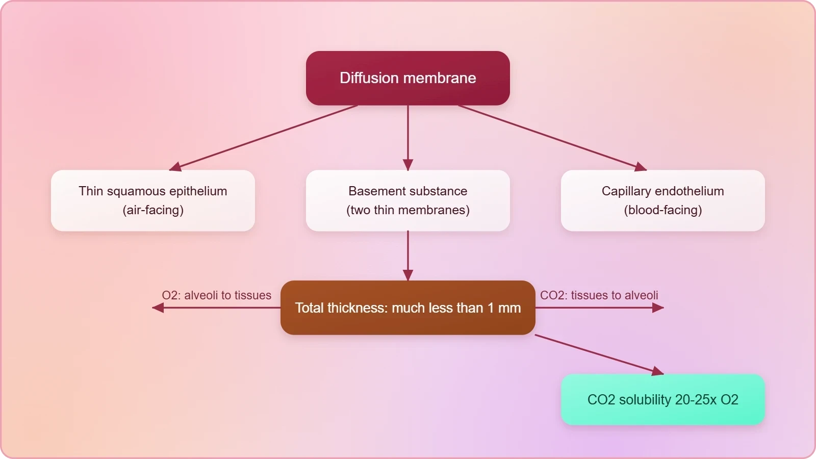
The diffusion membrane is defined by three major layers arranged in a precise sequence. First comes the thin squamous epithelium of the alveoli, the air-facing surface. Second is the endothelium of the alveolar capillaries, the blood-facing surface. Third, sandwiched between them, is the basement substance. This three-part arrangement is the structural core of the lesson, and each layer contributes to a barrier that is simultaneously strong enough to separate two very different environments and thin enough to permit rapid molecular traffic between them.

The Thin Squamous Epithelium of the Alveoli
The alveolar side of the membrane is formed by a thin squamous epithelium. Squamous cells are flattened, and here they form a delicate lining across which oxygen from the alveolar air begins its journey into the body. Because this epithelium is thin, it presents minimal resistance to diffusing molecules. The alveoli themselves are the terminal air sacs of the respiratory tract, and their epithelial lining is the first of the three layers that a gas molecule encounters when leaving the air space and entering the tissues of the lung.
This layer is not merely a passive wrapper. It defines the boundary between the external environment of the alveolar air and the internal environment of the body, and its thinness is a direct structural adaptation for diffusion. When we later consider respiratory disorders, the integrity of this epithelium becomes clinically important, but for now the essential point is simple: the air-facing layer is thin, flat, and continuous, exactly as a diffusion surface should be.
The Endothelium of Alveolar Capillaries
The blood-facing layer is the endothelium of the alveolar capillaries. Capillaries are the smallest blood vessels, and their walls are formed of endothelial cells. In the lung, these capillaries run in intimate contact with the alveoli, so that the endothelial layer lies immediately adjacent to the alveolar epithelium, separated only by the basement substance. A gas molecule leaving the alveolar air must therefore cross the epithelium, the basement substance, and the endothelium before it can enter the blood plasma and reach the red blood cells.
The endothelium is described as a single layer of endothelial cells, and this single-layer construction is central to the membrane's function. A multi-layered vessel wall would add distance and impede diffusion; a single layer keeps the barrier minimal. The capillary endothelium thus mirrors the alveolar epithelium in its thinness, and the two thin layers face each other across the basement substance, forming a paired boundary that gases cross in either direction depending on the prevailing gradient.
The Basement Substance Between the Layers
Between the squamous epithelium and the capillary endothelium lies the basement substance. It is composed of a thin basement membrane supporting the squamous epithelium and the basement membrane surrounding the single layer of endothelial cells of the capillaries. This middle layer is easy to overlook because it is not the surface that touches air or blood, yet it is essential: it provides structural support to both cellular layers and holds them in their correct relationship, ensuring that the diffusion pathway remains organised and continuous.
Composition and Supporting Role
The basement substance is best understood as two basement membranes brought together. One supports the squamous epithelium of the alveolus; the other surrounds the endothelial cells of the capillary. Together they form the connective middle of the diffusion membrane. This arrangement gives mechanical coherence to a barrier that must remain intact through millions of breathing cycles, while adding only a minimal amount to the total distance a gas molecule must travel. Support and thinness are achieved at the same time, which is precisely what an efficient diffusion surface requires.
Because the basement substance is thin, it does not become a bottleneck. The two basement membranes are described as thin, and their combined contribution to the overall thickness is small. The middle layer therefore performs a structural duty without compromising the functional purpose of the membrane. In the language of the lesson, it is the layer "in between them," the connective tissue that completes the three-part definition and makes the diffusion membrane a single coherent structure rather than two separate surfaces.
Why the Middle Layer Matters for Diffusion
Diffusion depends on distance, and every layer adds to the path a molecule must travel. The basement substance matters because it is deliberately thin, keeping the total pathway short. If this middle layer were thick or fibrous, it would slow the movement of oxygen and carbon dioxide regardless of how favourable the partial pressure gradients were. Its thinness is therefore not incidental but part of the design: the membrane is built so that support and permeability coexist, and the middle layer is the clearest illustration of that balance.
There is also an organisational logic here. The basement substance keeps the epithelium and endothelium aligned, so that the air space and the capillary lumen remain separated by a consistent, minimal distance along the exchange surface. This regularity means that gas exchange is not haphazard but uniform across the alveoli. The middle layer, though invisible in casual description, is what allows the two thin cellular layers to function as a coordinated diffusion barrier rather than as unrelated sheets.
Total Thickness and the Geometry of Exchange
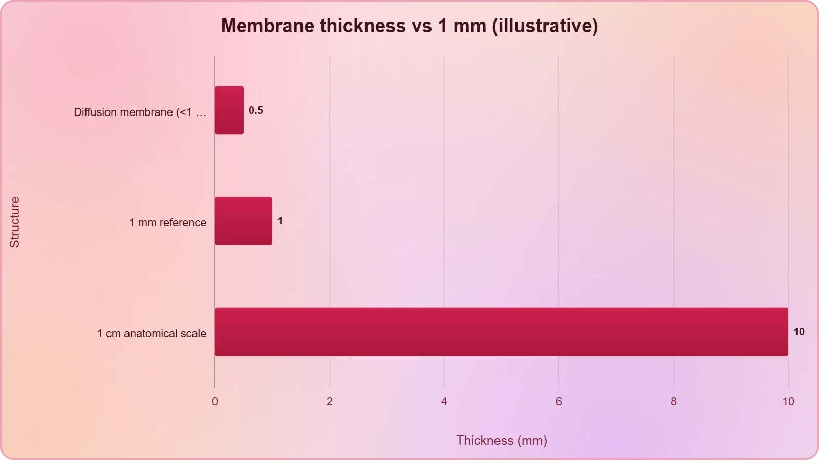
The defining quantitative fact about the diffusion membrane is that its total thickness is much less than a millimetre. This single statement carries the lesson's central argument. Diffusion is a distance-dependent process, so a barrier measured in fractions of a millimetre rather than in millimetres or centimetres is exceptionally well suited to rapid gas exchange. The three layers, each thin in itself, combine into a composite that remains extraordinarily shallow, and it is this shallow geometry that allows the partial pressure gradients established in the previous lesson to produce meaningful transfer of gases.
Reading the Order of Magnitude
The phrase "much less than a millimetre" is deliberately an order-of-magnitude statement rather than a precise measurement. It tells us that the barrier is small on the scale of human anatomy, where structures are often described in centimetres. For comparison, a millimetre is roughly the thickness of a fingernail; the diffusion membrane is far thinner than that. The lesson does not require a numerical value, only the recognition that the membrane is thin enough that distance does not limit diffusion under normal conditions.
This order-of-magnitude framing is useful because it directs attention to the functional consequence rather than to a single number. What matters is that the membrane is thin relative to the gradients available. When the barrier is this shallow, the rate of diffusion is governed chiefly by the partial pressure differences and by the solubility of the gases, not by the distance to be crossed. The geometry of the membrane therefore removes distance as a limiting factor and leaves the gradients to do their work.
Why Thinness Favours Both Gases
A thin membrane favours diffusion in both directions simultaneously. Oxygen moves from alveoli to tissues, and carbon dioxide moves from tissues to alveoli, and both journeys cross the same three-layered barrier. Because the barrier is thin, neither gas is disadvantaged by distance. The membrane does not need to be selective in the sense of favouring one direction; it simply needs to be short enough that both gradients can be expressed as actual molecular traffic. Thinness is thus a shared enabler for the two opposing flows.
This dual function is worth emphasising because it explains why the body's arrangement is described as favourable for both processes at once. The same structure that admits oxygen admits carbon dioxide, and the direction each gas takes is set by its own partial pressure gradient. The membrane's contribution is to keep the path short for both. In this sense the diffusion membrane is a neutral, efficient corridor rather than a gate that discriminates between the two gases.
Solubility of Carbon Dioxide and Its Consequence
The lesson draws an important comparison between the two gases. A gradient is present for carbon dioxide in the opposite direction to oxygen, that is, from tissues to blood and from blood to alveoli. Crucially, the solubility of carbon dioxide is 20-25 times higher than that of oxygen. As a result, the amount of carbon dioxide that can diffuse through the diffusion membrane per unit difference in partial pressure is much higher compared with that of oxygen. This asymmetry is a direct consequence of solubility and shapes how the two gases behave at the same membrane.

The 20-25 Fold Solubility Difference
The figure of 20-25 times is the quantitative heart of this comparison. For the same partial pressure difference, far more carbon dioxide can cross the membrane than oxygen, because carbon dioxide dissolves more readily. This does not mean carbon dioxide is more important; it means the membrane is more permeable to it per unit of driving pressure. The lesson uses this fact to explain why carbon dioxide exchange is so effective despite the relatively modest gradients that drive it, and it links solubility directly to the amount that can diffuse.
It is worth being precise about what the comparison claims. The statement is about the amount diffusing per unit difference in partial pressure, not about total quantities transported in blood. Solubility governs how readily a gas crosses the membrane for a given gradient, and on this measure carbon dioxide has a large advantage. The 20-25 fold figure should therefore be remembered as a permeability comparison at the diffusion membrane, a point that will matter again when transport in blood is discussed in later lessons.
Interpreting "Per Unit Difference in Partial Pressure"
The phrase "per unit difference in partial pressure" is a way of standardising the comparison. It asks: if oxygen and carbon dioxide each faced the same partial pressure difference across the membrane, how much of each would diffuse? Because carbon dioxide is 20-25 times more soluble, much more of it would cross. This standardisation isolates solubility as the variable and shows that the membrane's behaviour differs between the two gases even when the driving force is held equal.
Understanding this phrase prevents a common confusion. A larger amount of carbon dioxide diffusing per unit gradient does not imply that carbon dioxide gradients are larger; in fact, the gradients for the two gases differ in direction and magnitude. The point is comparative permeability. The lesson's wording is careful, and preserving that care helps you avoid overstating the claim. Solubility explains the per-unit advantage; the actual traffic depends on the gradients that exist in the body at any moment.
Why Every Factor in the Body Is Favourable
The lesson's conclusion is emphatic: because the membrane is much less than a millimetre thick, all the factors in our body are favourable for diffusion of oxygen from alveoli to tissues and of carbon dioxide from tissues to alveoli. This is the synthesis of the lesson. The three-layer structure provides a thin, continuous barrier; the gradients provide the driving force; and the solubility of carbon dioxide ensures that its transfer is efficient per unit of pressure difference. Together these conditions mean that gas exchange at the membrane is not constrained by the body's own design.
Bringing Structure and Gradient Together
Structure and gradient must be considered together. A steep gradient across a thick barrier would be wasted, and a thin barrier without a gradient would move nothing. The diffusion membrane is favourable precisely because it pairs a minimal thickness with the gradients established at the alveoli and tissues. The previous lesson supplied the gradients; this lesson supplies the structure that lets them act. The two lessons are complementary, and the phrase "all the factors are favourable" is the point at which they meet.
This meeting point also explains the direction of each flow. Oxygen moves from alveoli to tissues because its gradient points that way, and carbon dioxide moves from tissues to alveoli because its gradient points the opposite way. The membrane does not choose between them; it simply offers a short path to both. Favourability, in this context, means that neither gas faces a structural obstacle, so each follows its own gradient freely across the same three layers.
Preparing for Oxygen Transport
Having established that the membrane permits efficient diffusion, the next question is what happens to the gases once they have crossed. Oxygen enters the blood and must be carried to the tissues; carbon dioxide enters the blood from the tissues and must be carried to the alveoli. The next lesson takes up oxygen transport and the oxygen dissociation curve, where haemoglobin, partial pressures, and the sigmoid relationship between saturation and pO2 become central. The diffusion membrane is the gateway; transport is the road beyond it.
Keeping this sequence in mind helps you see why the membrane lesson comes where it does. Before asking how oxygen is carried, we must confirm that it can enter the blood at all, and under what structural conditions. The answer is that it enters readily, across a barrier much less than a millimetre thick, with every factor favourable. That conclusion licenses the study of transport that follows, and it also sets up the later discussion of respiratory disorders, where damage to this delicate barrier becomes significant.
Going Further
The core material establishes the three layers and the favourable thickness. A few well-established points of background can deepen the picture without altering the lesson's claims. These are offered separately so that the material itself remains clearly distinguished from supplementary context. They concern the relationship between membrane thickness and the speed of diffusion, and the nature of squamous epithelium as a single layer of flat cells. Both points reinforce, rather than revise, the lesson's central conclusion that the diffusion membrane is built for efficient gas exchange.
Thinner Membranes and Faster Diffusion
A thinner diffusion membrane allows gases to cross more quickly. This follows from the general behaviour of diffusion, in which the rate of transfer increases as the distance to be crossed decreases. The lesson's statement that the membrane is much less than a millimetre thick can therefore be read as a statement about speed as well as about structure: the shorter the path, the less time a molecule spends in transit. This background point is consistent with the material and helps explain why thickness is treated as such an important feature.
It is important to keep this as background rather than as a new claim about the body. The lesson does not quantify diffusion rates; it simply notes that the membrane's thinness makes conditions favourable. The general principle that thinner barriers permit faster diffusion supports that conclusion and gives you a way to reason about what would happen if the barrier thickened. It also anticipates why disorders affecting the membrane can impair exchange, a theme for later lessons.
The Squamous Epithelium as a Single Layer of Flat Cells
The squamous epithelium is a single layer of flat cells, which keeps the barrier thin. "Squamous" describes flattened cells, and a single layer of them forms a surface that is broad in extent but minimal in depth. This is precisely the geometry a diffusion surface requires: large area for exchange and short distance across. Recognising the epithelium as a single layer of flat cells makes the lesson's description of it as "thin" concrete rather than abstract, and it explains why this layer contributes so little to the total thickness.
This background detail also connects structure to function in a satisfying way. A single layer of flat cells cannot offer much mechanical bulk, but it does not need to; its role is to be a boundary, not a support structure. The basement substance supplies the support, and the endothelium supplies the blood-facing boundary. Each layer is specialised, and the squamous epithelium's specialisation is thinness. Together with the other two layers, it completes a membrane whose every feature points toward efficient diffusion.
Consolidating the Lesson
The diffusion membrane is a three-layered structure: the thin squamous epithelium of the alveoli, the endothelium of the alveolar capillaries, and the basement substance between them. Its total thickness is much less than a millimetre, and because of this, all the factors in the body are favourable for diffusion of oxygen from alveoli to tissues and carbon dioxide from tissues to alveoli. The solubility of carbon dioxide, 20-25 times higher than that of oxygen, means that more carbon dioxide can diffuse per unit difference in partial pressure. These are the facts to carry forward.
Key Terms Revisited
Review the terminology carefully. The squamous epithelium is the air-facing layer; the endothelium is the blood-facing layer; the basement substance is the middle layer, composed of a thin basement membrane supporting the squamous epithelium and the basement membrane surrounding the single layer of endothelial cells. The diffusion membrane is the composite of all three. The gradients run in opposite directions for the two gases, and solubility determines how much of each crosses per unit of partial pressure difference. Mastery of these terms is the foundation for the lessons that follow.
Notice how the lesson's order of ideas builds a case. It begins with the gradient for carbon dioxide, then introduces solubility, then describes the three layers, then states the total thickness, and finally concludes that all factors are favourable. This sequence is deliberate: each point supports the next, and the conclusion follows from the structure and the properties already established. Reconstructing this order in your own words is an effective way to check your understanding before moving on to transport.
Looking Ahead to Transport
The next lesson turns to the transport of oxygen and the oxygen dissociation curve. There you will meet haemoglobin, the red iron-containing pigment in red blood cells, and the sigmoid curve obtained when percentage saturation of haemoglobin with oxygen is plotted against pO2. The diffusion membrane lesson has shown that oxygen can enter the blood efficiently; the transport lesson will show how that oxygen is carried. Keeping the two lessons distinct but connected will help you see respiration as a continuous process rather than a set of isolated facts.
As you proceed, hold on to the central image of this lesson: a barrier of three thin layers, far less than a millimetre thick, across which two gases move in opposite directions, each driven by its own gradient and each favoured by the body's design. That image is the answer to the question of how external respiration is physically possible, and it is the foundation on which the study of internal transport is built.
Also Read
- No links available.
From our network :
- Trump Political Strategy How Geopolitical Stunts Serve As Media Diversions
- Mastering DB2 LUW v12 Tables: A Comprehensive Technical GuideComprehensive guide on DB2 LUW v12 tables. Explore CREATE TABLE syntax, BLU Acceleration, range partitioning, data types, and security features for IBM DB2 LUW v12.
- Vite 6/7 'Cold Start' Regression in Massive Module GraphsIn-depth analysis of Vite 6/7 cold start regressions in massive module graphs. Learn about barrel file issues, pre-bundling optimizations, and how to improve Vite dev server performance.
- EV 2.0: The Solid-State Battery Breakthrough and Global Factory ExpansionDiscover the impact of the Solid-State Battery Breakthrough on the EV market. Learn about CATL and BYD stock growth, global factory expansion in Europe, and the future of EV 2.0 technology.
- AI-Powered 'Precision Diagnostic' Replaces Standard GRE Score ReportsDiscover the new GRE Precision Diagnostic. Learn how AI-powered behavioral analytics and time-to-solve tracking are replacing standard GRE score reports to empower students.
- Mastering DB2 12.1 Instance Design: A Technical Deep Dive into Modern Database ArchitectureComprehensive guide to DB2 12.1 Instance Design. Explore architectural components, HADR setup, buffer pool tuning, and security policies for IBM DB2 12.1. Includes 10 functional SQL and Shell samples.
- 98% of Global MBA Programs Now Prefer GRE Over GMAT Focus EditionExplore why 98% of global MBA programs now prefer the GRE over GMAT Focus. Learn about the shorter GRE format, verbal logic importance, and strategic versatility for students.
- Analyzing Trump Deportation Numbers Insights Into The 2026 Immigration Crackdown
- 10 Physics Numerical Problems with Solutions for IIT JEEPrepare for IIT JEE with 10 physics numerical problems, complete with detailed solutions and conceptual explanations. Ideal for mastering kinematics, thermodynamics, electromagnetism, and optics.
RESOURCES
- Physiology, Pulmonary Ventilation and Perfusion - StatPearls - NCBIncbi.nlm.nih.govAccording to Fick's law of diffusion, diffusion of a gas across the alveolar membrane increases with: ... and gas exchange in the human lungs.…
- The impact of PM2.5 on the human respiratory system - PMC - NIHpmc.ncbi.nlm.nih.gov... respiratory tract with airflow and accumulate there by diffusion, damaging other parts of the body through air exchange in the lungs. What's more,…
- Mention the components of the diffusion membrane of human ...brainly.inMar 4, 2019 ... (iii) Basement substance between alveolar epithelium and endothelium. Note: Total thickness of respiratory membrane is about 0.2 mm which is ...
- Diffusion of gases through the alveolar membranederangedphysiology.comJan 10, 2024 ... molecular size (stable and predictable for respiratory gases); temperature (stable in most normal human lungs); fluid viscosity/chemical ...
- Pathophysiology of respiratory failure and physiology of gas ... - PMCpmc.ncbi.nlm.nih.govOct 15, 2020 ... This is achieved by exchanging these gases between blood and ambient air across the alveolar capillary membrane by the process…
- Single breath-hold measurement of pulmonary gas exchange and ...pubmed.ncbi.nlm.nih.govGas exchange across the respiratory membrane and gas diffusion in the ... Single breath-hold measurement of pulmonary gas exchange and diffusion in humans with ...
- Diffusion-Reaction in Branched Structures: Theory and Application ...link.aps.orgFeb 11, 2005 ... ... diffusion in air and transferred to blood through the alveolar membrane ... In the healthy human lungs, the value…
- Pulmonary effects of inhalation exposure to propylene glycol ethers ...pubmed.ncbi.nlm.nih.govDarkened blood color was previously observed following human inhalation exposure to PGEs, suggesting reduced blood oxygenation and gas diffusion in the lungs.
- Integrative structural modelling reveals the human Mic60-Mic19 ...nature.comSep 26, 2026 ... ... and the CJ membrane, severely restricting diffusion. In summary, our simulations suggest that the dynamic nature of the Mic60-Mic19 ...
- How the Lungs Work - The Respiratory System | NHLBI, NIHnhlbi.nih.govMar 24, 2022 ... The lungs are surrounded by the pleura, a membrane with two layers. ... Health and Human Services · National Institutes…
- Lung Diffusion - an overview | ScienceDirect Topicssciencedirect.comRespiration can be defined as “those processes concerned with gas exchange between an organism and its environment.” In human beings, the basic processes ...
- Gas Diffusion through the Fractal Landscape of the Lung: How Deep ...link.springer.comWe investigate oxygen transport to and across alveolar membranes in the human ... Inhaled Particle Deposition in the Human Respiratory System. References.
- Lung Diffusion Testing (DLCO) - Cleveland Clinicmy.clevelandclinic.orgFeb 8, 2024 ... A lung diffusion test (DLCO) is a type of pulmonary function test. It measures how much oxygen moves from your…
- unit 5 - human physiology - NCERTncert.nic.inFigure 14.1 Diagrammatic view of human respiratory system (sectional view of ... The diffusion membrane is made up of three major layers. (Figure 14.4) ...
- 39.1 Systems of Gas Exchange - Biology 2e | OpenStaxopenstax.orgMar 28, 2018 ... ... diffusion across the outer membrane ... The main structures of the human respiratory system are the nasal cavity, the…