Pulmonary capacities represent one of the most clinically consequential ideas in respiratory physiology, because they transform four separately measured volumes into composite values that describe how the lungs actually behave during rest, exertion, and disease. The four underlying volumes — tidal volume, inspiratory reserve volume, expiratory reserve volume, and residual volume — are measured one at a time, yet none of them alone tells a clinician how much air a patient can move in a single maximal effort or how much air remains trapped after the most forceful exhalation. Capacities solve that problem by addition, and each sum carries a distinct physiological meaning that maps onto a distinct diagnostic question.
This lesson, the eighth in a fifteen-part treatment of breathing and exchange of gases, builds directly on the volume definitions established earlier and prepares the ground for the study of partial pressures and diffusion that follows. The focus here is narrow and deliberate: how inspiratory capacity, expiratory capacity, functional residual capacity, vital capacity, and total lung capacity are derived from the four volumes, what each capacity measures in plain physiological terms, and why clinicians rely on these composite figures when they assess restrictive and obstructive patterns of lung disease. The arithmetic is simple; the interpretive logic is where the real learning lies.
What makes capacities worth studying carefully is that they are not merely larger numbers. Each one is anchored to a specific point in the breathing cycle — after a normal expiration, after a normal inspiration, after a forced expiration, at the end of a forced inspiration — and that anchor determines what the value reveals. A capacity measured from the wrong reference point, or misreported because its component volumes were confused, can send a diagnosis in entirely the wrong direction. The sections that follow therefore pair every formula with its physiological anchor and its clinical use, so that the definitions are understood rather than memorised.
On This Page
The Logic of Deriving Capacities from Measured Volumes
Spirometry yields volumes, but physiology and clinical medicine operate on capacities. The reason is practical: a physician cannot observe residual volume directly, and a patient cannot voluntarily empty the lungs completely, so the meaningful descriptors of lung function are built by combining what can be measured with what must be inferred. Adding volumes together produces quantities that correspond to real, observable events in the breathing cycle, such as the deepest possible breath or the air left behind after a quiet exhale. This is why the capacities, rather than the raw volumes, form the working vocabulary of pulmonary diagnosis.
Why Addition Produces Clinical Meaning
Each capacity is defined by a starting point and an endpoint in the respiratory cycle, and the sum of volumes between those two points describes a functional limit of the respiratory system. Inspiratory capacity, for instance, begins at the end of a normal expiration and ends at the peak of a maximal inspiration, so it captures the full reserve of inspiratory effort available from the resting position. The addition is not arbitrary bookkeeping; it converts separate measurements into a single number that answers a single question about respiratory capability, which is precisely what a clinician needs when comparing a patient against predicted values.
The four volumes that feed these sums were established in the preceding lesson and are used here without redefinition. Tidal volume is the air moved in a normal breath, inspiratory reserve volume is the additional air taken in beyond a normal inspiration, expiratory reserve volume is the additional air expelled beyond a normal expiration, and residual volume is the air that remains even after a forcible expiration, averaging 1100 mL to 1200 mL. Every capacity in this lesson is a specific combination of these four quantities, and no capacity introduces a new measurement of its own.
The Reference Points That Anchor Each Capacity
The reference point matters as much as the arithmetic. A capacity defined after a normal expiration describes the system at its resting equilibrium, while one defined after a forced inspiration describes the system at its absolute maximum. Functional residual capacity, for example, is measured after a normal expiration and therefore represents the baseline volume from which every breath begins; it is the air that stays in the lungs during the pause between breaths. Vital capacity, by contrast, is measured across the widest possible excursion, from full forced expiration to full inspiration, and therefore represents the total usable range of the lungs.
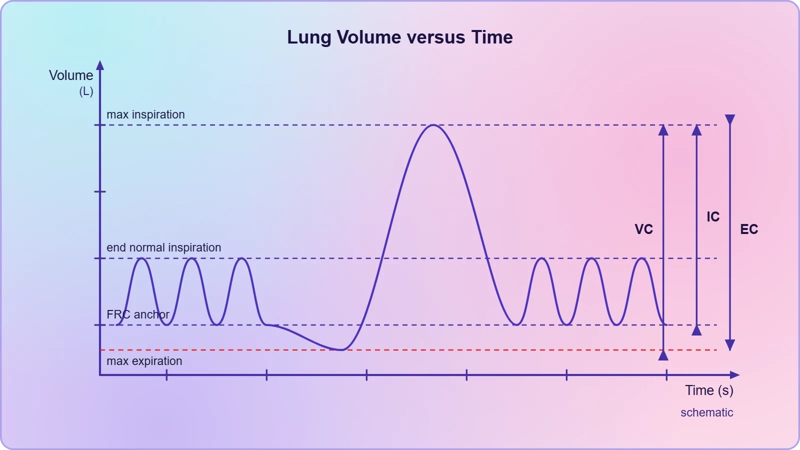
Understanding these anchors prevents a common error among learners, which is to treat capacities as interchangeable summaries of lung size. They are not. Two patients may share an identical total lung capacity while differing sharply in how that volume is partitioned between reserve and residual components, and that partition is often the diagnostic signal. The reference point tells you which partition a given capacity exposes, and the clinical sections below show how each exposed partition is read.
Inspiratory and Expiratory Capacities
The two capacities that describe voluntary movement of air in each direction are the simplest to grasp because they share the tidal volume as a common element. Inspiratory capacity measures the total volume a person can inspire after a normal expiration, and expiratory capacity measures the total volume a person can expire after a normal inspiration. Together they describe the working range of the respiratory apparatus in ordinary conditions, and both are directly observable at the bedside because neither depends on residual volume, which cannot be measured by simple spirometry.
Inspiratory Capacity: TV + IRV
Inspiratory capacity is defined as the total volume of air a person can inspire after a normal expiration, and it includes tidal volume and inspiratory reserve volume. In practical terms, it answers the question of how much additional air the lungs can draw in starting from the resting position at the end of a quiet exhale. A healthy adult at rest has a substantial inspiratory capacity because the inspiratory reserve volume is large relative to the tidal volume, giving the system considerable headroom for exertion, speech, or coughing when demand suddenly rises.
Clinically, a falling inspiratory capacity is one of the earlier signs that the lungs are becoming stiff or that the chest wall is restricting expansion, because the reserve available above the tidal breath shrinks before the resting breath itself changes much. The formula is compact and worth committing to memory:
Expiratory Capacity: TV + ERV
Expiratory capacity is the total volume of air a person can expire after a normal inspiration, and it includes tidal volume and expiratory reserve volume. It measures the expulsive reserve available after an ordinary breath in, which is the reserve recruited during forceful speech, singing, or the clearing of secretions. Because the expiratory reserve volume is typically smaller than the inspiratory reserve volume in a healthy resting adult, expiratory capacity is usually the smaller of the two directional capacities, though both vary with posture and body position.
The clinical interest in expiratory capacity lies in what happens when the expiratory reserve collapses, as it does when air trapping develops and the lungs sit at a higher resting volume. In that situation the patient has less air available to push out after a normal breath, and the expiratory capacity falls even though the tidal volume may be unchanged. The defining relation is:
Functional Residual Capacity and the Resting Baseline
Functional residual capacity occupies a special place among the pulmonary capacities because it describes the lungs at the moment of quiet equilibrium, when the inward recoil of the lung is balanced against the outward pull of the chest wall. It is the volume of air that will remain in the lungs after a normal expiration, and it includes expiratory reserve volume and residual volume. Because it contains residual volume, functional residual capacity cannot be obtained from a simple spirometer reading and requires indirect methods such as gas dilution or body plethysmography to estimate.
ERV + RV: The Air That Never Leaves
The composition of functional residual capacity explains its clinical importance. It is the sum of the air a person could still exhale if asked, which is the expiratory reserve volume, and the air that no amount of effort can expel, which is the residual volume. This means functional residual capacity is the true baseline from which every breath is drawn, and any process that raises it — air trapping, loss of elastic recoil, or expiratory flow limitation — signals that the lungs are sitting at an abnormally inflated resting position. The relation is written as:
Why the Baseline Matters in Diagnosis
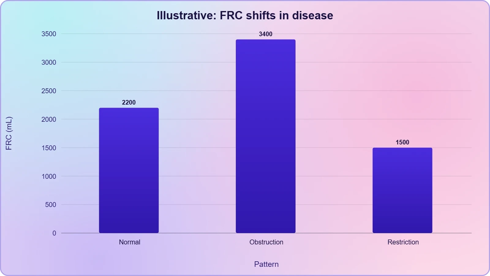
An elevated functional residual capacity is one of the hallmarks of obstructive physiology, because when small airways close prematurely during exhalation, air becomes trapped and the resting lung volume rises. A reduced functional residual capacity, by contrast, points toward restrictive conditions in which the lungs or chest wall cannot hold their normal resting volume, or toward situations in which the abdomen pushes upward and compresses the bases. Reading functional residual capacity alongside vital capacity therefore allows a clinician to distinguish between a lung that is overinflated at rest and one that is simply small.
This is also the capacity that connects most directly to the next lesson on gas exchange, because the air in the lungs at end-expiration is the gas mixture that participates in alveolar diffusion. Its composition and volume set the starting conditions for the partial pressure gradients that drive oxygen and carbon dioxide across the alveolar membrane, which is why an understanding of functional residual capacity precedes any serious discussion of diffusion.
Vital Capacity and Total Lung Capacity
The two largest capacities describe the extremes of respiratory excursion. Vital capacity is the maximum volume of air a person can breathe in after a forced expiration, or equivalently the maximum volume of air a person can breathe out after a forced inspiration, and it includes expiratory reserve volume, tidal volume, and inspiratory reserve volume. Total lung capacity is the total volume of air accommodated in the lungs at the end of a forced inspiration, and it includes residual volume, expiratory reserve volume, tidal volume, and inspiratory reserve volume, or simply vital capacity plus residual volume.
Vital Capacity: ERV + TV + IRV
Vital capacity is the most familiar of all the pulmonary capacities because it captures the full usable range of the lungs, from the deepest possible exhalation to the deepest possible inhalation. It deliberately excludes residual volume, which is why it can be measured with an ordinary spirometer and why it is so widely used in screening. A reduced vital capacity indicates that the lungs cannot expand fully or cannot empty fully, and the pattern of reduction, when compared with the other capacities, helps separate restrictive from obstructive disease. The formula is:
Because vital capacity depends on patient effort, a poor result must be interpreted with care; a submaximal effort can mimic restriction. Clinicians therefore look at the ratio of the volume expelled in the first second of a forced exhalation to the total vital capacity, a derived index that separates effort from obstruction. Vital capacity itself remains the denominator of that ratio, which is why its accurate measurement underpins so much of pulmonary function testing.
Total Lung Capacity: VC + RV
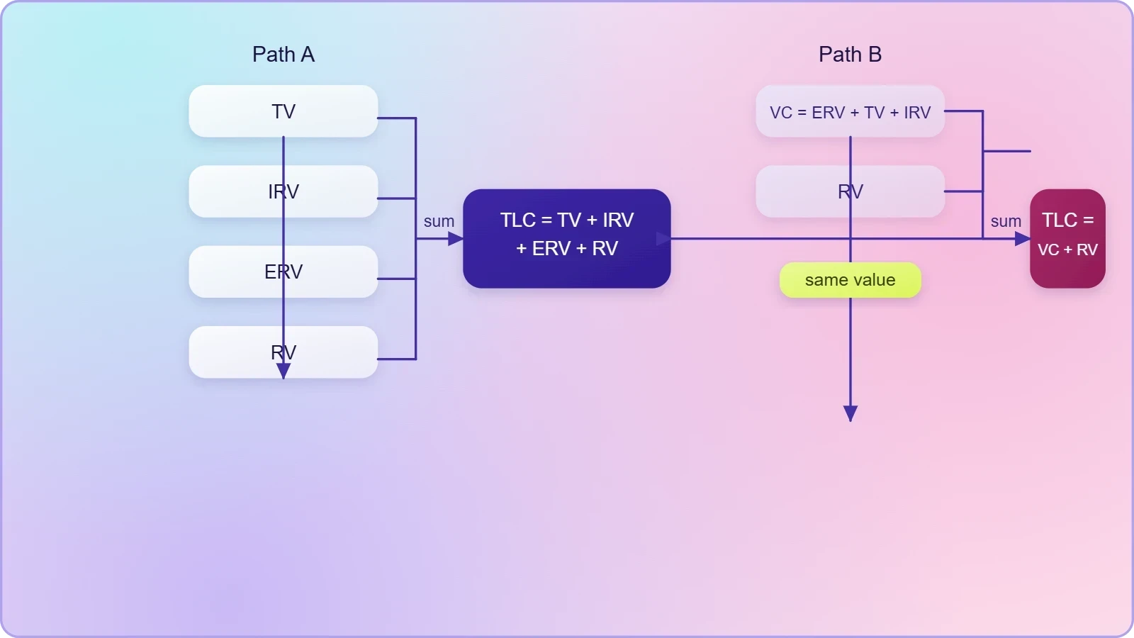
Total lung capacity is the sum of all four volumes and represents the absolute ceiling of lung inflation. It is the volume of air accommodated in the lungs at the end of a forced inspiration, and because it includes residual volume it cannot be measured by spirometry alone. In restrictive disease total lung capacity falls, reflecting a lung that is small or stiff; in obstructive disease it often rises, reflecting hyperinflation and air trapping. The two equivalent expressions are:
The equivalence of the two expressions is a useful check on understanding: if vital capacity is the sum of the three non-residual volumes, then adding residual volume to vital capacity must yield the total. Learners who can move fluidly between these two forms of the equation have grasped the architecture of the capacity system rather than merely memorised five formulas, and that fluency pays off when interpreting clinical data in which only some of the values are reported.
Reading Capacities in Clinical Diagnosis
The stated purpose of deriving these capacities is clinical diagnosis, and the diagnostic value comes from reading them as a set rather than in isolation. A single reduced vital capacity tells a clinician that something limits lung excursion but not what; pairing it with total lung capacity and functional residual capacity reveals whether the limitation arises from a lung that cannot expand or from one that cannot empty. This is why the capacities are taught together, and why the arithmetic of their derivation matters as much as the numbers themselves.
Patterns That Point to Restriction or Obstruction
In a restrictive pattern, the lungs are small or stiff, so vital capacity and total lung capacity both fall, and functional residual capacity typically falls as well because the resting volume is compressed. In an obstructive pattern, expiratory flow is limited and air becomes trapped, so residual volume and functional residual capacity rise, total lung capacity rises, and vital capacity may fall or remain near normal depending on how much hyperinflation compensates for the trapped air. The contrast between these two directions of change is the core diagnostic insight that the capacity system delivers.
These patterns are not absolute rules, and mixed disease can blur the picture, but the underlying logic holds: capacities that include residual volume reveal trapping and hyperinflation, while capacities that exclude it reveal the usable range of the lungs. A clinician who knows which capacities contain residual volume therefore knows immediately which values will be affected by air trapping and which will not, and that single piece of structural knowledge organises the entire interpretation.
Worked Example: Assembling the Capacities
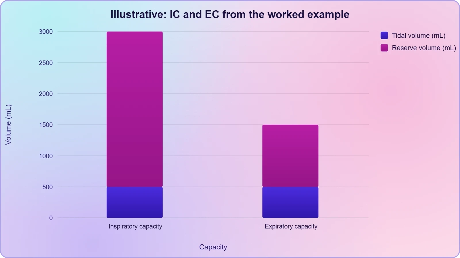
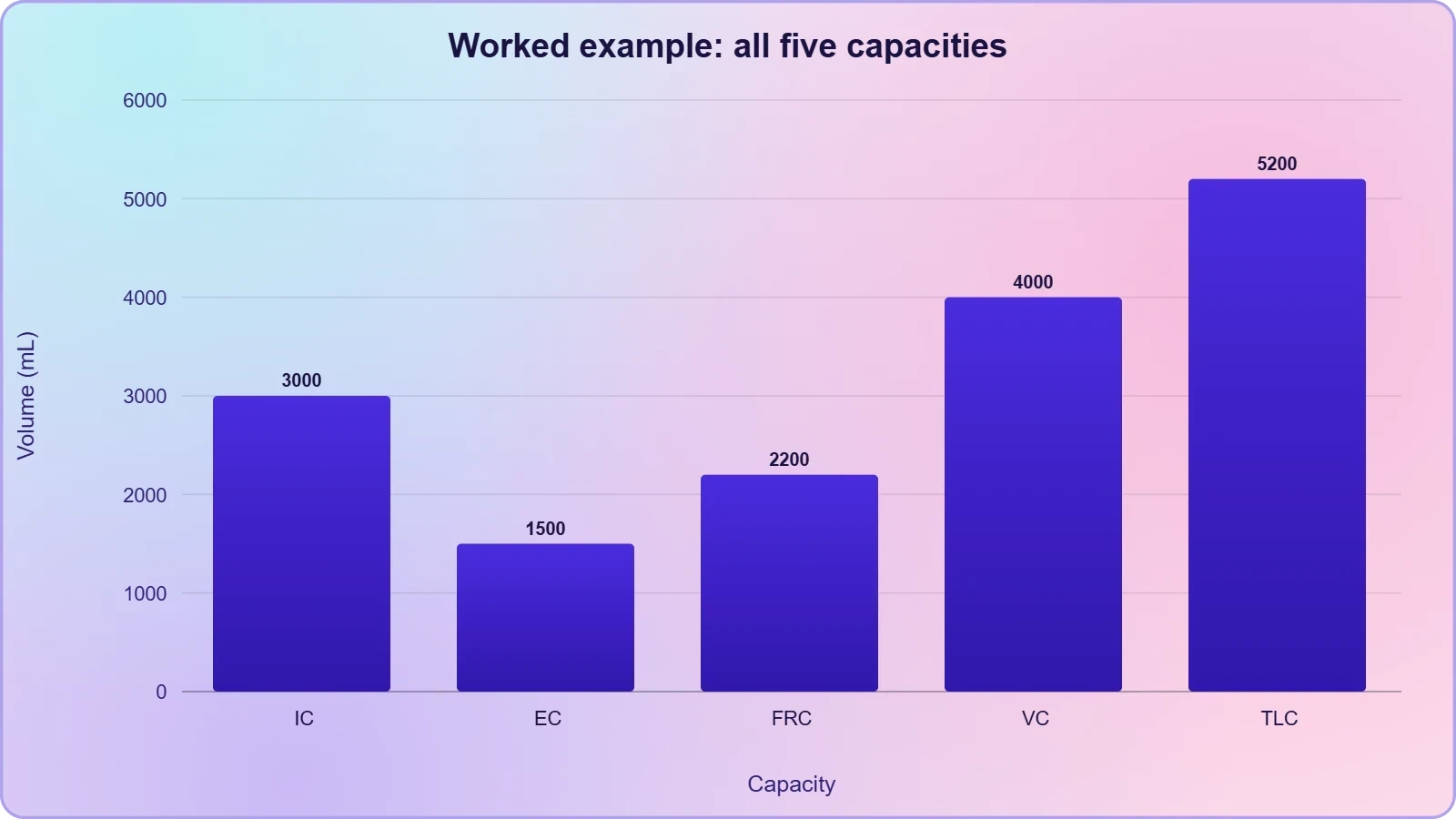
Consider a hypothetical set of measured volumes chosen for arithmetic convenience rather than clinical realism: a tidal volume of 500 mL, an inspiratory reserve volume of 2500 mL, an expiratory reserve volume of 1000 mL, and a residual volume of 1200 mL. From these four numbers every capacity can be assembled, and the exercise demonstrates how the formulas interlock. The code below performs the derivation and prints each capacity alongside its component volumes, which is a useful way to verify understanding before moving to the next lesson.
# Pulmonary capacities derived from measured respiratory volumes
TV  = 500    # tidal volume, mL
IRV = 2500   # inspiratory reserve volume, mL
ERV = 1000   # expiratory reserve volume, mL
RV  = 1200   # residual volume, mL

IC  = TV + IRV            # inspiratory capacity
EC  = TV + ERV            # expiratory capacity
FRC = ERV + RV            # functional residual capacity
VC  = ERV + TV + IRV      # vital capacity
TLC = RV + ERV + TV + IRV # total lung capacity

print("IC  =", IC,  "mL")
print("EC  =", EC,  "mL")
print("FRC =", FRC, "mL")
print("VC  =", VC,  "mL")
print("TLC =", TLC, "mL")
print("Check: VC + RV =", VC + RV, "mL (should equal TLC)")
Running this derivation yields an inspiratory capacity of 3000 mL, an expiratory capacity of 1500 mL, a functional residual capacity of 2200 mL, a vital capacity of 4000 mL, and a total lung capacity of 5200 mL. The final check confirms that vital capacity plus residual volume equals total lung capacity, which is the internal consistency test that every student should apply when working through such problems. If the check fails, a component volume has been misassigned, and the error is almost always a residual volume included in or omitted from the wrong capacity.
Going Further
Two observations extend the material without altering it. First, vital capacity can be described from either direction: it is the maximum volume of air a person can breathe in after a forced expiration, and equally the maximum volume of air a person can breathe out after a forced inspiration. Both descriptions yield the same sum of expiratory reserve volume, tidal volume, and inspiratory reserve volume, which is why the capacity is symmetric even though the effort is not. Recognising this symmetry helps when interpreting manoeuvres performed in different starting positions.
Second, every pulmonary capacity in this lesson is calculated by adding measured respiratory volumes rather than by direct measurement. This is a methodological point with practical consequences: the accuracy of a capacity is only as good as the accuracy of its components, and a capacity that includes residual volume inherits the difficulty of estimating residual volume. Learners preparing for the next lesson on partial pressures and diffusion should carry forward the idea that the lungs at end-expiration hold a defined volume of gas whose composition sets the stage for exchange across the alveolar membrane.
Consolidating the Capacity System
The five capacities form a coherent system rather than a list, and the coherence is what makes them usable at the bedside. Inspiratory capacity and expiratory capacity describe the voluntary reserves in each direction and exclude residual volume, so they are directly measurable. Functional residual capacity, vital capacity, and total lung capacity each incorporate residual volume in different ways, and each therefore carries information about the air that cannot be exhaled. Reading them together converts a set of arithmetic sums into a description of how the lungs are partitioned between usable and trapped air.
Common Errors and How to Avoid Them
The most frequent mistake is to include residual volume in a capacity that should not contain it, or to omit it from one that should. Inspiratory capacity and expiratory capacity never include residual volume; functional residual capacity always does; vital capacity never does; total lung capacity always does. A second common error is to confuse the starting point of a capacity with its endpoint, which leads to assigning the wrong reserve volume. Both errors are caught by the consistency check that vital capacity plus residual volume must equal total lung capacity.
A third error is interpretive rather than arithmetic: treating a normal total lung capacity as evidence that the lungs are healthy. A patient with severe air trapping can maintain a normal or even elevated total lung capacity while vital capacity falls, because the trapped air inflates the total without contributing to usable ventilation. The lesson for learners is that capacities must be read as a pattern, with attention to which values contain residual volume and which do not, rather than as isolated numbers judged against a single reference range.
Preparing for Gas Exchange
The capacities described here set the physical stage for the next lesson. The volume of air in the lungs at end-expiration, which is the functional residual capacity, is the gas reservoir from which oxygen diffuses into the blood and into which carbon dioxide diffuses out. Its composition, together with the partial pressures of oxygen and carbon dioxide in the alveoli, blood, and tissues, determines the direction and rate of diffusion. Understanding how much air sits in the lungs at rest is therefore a necessary preliminary to understanding what that air is made of and how it exchanges with the blood.
Learners moving forward should retain three anchors: the formulas for the five capacities, the distinction between capacities that include residual volume and those that do not, and the diagnostic directions in which each capacity shifts in restrictive and obstructive disease. With these in hand, the study of partial pressures and diffusion becomes an extension of the same physical picture rather than a separate topic, and the respiratory system can be understood as a single integrated mechanism for moving and exchanging gases.
Also Read
- No links available.
From our network :
- Trump Political Strategy How Geopolitical Stunts Serve As Media Diversions
- EV 2.0: The Solid-State Battery Breakthrough and Global Factory ExpansionDiscover the impact of the Solid-State Battery Breakthrough on the EV market. Learn about CATL and BYD stock growth, global factory expansion in Europe, and the future of EV 2.0 technology.
- Mastering DB2 12.1 Instance Design: A Technical Deep Dive into Modern Database ArchitectureComprehensive guide to DB2 12.1 Instance Design. Explore architectural components, HADR setup, buffer pool tuning, and security policies for IBM DB2 12.1. Includes 10 functional SQL and Shell samples.
- 10 Physics Numerical Problems with Solutions for IIT JEEPrepare for IIT JEE with 10 physics numerical problems, complete with detailed solutions and conceptual explanations. Ideal for mastering kinematics, thermodynamics, electromagnetism, and optics.
- Vite 6/7 'Cold Start' Regression in Massive Module GraphsIn-depth analysis of Vite 6/7 cold start regressions in massive module graphs. Learn about barrel file issues, pre-bundling optimizations, and how to improve Vite dev server performance.
- 98% of Global MBA Programs Now Prefer GRE Over GMAT Focus EditionExplore why 98% of global MBA programs now prefer the GRE over GMAT Focus. Learn about the shorter GRE format, verbal logic importance, and strategic versatility for students.
- Analyzing Trump Deportation Numbers Insights Into The 2026 Immigration Crackdown
- Mastering DB2 LUW v12 Tables: A Comprehensive Technical GuideComprehensive guide on DB2 LUW v12 tables. Explore CREATE TABLE syntax, BLU Acceleration, range partitioning, data types, and security features for IBM DB2 LUW v12.
- AI-Powered 'Precision Diagnostic' Replaces Standard GRE Score ReportsDiscover the new GRE Precision Diagnostic. Learn how AI-powered behavioral analytics and time-to-solve tracking are replacing standard GRE score reports to empower students.
RESOURCES
- Interstitial lung disease: a review of classification, etiology ... - PMCpmc.ncbi.nlm.nih.gov... pulmonary fibrosis (CAPACITY): two ... Presentation, diagnosis and clinical course of the spectrum of progressive-fibrosing interstitial lung diseases.
- Significance of pulmonary arterial pressure and diffusion capacity of ...pubmed.ncbi.nlm.nih.govShiga University of Medical Science, Department of Respiratory Medicine, Otsu, Shiga. ... Hypertension, Pulmonary / diagnosis*; Hypertension, Pulmonary / ...
- Diffusing Capacity of the Lungs for Carbon Monoxide - NCBI - NIHncbi.nlm.nih.govOct 6, 2024 ... ... capacity in newly diagnosed idiopathic pulmonary fibrosis. Sci Rep ... Pulmonary Function and Clinical Respiratory Physiology Committee ...
- Retrospective study of pulmonary function tests in patients ... - PubMedpubmed.ncbi.nlm.nih.govPulmonary Diffusing Capacity / physiology*; Pulmonary Emphysema / diagnosis; Pulmonary Emphysema / physiopathology*; Respiratory Function Tests*; Retrospective ...
- Chronic Obstructive Pulmonary Disease (COPD) - StatPearls - NCBIncbi.nlm.nih.govApr 15, 2026 ... COPD diagnosis requires spirometry: a postbronchodilator FEV1/FVC ratio less than 0.70 is the diagnostic criterion; clinical diagnosis without ...
- Pulmonary hypertension - Diagnosis and treatment - Mayo Clinicmayoclinic.orgApr 11, 2025 ... Once a diagnosis of pulmonary hypertension is confirmed, the condition is classified according to how the symptoms affect you and…
- Spirometry: Purpose, Procedure, Risks & Results - Cleveland Clinicmy.clevelandclinic.orgFeb 8, 2024 ... Determine your lung capacity. ... These tests supplement spirometry tests by helping your provider diagnose certain lung or breathing conditions.
- Revised Definition of Pulmonary Hypertension and Approach to ...ahajournals.orgApr 7, 2023 ... This clinical primer highlights key changes in diagnosis and approach to PH management, focusing on concepts that are likely to…
- Measuring Lung Function More Accurately and More Equitablyhms.harvard.eduMay 19, 2024 ... ... pulmonary function testing.” Shedding the legacy of race-based medical testing and diagnosis. Race is a notoriously unreliable indicator of ...
- Prognostic implication of 1-year decline in diffusing capacity ... - Naturenature.comApr 17, 2024 ... Clinical features, diagnosis, management, and outcomes of idiopathic pulmonary fibrosis in Korea: Analysis of the Korea IPF cohort (KICO) ...
- Interpreting pulmonary function tests: Recognize the pattern, and the ...ccjm.orgSpirometry cannot measure the residual volume or the total lung capacity. Page 4. 872 CLEVELAND CLINIC JOURNAL OF MEDICINE VOLUME 70 • NUMBER 10…
- Diagnosis of Idiopathic Pulmonary Fibrosis. An Official ATS/ERS ...thoracic.orgSep 1, 2018 ... Question 6: For Patients with. Newly Detected ILD of. Unknown Cause Who Are. Clinically Suspected of Having. IPF, Is Transbronchial…
- Clinical Guidelines for Diagnosis and Treatment of Botulism, 2021cdc.govMay 7, 2021 ... Conduct frequent, serial neurologic examinations, with an emphasis on cranial nerve palsies, swallowing ability, respiratory status, and ...