The human respiratory system is best understood as a continuous, branching corridor that begins at the face and ends in microscopic air sacs deep inside the chest. Every structure along that corridor exists for a reason: some passages warm, moisten and filter incoming air, others produce sound, still others guard the airway against food, and the final chambers present an enormous, blood-rich surface for gas exchange. Following the path of air in sequence — nostrils, nasal chamber, pharynx, larynx, trachea, bronchi, bronchioles and alveoli — is therefore not a memory exercise but a study in functional design, where each segment's anatomy explains its physiological role.
This lesson traces that route in detail, staying close to the standard terminology used in human physiology. It examines the paired external nostrils and the nasal chamber, the shared food-and-air passage of the pharynx, the cartilaginous sound box called the larynx with its protective epiglottis, the straight trachea dividing at the fifth thoracic vertebra, and the repeated branching of bronchi into bronchioles. It then describes the alveoli as thin, irregular-walled, vascularised bag-like structures, and explains how incomplete cartilaginous rings keep the conducting airways open without stiffening them against the neck's movements.
Because this lesson builds directly on the comparative survey of respiratory organs across animal groups, it assumes familiarity with the idea that different animals solve gas exchange in different ways. Here the focus narrows to one species and one route. The material deliberately stops at the alveoli and the branching network that constitutes the lungs; the pleural membranes, the thoracic chamber and the mechanics of breathing belong to the lesson that follows, so they are mentioned only where the airway anatomy requires it.
On This Page
The Upper Airway: Nostrils, Nasal Chamber and Pharynx
The journey of air begins above the upper lip, where a pair of external nostrils opens to the outside. These paired openings lead through the nasal passage into the nasal chamber, the first substantial chamber of the respiratory tract. Although the source material states this plainly, the arrangement already reveals a design principle: air is admitted through two separate portals rather than one, and it is admitted high on the face, above the mouth, so that breathing can continue while the mouth is occupied with food or speech.

From the nasal chamber, air passes into the pharynx. The material is careful here, noting that a portion of the pharynx is the common passage for food and air. This shared function is the anatomical reason the upper airway requires a guard further down, and it explains why the pharynx cannot be described as a purely respiratory structure. It is a crossroads, and everything that follows in the lesson — the larynx, the epiglottis, the glottis — exists in part because of that crossroads.
The Nasal Chamber as the First Conditioning Segment
The nasal chamber opens into the pharynx through the nasal passage, forming a continuous route from the exterior to the throat. In the sequence given by the material, the nasal chamber is simply the chamber between the nostrils and the pharynx, and that is the definition to retain. Its position at the very entrance of the tract means that all air drawn in through the nostrils passes through it before reaching any other structure, making it the first segment of the conducting portion of the respiratory system.
Keeping the material's order of ideas matters here. The nostrils open above the upper lips; the nasal passage leads to the nasal chamber; the nasal chamber opens into the pharynx. Each step is a named structure with a named successor, and the lesson's terminology should be reproduced in exactly that sequence. Students who can recite the chain in order, and attach one functional remark to each link, will find the later lessons on breathing mechanics far easier to follow.
The Pharynx as a Common Passage
The pharynx is described as a portion of which is the common passage for food and air. That single clause carries a great deal of physiological weight. Because the same space must serve two systems, the body needs a mechanism to route swallowed food away from the airway, and the material supplies that mechanism at the next station: the epiglottis, which can cover the glottis during swallowing. The pharynx therefore sets up the problem that the larynx solves.
It is worth noting what the material does not claim. It does not describe the pharynx as divided into named parts, nor does it assign it a role in gas exchange. Its function in this account is purely conductive and shared. The pharynx opens through the larynx region into the trachea, and that phrase — through the larynx region — is the bridge to the next section of the lesson, where the airway acquires cartilage, a voice, and a protective flap.
The Larynx: Cartilaginous Sound Box and Guardian of the Airway
The pharynx opens through the larynx region into the trachea. The material defines the larynx as a cartilaginous box which helps in sound production and hence is called the sound box. Two facts are packed into that definition: the larynx is built of cartilage, and its cartilaginous construction serves sound production. The nickname follows directly from the function, and the lesson should preserve both the structural description and the functional label, since examination questions frequently ask for either one.
Alongside sound production, the larynx performs a protective duty. During swallowing, the glottis can be covered by a thin elastic cartilaginous flap called the epiglottis, preventing the entry of food into the larynx. The glottis is the opening guarded by this flap, and the epiglottis is described precisely — thin, elastic, cartilaginous. These three adjectives are part of the material's terminology and should be retained whenever the epiglottis is defined, because they distinguish it from the incomplete cartilaginous rings found lower in the airway.
Sound Production and the Sound Box
Calling the larynx the sound box is not a metaphor to be softened; it is the material's own term. The cartilaginous box helps in sound production, and that is the reason for the name. In the order of ideas presented, sound production is mentioned before protection, so the lesson should introduce the larynx as a sound-producing structure first and then add its guarding role. This ordering keeps faith with the source and mirrors the way the airway is described from the pharynx downward.
The larynx sits at a strategic junction. Above it lies the pharynx, the common passage for food and air; below it lies the trachea, the straight tube of the lower airway. Anything that enters the trachea has passed the larynx, and anything that enters the larynx has passed the glottis. Understanding the larynx as a checkpoint between the shared upper passage and the dedicated lower airway clarifies why a protective flap is needed at precisely this location and not higher or lower.
The Epiglottis and the Glottis During Swallowing
The protective arrangement is elegantly simple. The glottis is the opening into the larynx, and the epiglottis is a thin elastic cartilaginous flap that can cover it during swallowing. The word can is significant: the material describes a capacity, an action available when needed, rather than a permanent seal. Between swallows the airway must remain open for breathing, so a flap that closed permanently would defeat its own purpose. The epiglottis is therefore a conditional guard, deployed at the moment food or drink threatens the laryngeal entrance.
This mechanism resolves the difficulty created by the pharynx. Because a portion of the pharynx serves as the common passage for food and air, the two streams must be separated at the point where they diverge. The epiglottis performs that separation by covering the glottis, directing the swallowed bolus toward the food passage and away from the larynx. The material states the outcome plainly — to prevent the entry of food into the larynx — and that phrase should anchor any explanation of the reflex.
The Trachea and the Bronchial Tree
Below the larynx, the airway becomes a dedicated tube. The trachea is a straight tube extending up to the mid-thoracic cavity, which divides at the level of the fifth thoracic vertebra into a right and left primary bronchi. The material gives both a length description and a precise level of division, and both should be preserved. The trachea is not a short connector but a substantial conduit reaching the middle of the chest before it splits, and the level of that split is fixed at a named vertebra.
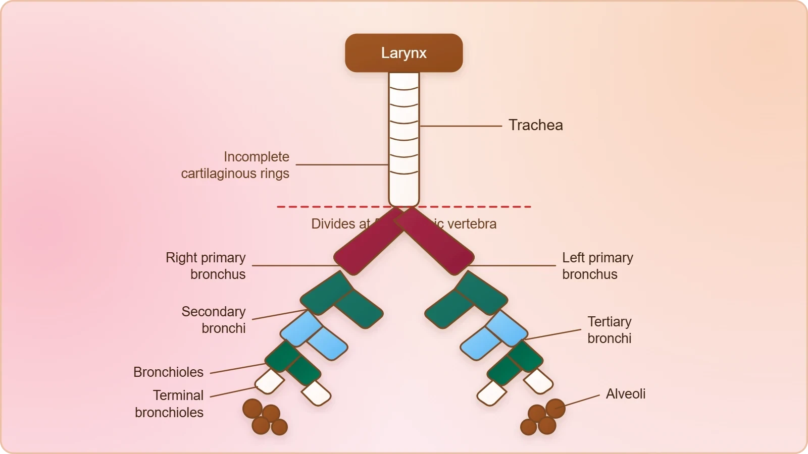
From the primary bronchi, the airway branches repeatedly. Each bronchus undergoes repeated divisions to form the secondary and tertiary bronchi and bronchioles, ending up in very thin terminal bronchioles. This is a genuine tree, narrowing at every fork, and the material names each generation in order: primary, secondary, tertiary, bronchioles, terminal bronchioles. The branching network of bronchi, bronchioles and alveoli together comprises the lungs, so the lungs are defined here not as two simple bags but as the whole arborisation within the chest.
Division at the Fifth Thoracic Vertebra
The level of division is one of the few numerical facts in this section, and it deserves emphasis. The trachea divides at the level of the fifth thoracic vertebra into a right and left primary bronchi. The number five, the word thoracic, and the pairing of right and left are all part of the material's account. A student who can state that the split occurs at the fifth thoracic vertebra, producing one primary bronchus for each lung, has captured the essential geometry of the lower airway.
The division produces two primary bronchi, one entering each lung. From that point onward the airway is paired and internal, and the subsequent branching occurs within the lung substance itself. The material's phrase — each bronchi undergoes repeated divisions — signals that the pattern is recursive: every branch gives rise to smaller branches, and the process continues until the finest conducting tubes are reached. This recursion is what creates the enormous number of terminal passages that ultimately supply the alveoli.
Cartilaginous Rings and the Support of the Airways
Support along the bronchial tree is provided by cartilage, but not of the enclosing kind. The tracheae, primary, secondary and tertiary bronchi, and initial bronchioles are supported by incomplete cartilaginous rings. The word incomplete is essential. A complete ring would encircle the tube entirely; an incomplete ring leaves a gap, usually occupied by softer tissue, allowing the tube to flex and the oesophagus behind it to expand during swallowing while still preventing the airway from collapsing.

The list of supported structures is equally important. Support extends from the trachea through the primary, secondary and tertiary bronchi and into the initial bronchioles, but no further. The terminal bronchioles, the finest conducting tubes, are not included in that list. This boundary marks a transition in the airway's construction: cartilage gives way to other tissues as the tubes become too small to house rings, and the material's careful wording allows the reader to locate exactly where that change occurs.
Bronchioles and the Alveoli
The branching continues beyond the bronchi into the bronchioles, and the finest of these are the terminal bronchioles, described as very thin. Each terminal bronchiole gives rise to a number of very thin, irregular-walled and vascularised bag-like structures called alveoli. This sentence is the climax of the lesson, because it introduces the structures where the conducting airway ends and the exchange surface begins. Every adjective in the description — thin, irregular-walled, vascularised, bag-like — is functional, and each one should be explained rather than merely memorised.
The branching network of bronchi, bronchioles and alveoli comprise the lungs. The material defines the lungs by their constituent network, not by any single enclosing structure, and we have two lungs. This definition is worth pausing over: the lung is the whole tree, from the largest bronchus to the smallest alveolar sac, and the alveoli are its terminal leaves. The pleural membranes that cover the lungs are mentioned in the material but belong to the next lesson, where the thoracic chamber and its coverings are treated in full.
Terminal Bronchioles and Their Alveolar Clusters
Each terminal bronchiole gives rise to a number of alveoli, so the relationship is one of multiplication: a single fine tube ends in a cluster of sacs rather than a single sac. This arrangement is what allows the airway to terminate in an enormous number of small chambers while keeping each conducting tube short. The material's phrase — a number of — signals multiplicity without committing to a count, and the lesson should respect that restraint rather than importing figures the source does not supply.
The terminal bronchioles are also the last structures in the material's list of cartilage-supported tubes. Beyond them, the airway wall is no longer stiffened by rings, which is consistent with the alveoli being described as thin and irregular-walled. A cartilaginous ring around a structure only micrometres thick would be a contradiction in terms. The transition from supported conducting tube to delicate exchange chamber is therefore built into the material's own sequence, and the student should be able to point to the exact sentence where it happens.
Why the Alveoli Are Thin, Irregular and Vascularised
Three adjectives carry the physiology. Thin walls shorten the distance across which gases must move. Irregular walls increase the surface available within a small volume. Vascularised means the alveoli are richly supplied with blood vessels, so that the air they contain is brought into intimate contact with the bloodstream. Together these features make the alveolus the site where the respiratory system meets the circulatory system, and the material's compact description is effectively a checklist of the requirements for efficient gas exchange.

The bag-like shape completes the picture. A bag encloses a volume of air and presents an inner surface to it; the thin, irregular wall of that bag is the barrier between air and blood. Because each terminal bronchiole supplies a number of such bags, and because the branching tree multiplies at every level, the total surface presented by the alveoli is vast relative to the size of the lungs. The material does not quantify this surface, but its description of thin, irregular, vascularised bags explains why the surface can be so large.
Cartilage, Protection and the Logic of the Airway
Reading the airway as a whole reveals a consistent logic. Cartilage appears at the larynx, where it forms a box for sound production, and again along the trachea and bronchi, where it forms incomplete rings that hold the tubes open. The epiglottis, a thin elastic cartilaginous flap, uses cartilage for flexibility rather than rigidity. The same tissue is thus deployed in three different ways along one continuous route, and the material's careful use of the words box, rings and flap reflects those distinct structural roles.
Protection is layered in the same way. The pharynx, as a common passage, creates the risk of food entering the airway; the epiglottis answers that risk by covering the glottis during swallowing. Lower down, the incomplete cartilaginous rings protect the airway from collapse while permitting movement. The material presents these safeguards in the order the air encounters them, so a reader moving from top to bottom of the tract meets each protective feature at the point where it is needed.
Incomplete Rings Versus the Epiglottic Flap
The contrast between the two cartilaginous devices is instructive. The epiglottis is a thin elastic flap that can cover the glottis; the rings are incomplete supports that keep tubes patent. One is mobile and conditional, the other is fixed and continuous. One guards an opening, the other maintains a lumen. Both are cartilaginous, yet their mechanical behaviour is almost opposite, and the material's precise vocabulary — flap for the epiglottis, rings for the airways — allows the distinction to be drawn without ambiguity.
This distinction also explains why the rings are described as incomplete rather than complete. A complete ring would be rigid in every direction, but the trachea lies in front of the oesophagus, which must expand when food passes. An incomplete ring, open at the back, allows that expansion while still preventing the airway from collapsing. The material does not spell out this reasoning, but it follows directly from the stated fact that the rings are incomplete, and it is a legitimate inference for an intermediate learner to make.
From Conducting Tubes to Exchange Surface
The airway can be divided by function into a conducting portion and an exchange portion. The nostrils, nasal chamber, pharynx, larynx, trachea, bronchi and bronchioles all conduct air; the alveoli exchange gases. The material's structure follows this division implicitly, since cartilage support is listed for the conducting tubes and stops at the terminal bronchioles, while the alveolar description introduces thinness and vascularisation instead. Recognising the boundary between the two portions is one of the most useful outcomes of this lesson.
That boundary is not abrupt. The terminal bronchioles are already very thin, and they give rise directly to the alveoli, so the transition is gradual. Nevertheless, the functional change is decisive: before the alveoli, the airway's task is to move air and keep the passage open; at the alveoli, the task becomes the transfer of gases between air and blood. The next lessons take up that transfer, along with the pleural coverings and the mechanics that move air along the entire route described here.
Going Further
The epiglottis behaves like a flap that closes over the airway during swallowing so that food travels down the food pipe instead. This everyday image matches the material's statement that the epiglottis can cover the glottis during swallowing to prevent the entry of food into the larynx. It also explains the familiar sensation of choking when swallowing and breathing compete for the same moment: the flap must be in the right position, and speech or laughter during a meal can disturb that timing.
Alveoli are tiny air sacs, and their very large number gives the lungs an exceptionally large surface area for gas exchange. The material describes each terminal bronchiole as giving rise to a number of alveoli, and the branching tree multiplies that number at every level. The result is a total exchange surface far greater than the outer surface of the lungs themselves, which is why such small organs can serve the whole body's need for oxygen uptake and carbon dioxide removal.
Estimating the Scale of the Exchange Surface
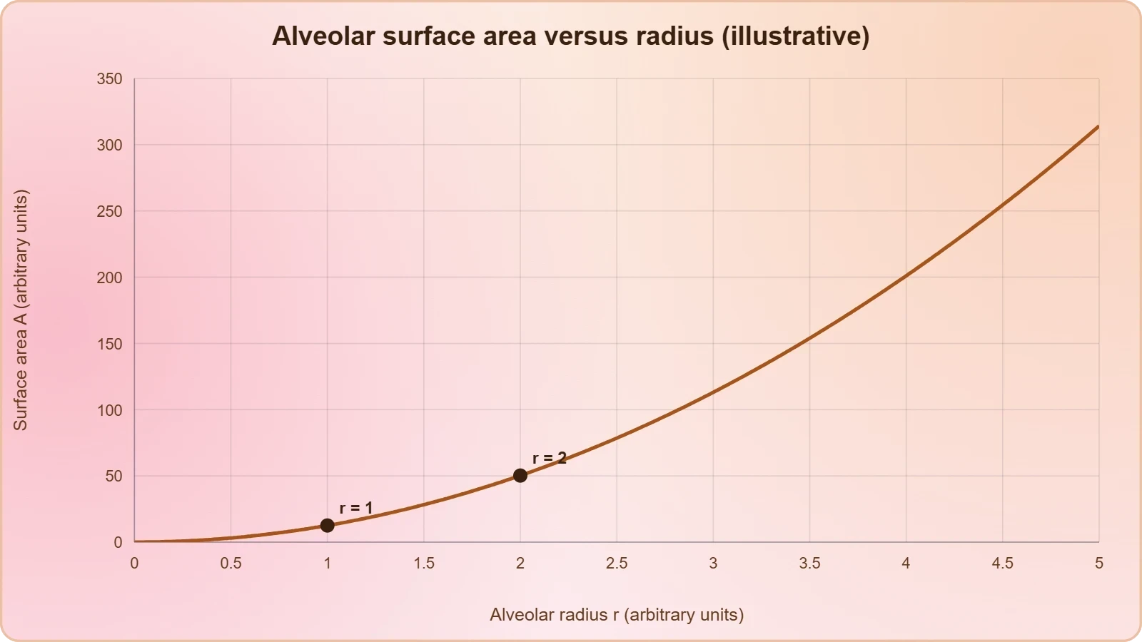
A simple model shows why the alveolar surface is so large. If each alveolus is treated as a sphere of radius ##r##, its surface area is ##A = 4\pi r^2##, and the total area for ##N## alveoli is:
Because ##N## is enormous while ##r## is very small, the product remains large. Halving the radius of each sac quarters its individual area, but the number of sacs that can fit in the same volume rises by a factor of eight, so the total area actually increases. This is the geometric reason the material's phrase a number of alveoli matters: multiplicity, not size, is what makes the exchange surface so extensive.
Reading the Airway as a Sequence
The most reliable way to retain this lesson is to rehearse the sequence aloud, attaching one fact to each station. Nostrils above the upper lips; nasal passage to the nasal chamber; nasal chamber to the pharynx, the common passage for food and air; pharynx through the larynx region into the trachea; larynx the cartilaginous sound box, its glottis covered by the epiglottis during swallowing; trachea dividing at the fifth thoracic vertebra into right and left primary bronchi; repeated divisions into secondary and tertiary bronchi and bronchioles; terminal bronchioles giving rise to alveoli.
Once the sequence is secure, the supporting details can be layered on: incomplete cartilaginous rings along the trachea, bronchi and initial bronchioles; alveoli thin, irregular-walled and vascularised; the branching network of bronchi, bronchioles and alveoli comprising the lungs. The pleural membranes and thoracic chamber, mentioned only in passing here, are the subject of the next lesson, where the mechanics of breathing and the volumes and capacities of the respiratory system are developed in full.
Checking Understanding Before Moving On
A short self-check consolidates the lesson. Can you name, in order, every structure air passes from the external nostrils to the alveoli? Can you state which structure is the common passage for food and air, and which flap protects the airway during swallowing? Can you give the level at which the trachea divides, and the point at which cartilaginous support ends? If each of these can be answered without reference to notes, the anatomical groundwork for the lessons on breathing mechanics and gas transport is firmly in place.
Two common errors are worth flagging. The first is to describe the cartilaginous rings as complete; the material specifies incomplete rings, and that word carries the functional reason for their shape. The second is to treat the lungs as simple sacs rather than as the branching network of bronchi, bronchioles and alveoli. Correcting both errors early prevents confusion when the pleural membranes, the thoracic chamber and the mechanics of ventilation are introduced in the lessons that follow.
Common Misconceptions to Avoid
Another frequent slip is to confuse the glottis with the epiglottis. The glottis is the opening; the epiglottis is the flap that can cover it. A related error is to say the epiglottis permanently seals the larynx, when the material states only that it can cover the glottis during swallowing. Finally, students sometimes extend cartilaginous support all the way to the alveoli; the material limits it to the trachea, primary, secondary and tertiary bronchi, and initial bronchioles, leaving the terminal bronchioles unsupported.
Each misconception arises from compressing a precise statement into a vague one. The remedy is to return to the exact wording: thin elastic cartilaginous flap, incomplete cartilaginous rings, very thin terminal bronchioles, thin irregular-walled vascularised bag-like alveoli. Precision in vocabulary is not pedantry in physiology; the adjectives carry the function, and a student who keeps them will find that the mechanics of breathing and the exchange of gases follow naturally from the structures described here.
Preparing for the Next Lesson
The airway described in this lesson ends at the alveoli, but the lungs do not float freely in the chest. They are covered by a double-layered pleura with pleural fluid between the layers, and the outer pleural membrane lies in close contact with the thoracic wall. Those facts are stated in the material but are developed in the next lesson, which treats the lungs, the pleura and the thoracic chamber together with the mechanics of breathing and the respiratory volumes and capacities.
Carrying forward a clear mental map of the airway will make that next lesson easier. When the thoracic chamber expands, the effect travels down the branching tree to the alveoli described here; when volumes are measured, the air being measured has passed through the nostrils, nasal chamber, pharynx, larynx, trachea and bronchi. The anatomy is the fixed scaffolding on which every subsequent physiological explanation is built, and it is worth knowing well.
Also Read
- No links available.
From our network :
- 10 Physics Numerical Problems with Solutions for IIT JEEPrepare for IIT JEE with 10 physics numerical problems, complete with detailed solutions and conceptual explanations. Ideal for mastering kinematics, thermodynamics, electromagnetism, and optics.
- Mastering DB2 LUW v12 Tables: A Comprehensive Technical GuideComprehensive guide on DB2 LUW v12 tables. Explore CREATE TABLE syntax, BLU Acceleration, range partitioning, data types, and security features for IBM DB2 LUW v12.
- EV 2.0: The Solid-State Battery Breakthrough and Global Factory ExpansionDiscover the impact of the Solid-State Battery Breakthrough on the EV market. Learn about CATL and BYD stock growth, global factory expansion in Europe, and the future of EV 2.0 technology.
- Analyzing Trump Deportation Numbers Insights Into The 2026 Immigration Crackdown
- Vite 6/7 'Cold Start' Regression in Massive Module GraphsIn-depth analysis of Vite 6/7 cold start regressions in massive module graphs. Learn about barrel file issues, pre-bundling optimizations, and how to improve Vite dev server performance.
- AI-Powered 'Precision Diagnostic' Replaces Standard GRE Score ReportsDiscover the new GRE Precision Diagnostic. Learn how AI-powered behavioral analytics and time-to-solve tracking are replacing standard GRE score reports to empower students.
- Trump Political Strategy How Geopolitical Stunts Serve As Media Diversions
- 98% of Global MBA Programs Now Prefer GRE Over GMAT Focus EditionExplore why 98% of global MBA programs now prefer the GRE over GMAT Focus. Learn about the shorter GRE format, verbal logic importance, and strategic versatility for students.
- Mastering DB2 12.1 Instance Design: A Technical Deep Dive into Modern Database ArchitectureComprehensive guide to DB2 12.1 Instance Design. Explore architectural components, HADR setup, buffer pool tuning, and security policies for IBM DB2 12.1. Includes 10 functional SQL and Shell samples.
RESOURCES
- Respiratory System: Organs, Facts, Anatomy & Functionmy.clevelandclinic.orgApr 9, 2024 ... Your upper respiratory tract brings air into your body and helps move it toward your lungs. It adds moisture to…
- Conducting Passages - SEER Training Modulestraining.seer.cancer.govThe lower respiratory tract consists of the trachea, bronchial tree, and lungs. These tracts open to the outside and are lined with mucous membranes.…
- Airways: Respiratory Tract, Parts, Anatomy & Functionmy.clevelandclinic.orgApr 2, 2024 ... Your airways are a series of passages that air travels through to move to and from your lungs. Parts include…
- "Pills" and the air passages - PubMedpubmed.ncbi.nlm.nih.govHumans; Iatrogenic Disease; Pharmaceutical Preparations / administration ... Respiratory System / drug effects*; Tablets / adverse effects*. Substances.
- How the Lungs Work - The Respiratory System | NHLBI, NIHnhlbi.nih.govMar 24, 2022 ... Nose and linked air passages called the nasal cavity and sinuses. Larynx (voice box); Trachea (windpipe); Tubes called bronchial tubes.…
- Plugs of the Air Passages: A Clinicopathologic Review - PMC - NIHpmc.ncbi.nlm.nih.govAlthough mucus is a normal product of the tracheobronchial tree, some diseases of the respiratory tract are characterized by unusually thick (inspissated) ...
- Anatomy, Airway - StatPearls - NCBI Bookshelfncbi.nlm.nih.govSep 15, 2025 ... The airway comprises the passages of the respiratory tract that conduct air from the external environment to the alveoli.
- Anatomical Details of the Rabbit Nasal Passages and Their ... - PMCpmc.ncbi.nlm.nih.gov... Passages and Their Implications in Breathing, Air Conditioning, and Olfaction ... and vapor dosimetry in the respiratory tracts of rat, monkey, and human.
- Heat and Water Transport in the Human Respiratory Systemlink.springer.comAn understanding of heat and water movement from the underlying blood vessels to and from the air flowing in the respiratory passageways, the problem…
- Parasites of the Air Passages - ScienceDirect.comsciencedirect.comInfective filariform larvae can penetrate the skin and infect human hosts. The larvae migrate through the soft tissues and enter the lungs via the…
- Tips to Keep Your Lungs Healthy | American Lung Associationlung.orgJun 22, 2026 ... Cigarette smoke can narrow the air passages and make breathing more difficult. ... An illustration of human lungs with colorful…
- Understanding RRP - THE VOICE FOUNDATIONvoicefoundation.org... air passageway (respiratory tract); these tumors are caused by infection with HPV. These tumors often have a cauliflower-like appearance, and either have a ...
- Wheezing: MedlinePlus Medical Encyclopediamedlineplus.govMay 3, 2026 ... Wheezing is a high-pitched whistling sound during breathing. It occurs when air moves through narrowed breathing tubes in the lungs.
- How your lungs work | Canadian Lung Associationlung.caThe air we inhale travels through the trachea, the bronchi and bronchioles to reach the alveoli. The alveoli is where oxygen is transferred from…
- Asthma - World Health Organization (WHO)who.intApr 28, 2026 ... ... and other sources of air pollution, as well as viral respiratory infections. ... bronchodilators (such as salbutamol), that open…