The frog is a small animal with a demanding physiological brief: it must move oxygen, nutrients, hormones and waste through a body that alternates between pond water and dry land, often within the same hour. Two systems carry that burden. The vascular system, a well-developed closed network of heart, blood vessels and blood, drives every exchange by pumping; the excretory system, built from a pair of kidneys, ureters, cloaca and urinary bladder, filters the chemical debris of metabolism and expels it. Together they form the internal logistics of the frog, and reading them side by side reveals how tightly structure is matched to habit.
This lesson, the fourth of five in the course on structural organisation in animals, builds directly on the digestive and respiratory systems already studied. Food absorbed through the villi and oxygen taken in by lungs, skin and buccal cavity do not travel by themselves; they require a transport architecture, and that architecture is the subject here. Equally, the nitrogenous waste generated by protein metabolism must be collected, concentrated and released, which is the work of the excretory organs. The treatment below follows the sequence of the chapter: the closed vascular plan, the three-chambered heart, the portal systems, the lymphatic system, and finally the excretory pathway that makes the frog ureotelic.
Precision matters in this material because several frog features are easy to confuse with those of mammals. The heart has three chambers, not four. Red blood cells are nucleated. Two portal systems, hepatic and renal, exist where a mammal has essentially one. The ureters of the male frog double as urinogenital ducts, while in the female the ureters and oviducts open separately into the cloaca. Each of these details is examinable and each carries functional meaning. By the end of the lesson, the frog should read as an integrated organism whose circulation and excretion are tuned to amphibian life.
On This Page
The Closed Vascular Plan and the Place of Lymph
The vascular system of the frog is described as well-developed and of the closed type, meaning that blood remains within vessels for the whole of its circuit and is propelled by a muscular pump rather than bathing the tissues in an open cavity. The system comprises three components: the heart, the blood vessels and the blood itself. Arteries, forming the arterial system, carry blood away from the heart to all parts of the body, while veins, forming the venous system, collect blood from the different regions and return it. This division of labour between efferent and afferent channels is the structural foundation on which every subsequent detail rests.
Heart, Vessels and the Course of Blood
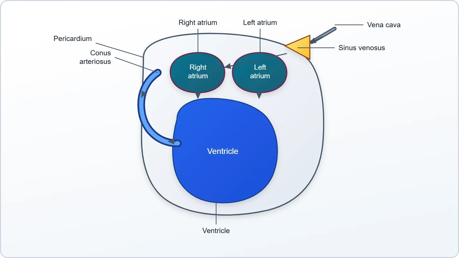
The heart is a muscular structure situated in the upper part of the body cavity and is enclosed by a membrane called the pericardium. It possesses three chambers: two atria and one ventricle. A triangular structure, the sinus venosus, joins the right atrium and receives blood through the major veins known as the vena cava. On the ventral side, the ventricle opens into a sac-like conus arteriosus, from which blood passes into the arterial system. This arrangement means that venous blood is gathered before it enters the heart and that outflow is funnelled through a single muscular channel, a design quite unlike the four-chambered mammalian heart.
The blood itself is composed of plasma and cells. The cells are of three kinds: RBC or erythrocytes, WBC or leucocytes, and platelets. Frog erythrocytes are nucleated and contain the red pigment haemoglobin, a feature that distinguishes amphibian blood from the mature mammalian condition in which red cells lose their nucleus. The blood carries nutrients, gases and water to their respective sites during circulation, and the movement of this fluid is achieved entirely by the pumping action of the muscular heart. Every exchange described in the respiratory lesson, and every nutrient absorbed in the digestive lesson, depends on this continuous transport.
The Lymphatic System and Its Distinction from Blood
Frogs possess a lymphatic system in addition to the blood vascular system. It consists of lymph, lymph channels and lymph nodes. The lymph is explicitly different from blood: it lacks a few proteins and lacks RBCs. This distinction is worth holding onto, because it explains why lymph cannot substitute for blood in gas transport. Without erythrocytes and their haemoglobin, lymph carries no respiratory pigment, and without the full complement of plasma proteins its composition is leaner. It is a drainage and immune network rather than a circulatory substitute, and the chapter treats it as a companion to, not a replacement for, the closed vascular circuit.
Placing the two systems side by side clarifies the logic of vertebrate organisation. The blood vascular system is a high-pressure, corpuscle-rich transport line; the lymphatic system is a lower-profile collection route that returns fluid and supports defence. In the frog, both are present and both are named in the material, but only the blood system is described as closed and pumped by the heart. Students should be able to state the three components of each system and to explain, in one sentence, why lymph differs from blood. That single comparison anchors much of the examination weight attached to this section.
The Three-Chambered Heart and the Portal Systems
The heart's three chambers, two atria and one ventricle, receive blood from two directions and discharge it through a single outlet. The sinus venosus, a triangular structure, joins the right atrium and gathers blood through the vena cava; the ventricle opens into the conus arteriosus on the ventral side. This arrangement means that oxygenated blood returning from the lungs and skin, and deoxygenated blood returning from the body, both enter the heart before being ejected. The single ventricle therefore handles mixed blood, a design adequate for an amphibian's relatively low metabolic demand and central to understanding frog circulation.
Why Three Chambers Serve an Amphibian
Two atria allow the heart to receive pulmonary and systemic returns separately, while one ventricle performs the ejection. The separation is therefore partial rather than complete, and the ventricle contracts to push blood into the conus arteriosus and onward into the arteries. For a frog, this is sufficient: metabolic rate is modest, and much gas exchange occurs across the skin, so the demand for perfectly segregated high-oxygen blood is lower than in a bird or mammal. The three-chambered heart is not a deficient four-chambered heart; it is a solution matched to the animal's physiology and habit.
The pericardium, the membrane covering the heart, provides a protective envelope for this muscular organ situated in the upper part of the body cavity. Students should be able to trace the path of blood in sequence: vena cava to sinus venosus to right atrium, atrium to ventricle, ventricle to conus arteriosus, and thence into the arterial system. Reversing that sequence describes the venous return. Mastery of this route is the single most reliable predictor of success on questions about frog circulation, because nearly every examination item is a variation on it.
Hepatic and Renal Portal Systems
Frogs possess special venous connections that are absent or much reduced in mammals. The first is the hepatic portal system, a venous link between the liver and the intestine; the second is the renal portal system, a venous link between the kidney and the lower parts of the body. A portal system, in general terms, is a route in which blood passes through two successive capillary beds before returning to the heart. In the frog, these two named systems ensure that blood from the gut is processed by the liver and blood from the hind limbs is screened by the kidney before rejoining the general circulation.
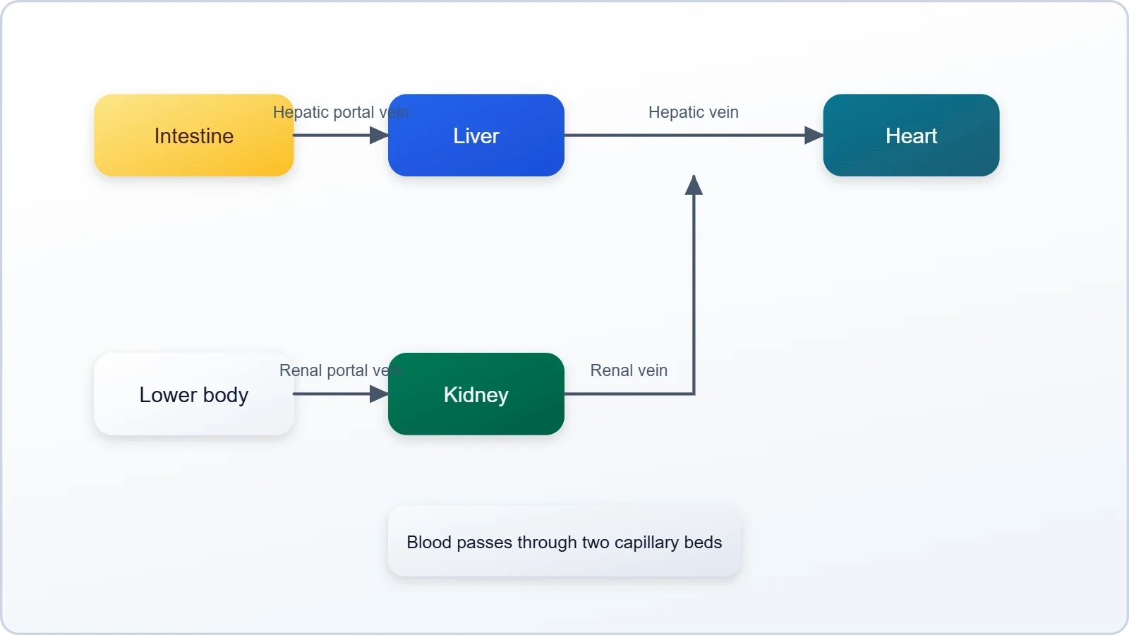
The functional logic is elegant. Blood arriving from the intestine carries absorbed nutrients, and routing it through the liver allows that organ to regulate what enters the systemic circulation. Blood from the lower body passes through the kidney, where the excretory tissue has an opportunity to remove nitrogenous waste before the blood returns to the heart. The renal portal system therefore links the vascular lesson directly to the excretory lesson that follows, and students should notice that connection. The two portal systems are named, located and functionally explained in the material, and each is a standard examination point.
The Excretory System: Kidneys, Ureters, Cloaca and Bladder
The elimination of nitrogenous wastes is carried out by a well-developed excretory system consisting of a pair of kidneys, ureters, cloaca and urinary bladder. The kidneys are compact, dark red, bean-like structures situated a little posteriorly in the body cavity on both sides of the vertebral column. Each kidney is composed of several structural and functional units called uriniferous tubules or nephrons. Excretory wastes are carried by blood into the kidney, where they are separated and excreted. The system is therefore a filtration and collection pathway, and its parts should be learned in that order rather than as an unconnected list.
Kidney Structure and the Nephron
The nephron, or uriniferous tubule, is the working unit of the kidney, and the material states that each kidney is composed of several such units. This is the same organisational principle seen throughout the course: cells form tissues, tissues form organs, and organs contain repeating functional units. The kidney's dark red colour reflects its rich blood supply, which is consistent with its role in receiving waste-laden blood from the renal portal system and from the general circulation. Positioned against the vertebral column, the paired kidneys sit where they can intercept blood returning from the lower body before it reaches the heart.
Students should be able to describe the kidney in three registers: its position, its appearance and its internal composition. Position: a little posteriorly in the body cavity, on both sides of the vertebral column. Appearance: compact, dark red and bean-like. Composition: several uriniferous tubules or nephrons. This three-part description is exactly the kind of structured answer that earns full marks, and it mirrors the descriptive style used for the heart earlier in the lesson. The parallel between the two organs, each a muscular or glandular mass with repeating units and a defined position, is worth noting.
Ureters, Cloaca and the Urinary Bladder
Two ureters emerge from the kidneys in the male frog, and these ureters act as a urinogenital duct that opens into the cloaca. In the female, the ureters and oviduct open separately in the cloaca. This sex difference is a favourite examination question and should be memorised precisely: in the male, one duct serves both urinary and genital functions; in the female, the two ducts remain distinct. The thin-walled urinary bladder is present ventral to the rectum, which also opens into the cloaca. The cloaca thus functions as a common chamber receiving multiple ducts, and the bladder stores urine before release.
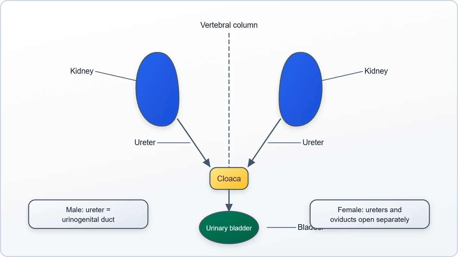
The term urinogenital duct deserves attention because it explains why the male frog's anatomy is described differently from the female's. A single channel carrying both urine and reproductive products is an economy of structure, and it is consistent with the reproductive system described in the next lesson, where vasa efferentia from the testes enter the kidney and open into Bidder's canal. For the present lesson, the essential point is the contrast between male and female duct arrangements. Students who can state that contrast cleanly, and locate the bladder ventral to the rectum, will handle the excretory questions with confidence.
Ureotelism and the Chemistry of Waste
The frog excretes urea and is therefore described as a ureotelic animal. This single term carries a great deal of physiology. Nitrogenous waste arises from the breakdown of proteins and nucleic acids, and different animals dispose of it in different chemical forms. The frog's choice of urea, rather than ammonia, reflects its amphibian habit: urea is far less toxic than ammonia and can be held in the body, and in the bladder, for a period before release. The excretory system is thus not merely a filter but a chemical strategy, and the term ureotelic is the label for that strategy.
From Blood to Bladder: The Excretory Route
The material states plainly that excretory wastes are carried by blood into the kidney, where they are separated and excreted. This gives a clean four-step route: waste enters the blood, blood reaches the kidney, the kidney separates the waste, and the waste is excreted. The renal portal system described earlier supports this route by directing blood from the lower body through the kidney. Once separated, the waste travels as urine through the ureters to the cloaca, and the thin-walled urinary bladder, lying ventral to the rectum, provides a storage chamber before final release.
It is worth connecting this route to the respiratory and digestive lessons. Protein-rich food digested in the intestine and absorbed through the villi ultimately yields nitrogenous waste, and the blood that carries absorbed nutrients also carries the waste products of their metabolism. The circulatory and excretory systems are therefore partners in a single cycle of intake, transport, processing and elimination. When students are asked to explain why the frog is ureotelic, the strongest answers link the chemistry of urea to the animal's semi-aquatic life and to the storage function of the urinary bladder.
Reading the Two Systems Together
Circulation and excretion are usually taught as separate chapters, but in the frog they are visibly interlocked. The renal portal system routes blood from the lower body through the kidney, so the excretory organ sits directly on a venous return path. The blood that carries nutrients and gases also carries nitrogenous waste, and the same pumping heart that distributes oxygen delivers that waste to the nephrons. Recognising this overlap turns two lists of organs into one integrated account of how a frog maintains its internal environment while moving between water and land.
Common Points of Confusion
Several errors recur in student answers. The heart is said to have four chambers when it has three. The sinus venosus is placed on the left atrium when it joins the right. The conus arteriosus is described as an inlet when it is an outlet from the ventricle. The frog is called ammonotelic when it is ureotelic. The ureters of the male are said to open separately when they act as a urinogenital duct. Each of these mistakes is a small slip with a large cost, and each is corrected by returning to the precise wording of the material and the tables above.
A second cluster of confusions concerns the lymphatic system. Students sometimes describe lymph as a second blood supply, but the material is explicit that lymph lacks a few proteins and lacks RBCs. It is a distinct fluid in a distinct network of lymph, lymph channels and lymph nodes. Similarly, the portal systems are sometimes merged into one; they are two, hepatic and renal, with different connections and different functions. Keeping these distinctions sharp is the difference between a passable answer and an excellent one, and the effort required is modest.
Going Further
A closed circulatory system keeps blood within vessels, which allows blood pressure and flow to be regulated more precisely. Because the fluid never spills into an open cavity, the heart can generate sustained pressure and vessels can adjust their diameter to direct blood where it is needed. In the frog, this precision supports the dual respiratory strategy: blood can be routed toward the skin or the lungs according to the animal's situation. The closed plan is therefore not a mere anatomical label but the condition that makes variable, controlled distribution possible across the body.
Ureotelic animals excrete nitrogenous waste mainly as urea, which is less toxic than ammonia and can be concentrated in urine. This chemical choice allows waste to be stored for a time rather than flushed away continuously, an advantage for an animal that may spend hours away from water. The thin-walled urinary bladder of the frog fits this strategy neatly, providing a reservoir before release through the cloaca. Together, the closed vascular plan and ureotelism describe an animal whose internal chemistry is adapted to a life divided between pond and land.
Consolidation Questions
Test understanding with a short set of prompts. Name the three chambers of the frog heart and state which structure joins the right atrium. Distinguish the hepatic portal system from the renal portal system in one sentence each. List the four organs of the excretory system in the order waste travels through them. Explain why the male frog's ureters are called a urinogenital duct while the female's are not. State what ureotelic means and name the waste product concerned. Finally, explain in two sentences why lymph cannot replace blood in gas transport, referring to its composition.
Answers should be written in full sentences and should use the material's own terminology. Where a comparison is requested, a two-column table is often the clearest format, and the tables in this lesson can serve as models. Where a definition is requested, the definition should be stated before any elaboration. These habits of precision are what the chapter rewards, and they carry forward into the next lesson, where the control and coordination systems, sense organs and reproductive organs complete the account of the frog's structural organisation.
Preparing for Control, Coordination and Reproduction
The vascular and excretory systems close the loop on transport and waste, but a frog also needs to sense its surroundings, coordinate its organs and reproduce. The next lesson takes up the neural system, with its central, peripheral and autonomic divisions, the endocrine glands, the sense organs and the well-organised male and female reproductive systems. Several threads from the present lesson will reappear there: the male urinogenital duct connects to the reproductive anatomy, and the blood supply described here serves every gland and gonad discussed next. The two lessons together complete the internal anatomy of the frog.
What to Carry Forward
Three ideas deserve to travel with the student into the final lesson. First, the frog's organs are arranged as repeating units within larger structures, whether nephrons in the kidney or chambers in the heart. Second, transport and elimination are linked by the blood, which carries both nutrients and waste. Third, structural details carry functional meaning, so a three-chambered heart, a pair of portal systems and a ureotelic excretory strategy are all answers to the same question: how does this animal live where it lives? Holding these ideas makes the remaining material easier to place.
Revision should be active rather than passive. Sketch the heart and label the sinus venosus, atria, ventricle and conus arteriosus. Draw the excretory organs and trace the route from kidney to ureter to cloaca to bladder. Write out the two portal systems from memory and check them against the tables. Then attempt the consolidation questions without notes. This cycle of recall, correction and re-recall is far more effective than rereading, and it prepares the ground for the neural, endocrine and reproductive material that follows in the concluding lesson of the course.
Also Read
- No links available.
From our network :
- Mastering DB2 LUW v12 Tables: A Comprehensive Technical GuideComprehensive guide on DB2 LUW v12 tables. Explore CREATE TABLE syntax, BLU Acceleration, range partitioning, data types, and security features for IBM DB2 LUW v12.
- Mastering DB2 12.1 Instance Design: A Technical Deep Dive into Modern Database ArchitectureComprehensive guide to DB2 12.1 Instance Design. Explore architectural components, HADR setup, buffer pool tuning, and security policies for IBM DB2 12.1. Includes 10 functional SQL and Shell samples.
- Trump Political Strategy How Geopolitical Stunts Serve As Media Diversions
- Vite 6/7 'Cold Start' Regression in Massive Module GraphsIn-depth analysis of Vite 6/7 cold start regressions in massive module graphs. Learn about barrel file issues, pre-bundling optimizations, and how to improve Vite dev server performance.
- 98% of Global MBA Programs Now Prefer GRE Over GMAT Focus EditionExplore why 98% of global MBA programs now prefer the GRE over GMAT Focus. Learn about the shorter GRE format, verbal logic importance, and strategic versatility for students.
- AI-Powered 'Precision Diagnostic' Replaces Standard GRE Score ReportsDiscover the new GRE Precision Diagnostic. Learn how AI-powered behavioral analytics and time-to-solve tracking are replacing standard GRE score reports to empower students.
- EV 2.0: The Solid-State Battery Breakthrough and Global Factory ExpansionDiscover the impact of the Solid-State Battery Breakthrough on the EV market. Learn about CATL and BYD stock growth, global factory expansion in Europe, and the future of EV 2.0 technology.
- 10 Physics Numerical Problems with Solutions for IIT JEEPrepare for IIT JEE with 10 physics numerical problems, complete with detailed solutions and conceptual explanations. Ideal for mastering kinematics, thermodynamics, electromagnetism, and optics.
- Analyzing Trump Deportation Numbers Insights Into The 2026 Immigration Crackdown
RESOURCES
- Resources will be updated soon.