Mutation is the engine of biological novelty and, simultaneously, one of the most consequential sources of human disease. Where recombination reshuffles existing genetic material, mutation actually alters the DNA sequence itself, changing the genotype and, frequently, the phenotype of an organism. This distinction matters enormously: recombination works with the cards already in the deck, while mutation writes new cards. Every heritable variation that natural selection has ever acted upon began as a mutation, and every inherited disorder traced through a family pedigree ultimately originates in a change to the genetic material carried in a chromosome.
This lesson examines mutation in three connected registers. First, the molecular anatomy of mutation: how single base substitutions, insertions, deletions, and chromosomal rearrangements arise and what they do to the reading frame of a gene. Second, the causes: the chemical and physical agents, collectively called mutagens, that raise mutation rates, together with the DNA repair systems that normally keep those rates astonishingly low. Third, the consequences: how particular mutation types map onto specific human disorders, how somatic mutations drive cancer through oncogenes and tumour suppressor genes, and why mutation is simultaneously destructive and creative.
Because the previous lesson established how sex is determined in humans and honey bees, the chromosomal machinery of inheritance is already familiar territory. This lesson builds directly on that foundation, treating the chromosome as a physical object that can break, duplicate, or mis-segregate, and treating the gene as a sequence that can be edited letter by letter. It also prepares the ground for the next lesson on pedigree analysis and Mendelian disorders, where the inheritance patterns of mutated alleles are traced across generations.
On This Page
The Molecular Anatomy of Mutation
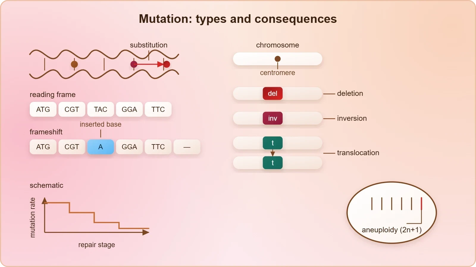
A mutation is any alteration in the DNA sequence of an organism, and the term covers an enormous range of scales. At the smallest scale, a single base pair may be exchanged for another. At the largest, whole segments of chromosomes may be lost, gained, inverted, or translocated to a different chromosome entirely. Between these extremes lie insertions and deletions of a few bases, which can be far more damaging than their size suggests because they shift the reading frame of a gene. Understanding mutation therefore requires thinking simultaneously at the level of the nucleotide and at the level of the chromosome.
The reference material captures this duality precisely. Loss (deletion) or gain (insertion or duplication) of a DNA segment alters the chromosome, and since genes are located on chromosomes, such alterations produce abnormalities or aberrations. Separately, mutation can arise from a change in a single base pair, which is termed a point mutation, with sickle cell anaemia as the classical example. Deletions and insertions of base pairs cause frameshift mutations. These categories are not merely descriptive labels; each produces a characteristic molecular outcome and a characteristic clinical signature.
Base Substitutions: Transitions and Transversions
Point mutations come in two chemically distinct flavours. A transition replaces a purine with a purine (adenine with guanine, or guanine with adenine) or a pyrimidine with a pyrimidine (cytosine with thymine, or thymine with cytosine). A transversion swaps a purine for a pyrimidine or vice versa. Transitions are more common in nature, partly because the deamination of cytosine produces uracil, which is read as thymine, generating a C-to-T transition. Within a coding sequence, a substitution may be silent, missense, or nonsense, depending on whether the new codon specifies the same amino acid, a different one, or a stop signal.
The sickle cell case illustrates a missense substitution with dramatic physiological reach. A single base substitution at the sixth codon of the beta globin gene changes GAG to GUG, replacing glutamic acid with valine at the sixth position of the beta chain. The mutant haemoglobin polymerises under low oxygen tension, deforming the red blood cell from a biconcave disc into an elongated sickle shape. One letter of DNA, one amino acid, one altered protein conformation, and a whole disease phenotype: the chain of causation is short and unforgiving.
Frameshift Mutations and Reading Frames
Because the genetic code is read in non-overlapping triplets, insertions or deletions that are not multiples of three destroy the reading frame from the point of the lesion onward. Every codon downstream is misread, and a premature stop codon usually appears within a short distance. The resulting protein is truncated and almost always non-functional. By contrast, an in-frame deletion of three bases removes exactly one amino acid and may leave the protein partly functional, which is why the position and length of a deletion matter as much as its existence.

Cystic fibrosis offers a textbook illustration of a small deletion with catastrophic consequences. The most common disease-causing allele, delta-F508, deletes three nucleotides and thereby removes phenylalanine at position 508 of the CFTR protein. Because the deletion is in frame, the protein is synthesised but misfolds and is degraded before reaching the membrane. Huntington's disease illustrates the opposite logic: expansion of a CAG trinucleotide repeat beyond a threshold produces an elongated polyglutamine tract, and the age of onset correlates inversely with repeat length. Expansion disorders are unstable across generations, a phenomenon called anticipation.
Chromosomal Aberrations and Aneuploidy
Mutations at the chromosomal scale operate by different rules. Because one DNA helix runs continuously from one end to the other in each chromatid, in a highly supercoiled form, the loss or gain of a segment alters the chromosome as a physical entity. Such alterations are called chromosomal aberrations, and they are commonly observed in cancer cells. Four structural classes dominate: deletions, duplications, inversions, and translocations. Each can disrupt genes at the breakpoint or alter gene dosage by changing copy number, and translocations can fuse two genes into a chimeric sequence with novel properties.
Numerical aberrations arise from failures in the mechanics of cell division. Failure of segregation of chromatids during the cell division cycle results in the gain or loss of a chromosome, a condition called aneuploidy. Down's syndrome results from the gain of an extra copy of chromosome 21, while Turner's syndrome results from the loss of an X chromosome in human females. A more drastic failure of cytokinesis after telophase produces an increase in a whole set of chromosomes, a phenomenon known as polyploidy, which is often seen in plants and is a major route to new plant species.

Structural Rearrangements in Detail
Inversions reverse a chromosomal segment and can be pericentric or paracentric depending on whether the centromere lies inside the inverted region. Carriers of inversions are usually phenotypically normal, but crossing over within the inverted loop during meiosis generates recombinant chromatids that are unbalanced and often inviable, which is why inversion heterozygotes show reduced fertility. Translocations exchange segments between non-homologous chromosomes; a reciprocal translocation carrier may be healthy yet produce a high proportion of unbalanced gametes, a recurrent theme in clinical cytogenetics.
Duplications are especially interesting from an evolutionary standpoint. A duplicated gene is released from selective constraint because one copy continues to perform the original function, allowing the second copy to accumulate mutations and diverge. This process, called neofunctionalisation when the duplicate acquires a new role, is thought to underlie the expansion of globin gene families and olfactory receptor repertoires. The globin clusters on chromosomes 16 and 11, with their tandem alpha and beta genes, are living records of ancient duplication events.
Counting Chromosomes: A Worked Example
The total number of chromosomes in a normal human cell is 46, comprising 23 pairs, of which 22 pairs are autosomes and one pair is sex chromosomes. Aneuploidy can be expressed as a simple deviation from this diploid number. If ##n## denotes the haploid number, then the normal diploid count is ##2n = 46##, and a trisomic cell carries ##2n + 1 = 47## chromosomes while a monosomic cell carries ##2n - 1 = 45##. Down's syndrome is therefore a trisomy 21 with 47 chromosomes, and Turner's syndrome is a 45,X monosomy.
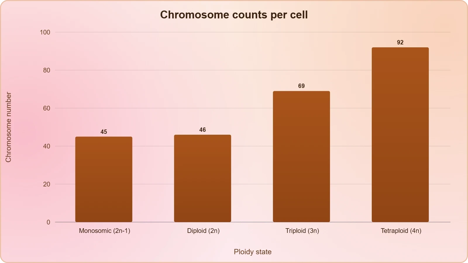
Polyploidy extends the same arithmetic to whole sets. A triploid cell carries ##3n = 69## chromosomes and a tetraploid cell carries ##4n = 92##. In plants, polyploidy is frequently tolerated and sometimes advantageous, producing larger fruits and flowers; in humans, triploidy is lethal. This contrast illustrates a general principle: the phenotypic consequence of a chromosomal change depends as much on the developmental context of the organism as on the change itself.
Mutagens, DNA Repair, and the Ames Test
Mutation rates in nature are kept low by two opposing forces: the intrinsic chemistry of DNA and the surveillance of repair enzymes. Yet many chemical and physical factors induce mutations, and these are referred to as mutagens. Ultraviolet radiation is a familiar physical mutagen; it creates pyrimidine dimers that distort the helix and block replication. Chemical mutagens include alkylating agents that add bulky groups to bases, base analogues that mimic normal nucleotides and mispair during replication, and intercalating agents that slip between base pairs and provoke frameshifts.
Cells defend themselves with an elaborate repair apparatus. Direct reversal enzymes such as photolyase remove UV dimers in many organisms, while base excision repair removes small damaged bases and nucleotide excision repair excises a short stretch of DNA around a bulky lesion. Mismatch repair corrects replication errors that escape proofreading, and double-strand breaks are repaired by homologous recombination or non-homologous end joining. When these systems fail, mutation rates rise sharply, a state known as a mutator phenotype, which is a common feature of cancer cells.
Detecting Mutagens: The Ames Test
The Ames test exploits a histidine-requiring strain of Salmonella typhimurium that cannot grow on histidine-free medium. When the bacteria are exposed to a test compound, any mutation that restores histidine synthesis allows the cells to form colonies. The number of revertant colonies is a direct index of mutagenic potency. Because many chemicals are only mutagenic after metabolic activation, the test is usually run with and without a liver extract (the S9 fraction), which supplies the activating enzymes.
The Ames test is a beautiful example of a biological assay built on a mutation principle. Its logic can be summarised as a ratio of revertants:
An index substantially above one signals mutagenicity. The test is fast, inexpensive, and highly predictive of carcinogenicity, which is why it remains a standard first screen in toxicology. Its limitation is equally instructive: it detects point mutations in bacteria, so it may miss chromosomal aberrations or mutations specific to mammalian cells.
From Mutation to Mendelian Disorder
The clinical payoff of mutation theory is that specific mutation types map onto specific inherited disorders, and the mapping is often remarkably precise. Sickle cell anaemia, as established, is an autosome-linked recessive trait transmitted when both partners are carriers. The disease is controlled by a single pair of alleles, HbA and HbS. Of the three possible genotypes, only homozygous HbS HbS individuals show the diseased phenotype, while heterozygous HbA HbS individuals appear unaffected but carry the disease with a fifty per cent probability of transmitting the mutant gene to each child.
Thalassemia provides a revealing contrast. It is also an autosome-linked recessive blood disease, but the defect may arise from either mutation or deletion, and it reduces the rate of synthesis of one of the globin chains. Alpha thalassemia is controlled by two closely linked genes, HBA1 and HBA2, on chromosome 16 of each parent, and the more genes affected, the less alpha globin produced. Beta thalassemia is controlled by a single gene, HBB, on chromosome 11. Crucially, thalassemia is a quantitative problem of synthesising too few globin molecules, whereas sickle cell anaemia is a qualitative problem of synthesising an incorrectly functioning globin.
Recessive, Dominant, and X-Linked Signatures
Phenylketonuria illustrates an inborn error of metabolism inherited as an autosomal recessive trait. The affected individual lacks the enzyme that converts phenylalanine into tyrosine, so phenylalanine accumulates and is converted into phenylpyruvic acid and other derivatives. Accumulation of these in the brain results in mental retardation, and they are excreted through urine because of poor absorption by the kidney. Colour blindness, by contrast, is a sex-linked recessive disorder caused by a defect in either the red or green cone of the eye, arising from mutation in genes on the X chromosome.
The population genetics of X-linked recessives follows directly from the chromosome count. Colour blindness occurs in about eight per cent of males but only about 0.4 per cent of females, because males have only one X chromosome while females have two. The son of a carrier woman has a fifty per cent chance of being colour blind; the mother herself is unaffected because the recessive gene is suppressed by her matching dominant normal allele. A daughter will not normally be colour blind unless her mother is a carrier and her father is colour blind. Haemophilia follows the same logic, transmitted from unaffected carrier females to some male progeny.
Pedigree Logic as a Diagnostic Tool
Because controlled crosses cannot be performed in humans, the study of family history across several generations, called pedigree analysis, provides the alternative. In pedigree analysis the inheritance of a particular trait is represented in a family tree over generations, and standard symbols distinguish males, females, affected individuals, carriers, and deceased family members. By inspecting the pattern, one can determine whether a trait is dominant or recessive and whether it is autosomal or sex-linked. A trait that appears in every generation suggests dominance; a trait that skips generations and appears among the offspring of unaffected parents suggests recessiveness.
The family pedigree of Queen Victoria is the most celebrated case study in the literature: as a carrier of haemophilia, she transmitted the disease to a number of her descendants across European royal houses. The pattern is diagnostic. Affected individuals are almost exclusively male, the trait passes through unaffected females, and no affected father transmits the disease to his sons, because he contributes a Y chromosome to them. These three observations together identify an X-linked recessive mode of inheritance with near certainty.
Somatic Mutation, Cancer, and the Two Faces of Variation
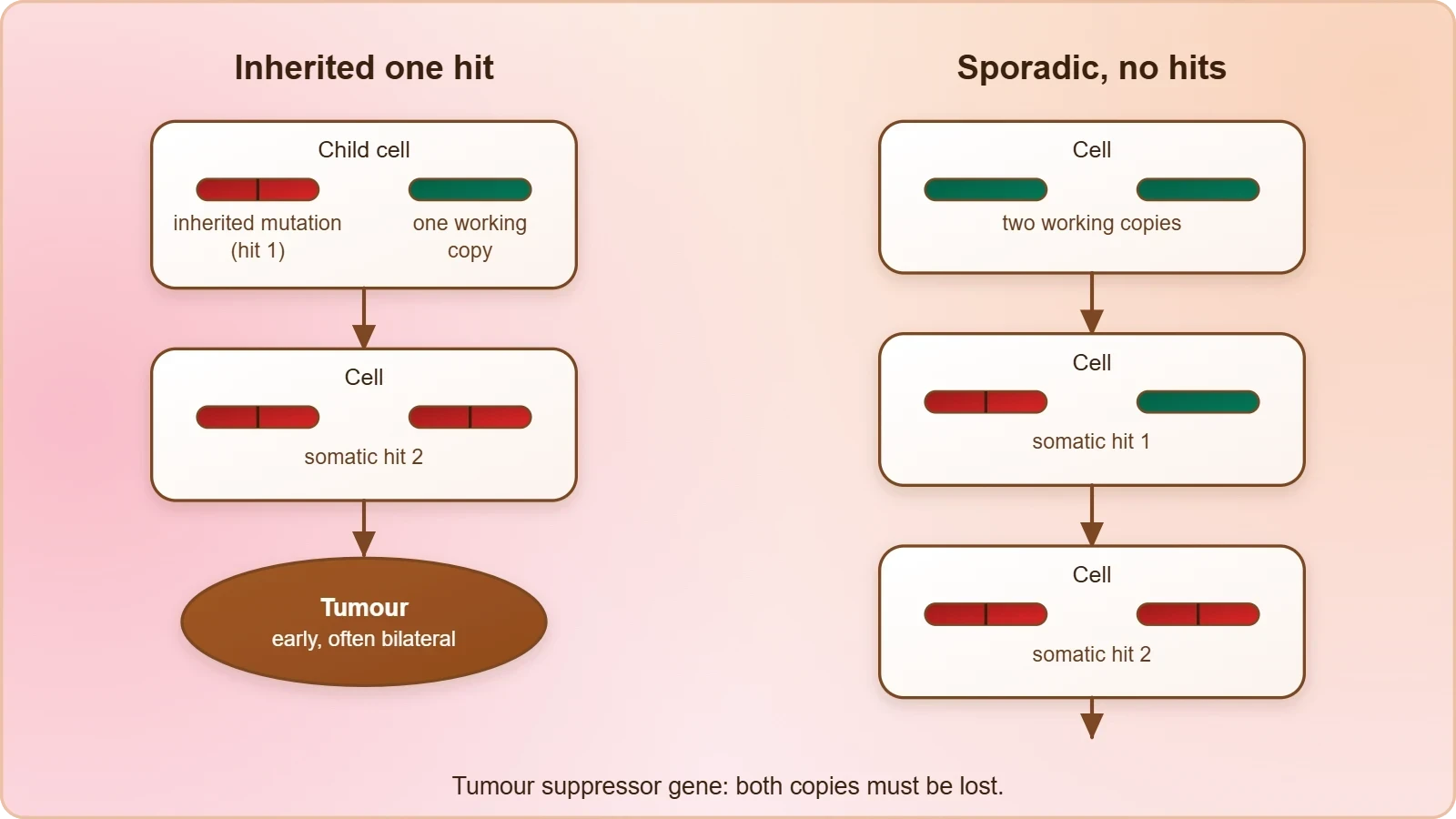
Not all mutations are inherited. Mutations in germ cells are transmitted to offspring and are the raw material of evolution, but mutations in somatic cells affect only the individual in which they arise. Somatic mutation is the central mechanism of cancer. A single mutation is rarely sufficient; malignancy typically requires the sequential accumulation of mutations in two classes of genes. Oncogenes are activated by gain-of-function mutations that make the encoded protein constitutively active, while tumour suppressor genes are inactivated by loss-of-function mutations, usually requiring both alleles to be disabled.
The two-hit hypothesis, formulated by Alfred Knudson for retinoblastoma, explains why inherited cancer syndromes often present earlier and more severely than sporadic cases. A child who inherits one defective copy of a tumour suppressor gene needs only one additional somatic mutation to lose both functional alleles, whereas a person born with two normal copies needs two independent somatic hits. The same logic applies to the chromosomal aberrations commonly observed in cancer cells, which frequently include translocations that activate oncogenes or deletions that remove suppressor loci.
Beneficial, Neutral, and Harmful Outcomes
Mutation is often described as an error, but the framing is incomplete. Most mutations are neutral, falling in non-coding regions or producing synonymous changes. Some are harmful, disrupting protein function or regulation. A minority are beneficial, and these are the ones natural selection preserves. The persistence of sickle cell trait in malaria-endemic regions is the classic demonstration: heterozygous carriers enjoy substantial resistance to Plasmodium falciparum, so a mutation that is lethal in homozygotes is maintained at high frequency by heterozygote advantage.
This balance explains why mutation is simultaneously a source of disease and a source of genetic variation. Without mutation, populations could not adapt to changing environments; with too much mutation, genomes would collapse under mutational load. The equilibrium is maintained by repair enzymes, by selection, and by the sheer redundancy of the genetic code. Mutation is therefore best understood not as a malfunction but as an unavoidable consequence of chemistry, managed by biology and exploited by evolution.
Common Misconceptions Worth Correcting
Several errors recur in student answers. Mutation is not directed by need; bacteria do not mutate in response to antibiotics, they are selected by them. Mutation is not synonymous with harm; the majority are neutral and some are advantageous. A point mutation is not always a missense mutation; it may be silent or nonsense. And chromosomal aberration is not the same as gene mutation; the former alters chromosome structure or number, the latter alters the nucleotide sequence. Keeping these categories distinct is essential for accurate pedigree interpretation in the next lesson.
Quantifying Mutation and Modelling Its Effects
Mutation can be treated quantitatively, and doing so sharpens intuition about why repair matters. Suppose a genome of ##N## base pairs is replicated with a per-base error probability ##\mu##. The expected number of new mutations per replication is simply ##N\mu##. For a human genome of roughly three billion base pairs and an uncorrected error rate near ##10^{-5}##, the expected count would be enormous; proofreading and mismatch repair reduce the effective rate to about ##10^{-9}##, bringing the expected number down to a handful per generation. The arithmetic makes the value of repair vivid.
The same logic applies to the probability that a specific codon is hit. If a gene contains ##L## codons, the probability that a given replication introduces a mutation somewhere in its coding sequence is approximately ##3L\mu##, assuming uniform mutation probability. For a gene of a thousand codons and a rate of ##10^{-9}##, this is about ##3 \times 10^{-6}## per replication, which is why most individual genes are not mutated in any given generation but why, across a population, every gene is mutated somewhere.
A Short Computational Illustration
Programming makes these calculations concrete and lets students explore how mutation rates and repair efficiencies interact. The snippet below simulates point mutations across a sequence and reports the resulting substitution spectrum, distinguishing transitions from transversions. It is deliberately simple so that the logic of the classification is visible in the code itself.
import random

bases = "ACGT"
purines = {"A", "G"}

def classify(original, mutated):
    if original == mutated:
        return "unchanged"
    if (original in purines) == (mutated in purines):
        return "transition"
    return "transversion"

def simulate(sequence, rate):
    counts = {"transition": 0, "transversion": 0, "unchanged": 0}
    for base in sequence:
        if random.random() < rate:
            new_base = random.choice([b for b in bases if b != base])
            counts[classify(base, new_base)] += 1
        else:
            counts["unchanged"] += 1
    return counts

seq = "".join(random.choice(bases) for _ in range(10000))
print(simulate(seq, 0.01))
Running this simulation with a mutation rate of one per cent produces roughly twice as many transitions as transversions, because for any given base there is exactly one transition partner but two transversion partners. This simple combinatorial asymmetry explains a pattern observed in real mutation spectra and in disease-causing variants catalogued in clinical databases. Students who run the code and then compare the output with published spectra gain an appreciation for how theoretical models connect to empirical data.
Mutation, Variation, and the Next Lesson
The conceptual arc of this lesson runs from the nucleotide to the population. A single base substitution can alter one amino acid and produce sickle cell anaemia; a three-base deletion can remove one residue and produce cystic fibrosis; a trinucleotide expansion can lengthen a protein tract and produce Huntington's disease; a failure of chromatid segregation can add a chromosome and produce Down's syndrome. Each of these is a mutation, and each illustrates a different point on the spectrum from molecular lesion to clinical phenotype.
Mutation is also the ultimate source of the variation that the previous lessons described. Recombination reshuffles alleles, but mutation creates them. The pedigree analysis that follows in the next lesson depends entirely on the existence of mutated alleles whose inheritance can be traced through families. Without mutation there would be no variant alleles, no recessive carriers, no dominant pedigrees, and no genetic disease. Mutation is thus both the origin of the variation that genetics studies and the origin of the disorders that clinical genetics seeks to understand.
Consolidating the Lesson: Key Takeaways
The material in this lesson can be compressed into a small set of durable propositions. Mutation is an alteration of DNA sequence that changes genotype and often phenotype, and it is, alongside recombination, the second great source of variation. Point mutations change single base pairs, with sickle cell anaemia as the classical example. Insertions and deletions that are not multiples of three cause frameshift mutations. Chromosomal aberrations arise from loss or gain of DNA segments and are commonly observed in cancer cells. Mutagens are the chemical and physical factors, including ultraviolet radiation, that induce these changes.
Beyond the reference material, the lesson has added the molecular vocabulary of transitions and transversions, the distinction between silent, missense, and nonsense substitutions, and the clinical logic that connects specific lesions to specific syndromes. It has introduced DNA repair as the counterweight to mutagenesis, the Ames test as a practical assay for mutagenicity, and the two-hit model as the explanation for why inherited cancer syndromes present early. It has also insisted that mutation is not inherently harmful: most mutations are neutral, some are beneficial, and all are the raw material of evolution.
Self-Assessment Questions
Test your understanding with the following. First, distinguish a transition from a transversion and explain why transitions are more frequent. Second, explain why a deletion of four bases is usually more damaging than a deletion of three. Third, describe the molecular lesion in sickle cell anaemia and trace its consequences from codon to cell shape. Fourth, explain why colour blindness is far more common in males than in females. Fifth, outline the Ames test and state its principal limitation. Sixth, distinguish an oncogene from a tumour suppressor gene and explain Knudson's two-hit hypothesis.
For each question, aim to write a paragraph that moves from the molecular level to the phenotypic level without skipping steps. The discipline of tracing causation explicitly is what distinguishes a strong genetics answer from a list of remembered facts. In the next lesson, these same skills will be applied to pedigree analysis and Mendelian disorders, where the inheritance of mutated alleles is reconstructed from family trees rather than from molecular data.
Looking Ahead to Pedigree Analysis
The transition to the next lesson is natural. Having established what mutations are and how they produce disorders, the question becomes how those disorders move through families. Pedigree analysis answers that question by representing inheritance as a family tree over generations, using standard symbols to distinguish affected individuals, carriers, and unaffected relatives. The patterns established here, autosomal recessive for sickle cell anaemia and phenylketonuria, X-linked recessive for haemophilia and colour blindness, autosomal dominant for myotonic dystrophy, become the diagnostic signatures that pedigree charts reveal.
Students who master the mutation categories in this lesson will find pedigree interpretation far easier, because each pedigree pattern is ultimately a consequence of a particular kind of mutation segregating in a particular way. The recessive disorders require two mutant alleles and therefore appear among the offspring of unaffected carriers; the X-linked disorders appear predominantly in males because they carry only one X chromosome. Mutation theory and pedigree logic are two views of the same underlying biology, and together they form the analytical core of human genetics.
Also Read
- No links available.
From our network :
- Where to Put Your Wireless Router for Better CoverageA practical, physics-grounded guide to router placement: why the centre of the home beats the corner, how walls and metal sap signal, and how to run a simple survey that proves the improvement.
- 98% of Global MBA Programs Now Prefer GRE Over GMAT Focus EditionExplore why 98% of global MBA programs now prefer the GRE over GMAT Focus. Learn about the shorter GRE format, verbal logic importance, and strategic versatility for students.
- Trump Political Strategy How Geopolitical Stunts Serve As Media Diversions
- Analyzing Trump Deportation Numbers Insights Into The 2026 Immigration Crackdown
- 10 Physics Numerical Problems with Solutions for IIT JEEPrepare for IIT JEE with 10 physics numerical problems, complete with detailed solutions and conceptual explanations. Ideal for mastering kinematics, thermodynamics, electromagnetism, and optics.
- EV 2.0: The Solid-State Battery Breakthrough and Global Factory ExpansionDiscover the impact of the Solid-State Battery Breakthrough on the EV market. Learn about CATL and BYD stock growth, global factory expansion in Europe, and the future of EV 2.0 technology.
- Do You Need a New Router or Is Your Internet Plan the Problem?A practical comparison of the signs that point to router limits versus signs your broadband speed or provider is the bottleneck, so you can decide what to replace before spending money.
- How to Print Cheaply: Cutting the Cost per PageA practical, measured guide to lowering what each printed page costs — duplex and draft modes, third-party ink, yield mathematics, and knowing when a new printer beats refills.
- AI-Powered 'Precision Diagnostic' Replaces Standard GRE Score ReportsDiscover the new GRE Precision Diagnostic. Learn how AI-powered behavioral analytics and time-to-solve tracking are replacing standard GRE score reports to empower students.
RESOURCES
- Types of mutations and their notations (article) - Khan Academykhanacademy.orgDepending on the size of the deletion and the genes affected, this can cause severe developmental or functional consequences. Translocation (single-chromosome): ...
- Types of mutations - Understanding Evolution - UC Berkeleyevolution.berkeley.educhange a codon to one that encodes the same amino acid and causes no change in the protein produced. These are called silent mutations.…
- Mutations (Updated) - YouTubeyoutube.comJul 31, 2019 ... ) about a plant species and mutations was published and furthers ... mutation -not having symptoms. 8:07 · Go to…
- TP53 Mutations in Human Cancers: Origins, Consequences ... - PMCpmc.ncbi.nlm.nih.govTumor spectrum in individuals with a germline TP53 mutation. The proportion of specific tumor types among all tumors reported in confirmed TP53 germline ...
- What Is a Genetic Mutation? Definition & Types - Cleveland Clinicmy.clevelandclinic.orgMay 24, 2022 ... If part of your DNA sequence is in the wrong place, isn't complete or is damaged, you might experience symptoms…
- The dark side of centromeres: types, causes and consequences of ...nature.comOct 18, 2018 ... If not solved correctly, the ensuing replication termination sites can be the source of DNA breaks as observed in the…
- Paternal Age Effect Mutations and Selfish Spermatogonial Selectionsciencedirect.comFeb 10, 2012 ... Paternal Age Effect Mutations and Selfish Spermatogonial Selection: Causes and Consequences for Human Disease ... Thanatophoric dysplasia (types I ...
- Mutations types and causes | PPTX - Slideshareslideshare.netMUTATION • A mutation occurs when DNA or it's gene is damaged/ altered in a.
- PRESENILIN 1 Mutations Causing Early-Onset Familial Alzheimer's ...pubmed.ncbi.nlm.nih.govThe molecular consequences of heterozygosity for these mutation types, and how they cause completely different diseases, remains largely unexplored.
- Mutation and its types - ADP Collegeadpcollege.ac.inIf the numbers of gene replicas are non-equivalent on the homologous chromosomes, they may cause different types of phenotypic effects over the organisms. 3.
- Mutation, Repair and Recombination - Genomes - NCBI Bookshelfncbi.nlm.nih.govThis definition is important because it distinguishes mutagens from other types of environmental agent that cause damage to cells in ways other than by…
- Mutation – A Genetic Change - BYJU'Sbyjus.comJul 26, 2019 ... Causes of Mutations ... The mutation leads to genetic variations among species. Positive mutations are transferred to successive generations. E.g. ...
- Origins and functional consequences of somatic mitochondrial DNA ...elifesciences.orgOct 1, 2014 ... Several possible explanations could underpin these differences across tumor types. It could be that the mutation rates differ across cell ...
- Adaptive Value of High Mutation Rates of RNA Viruses - ASM Journalsjournals.asm.orgAdaptive Value of High Mutation Rates of RNA Viruses: Separating Causes from Consequences ... Mutation Types · Quasispecies Dynamics · Selection and Fitness ...
- Mechanisms of Mutation - UT Health San Antonioww2.uthscsa.eduThis review will focus on the types, causes, and consequences of mutation. Types of Mutation. The following table displays schematics illustrating the ...