Microorganisms constitute the invisible majority of life on Earth, and their story is one of the most consequential in all of biology. Long before plants and animals diversified, microbes were already shaping the chemistry of the planet, and they continue to govern processes that range from the digestion of a meal to the treatment of municipal wastewater. This opening lesson of the course establishes the conceptual foundation for everything that follows: what microbes are, where they live, how scientists classify and study them, and why the widespread assumption that they are simply agents of disease is one of the most misleading ideas in popular science. By the end, learners should be able to distinguish harmful from beneficial microbes with confidence and appreciate the sheer scale of microbial influence on human welfare.
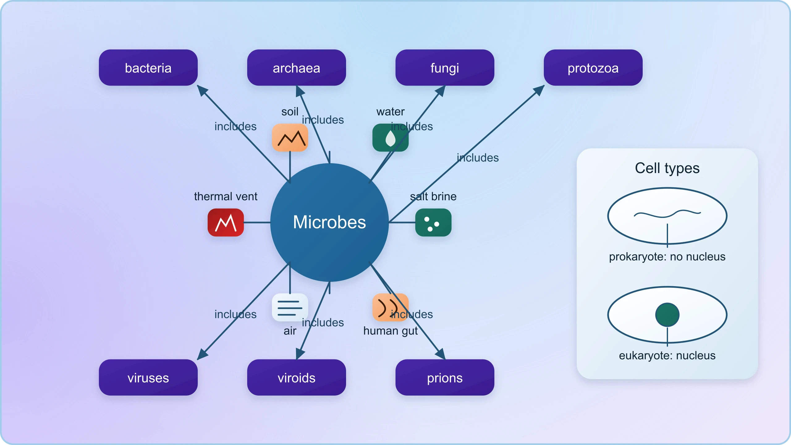
The reference material for this lesson makes a deceptively simple point: besides macroscopic plants and animals, microbes are the major components of biological systems on this earth. That sentence deserves careful unpacking. It means that the visible living world is a minority report, and that the ecological and biochemical machinery sustaining life is largely microbial. Microbes are present everywhere — in soil, water, air, inside our bodies and those of other animals and plants — and they persist even at sites where no other life-form could possibly exist, such as deep inside geysers and thermal vents where temperatures may reach 100°C, deep in the soil, under layers of snow several metres thick, and in highly acidic environments. This ubiquity is not incidental; it is the direct consequence of extraordinary metabolic versatility.
This lesson also sets the intellectual stage for the eleven lessons that follow. It introduces the diversity of microorganisms, their ubiquity, and the crucial distinction between harmful and beneficial microbes, preparing learners for detailed treatments of household products, industrial fermentation, sewage treatment, biogas production, biocontrol and biofertilisers. Because the course builds cumulatively, the concepts established here — the taxonomic groups, the prokaryote–eukaryote divide, the idea of the microbiome, and the historical arc from germ theory to modern microbial ecology — will be assumed knowledge in every subsequent lesson. Learners should treat this as the map before the territory.
On This Page
- The Invisible Majority: Defining and Classifying Microorganisms
- Ubiquity and Abundance: Where Microbes Live and How Many
- Studying the Unseen: Culture, Colonies and the Germ Theory
- Harmful and Beneficial: Correcting the Great Misconception
- Microbial Diversity in Numbers: Scale, Growth and Measurement
- From Diversity to Application: Preparing for the Lessons Ahead
The Invisible Majority: Defining and Classifying Microorganisms
Defining a microorganism is less straightforward than it appears, because the category is functional rather than evolutionary. Microbes are simply organisms too small to be seen clearly with the naked eye, which means the term gathers together groups that are only distantly related. The reference material lists them plainly: protozoa, bacteria, fungi, and microscopic animal and plant viruses, viroids, and also prions that are proteinaceous infectious agents. Notice that this list crosses every major boundary of biology. Bacteria are prokaryotes, fungi and protozoa are eukaryotes, viruses are not cells at all, viroids are naked RNA molecules, and prions are infectious proteins with no nucleic acid whatsoever. The word "microbe" is therefore a convenience of scale, not a statement of kinship.
Prokaryotic and Eukaryotic Microbes
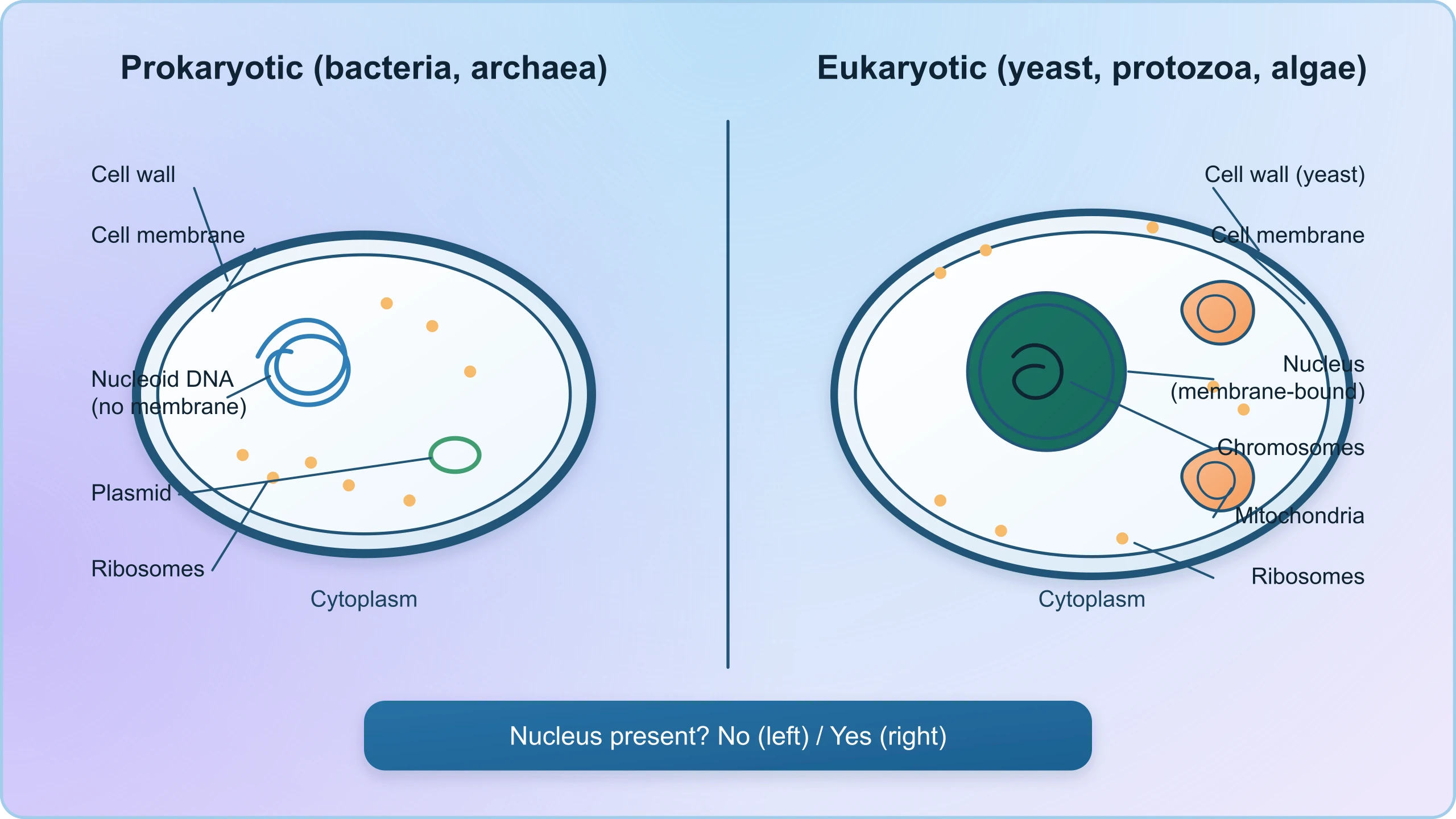
The most important structural distinction in this lesson is between prokaryotic and eukaryotic microbes. Prokaryotes, which include all bacteria and the archaea, lack a membrane-bound nucleus and other organelles; their genetic material sits in a nucleoid region, and their ribosomes differ sufficiently from ours to be a target for antibiotics. Eukaryotic microbes — fungi, protozoa and microscopic algae — possess a true nucleus, mitochondria and internal membrane systems, and are therefore biochemically closer to human cells. This distinction matters practically: it explains why antibacterial drugs can be selective, and why antifungal medicines are harder to design without side effects, since fungal cells resemble our own.
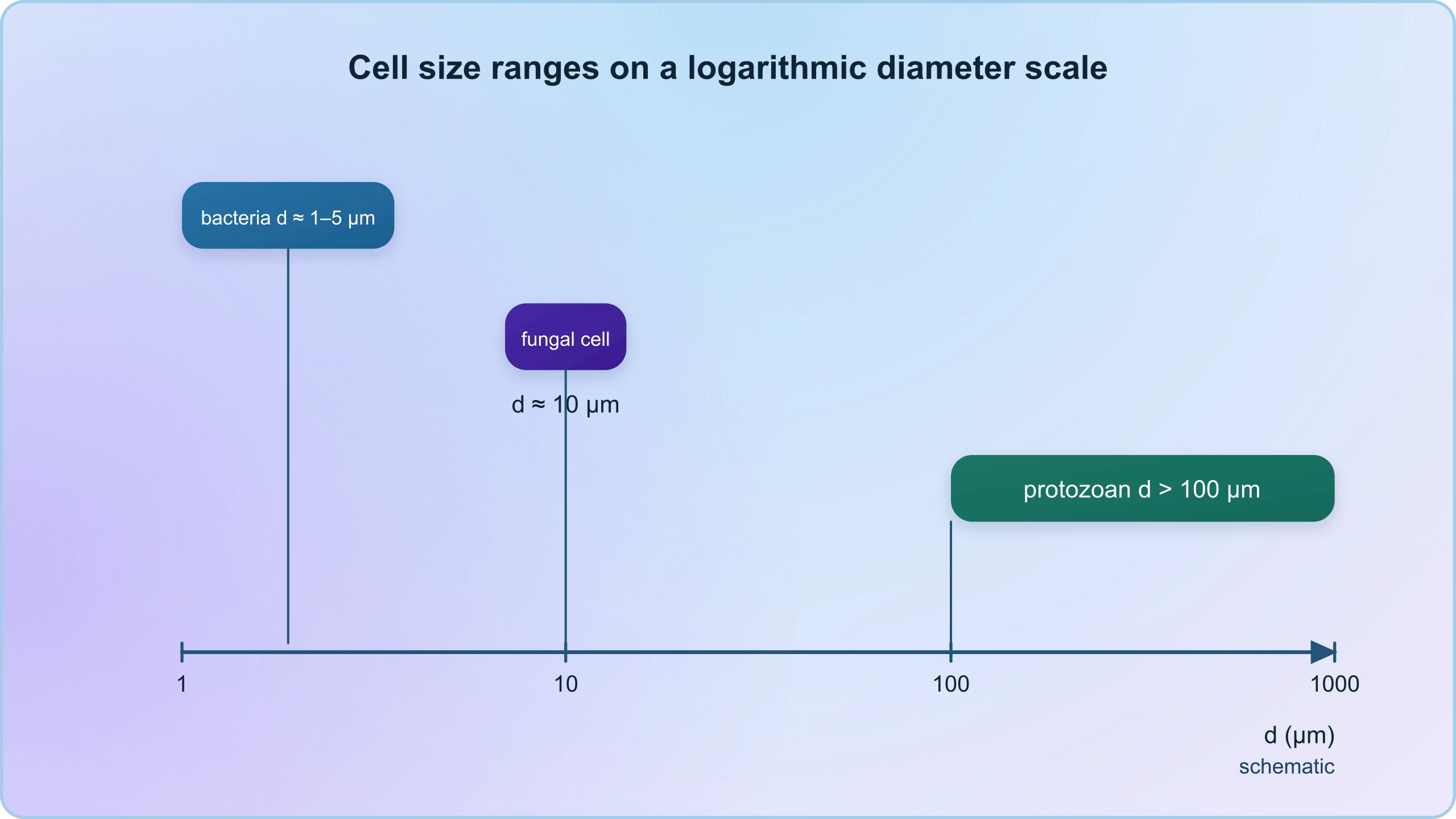
A useful way to anchor the difference is through scale and organisation. A typical bacterium might measure one to five micrometres, while a yeast cell is roughly five to ten micrometres and a protozoan can exceed one hundred. The size relationship can be expressed simply as ##d \approx 1\text{–}5\ \mu m## for most bacteria, compared with ##d \approx 10\ \mu m## for a typical fungal cell. These dimensions explain why the colonies shown in culture plates become visible only after millions of cells accumulate, and why magnification of 1500X is needed to resolve individual rod-shaped or spherical bacteria, while viruses require magnifications of about 1,00,000 to 1,50,000X.
Archaea: The Extremophile Third Domain
Archaea deserve separate attention because they are frequently folded into "bacteria" in casual speech, which is a serious error. Molecular phylogeny revealed that archaea form a distinct domain, as different from bacteria as both are from eukaryotes. Their distinguishing feature for our purposes is habitat: thermophiles thrive at temperatures near or above 100°C in thermal vents and geysers, halophiles flourish in saturated salt brines, and methanogens produce methane in anaerobic environments such as swamp sediments, rice paddies and the rumen of cattle. The reference material's observation that microbes live where no other life-form could possibly exist is, in large part, a description of archaeal ecology.
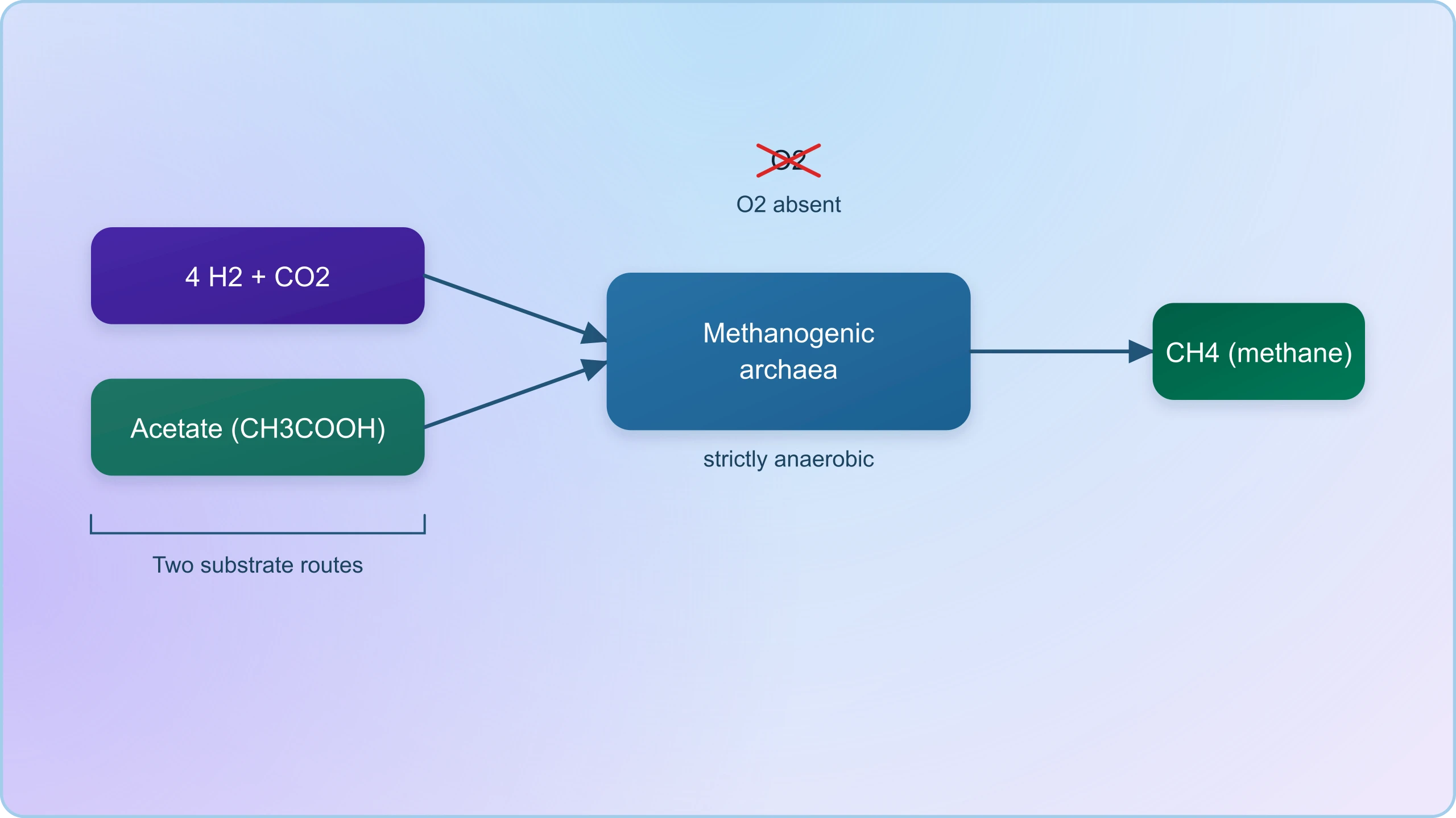
Methanogens are especially relevant to this course because they return in the lesson on biogas production. These archaea convert hydrogen and carbon dioxide, or acetate, into methane through anaerobic respiration, a process that can be summarised as ##\text{CO}_2 + 4\text{H}_2 \rightarrow \text{CH}_4 + 2\text{H}_2\text{O}##. Understanding that this is an archaeal process rather than a bacterial one corrects a common textbook shorthand and prepares learners for the metabolic detail in later lessons. It also illustrates a broader principle: microbial diversity is not merely taxonomic curiosity but the source of the biochemical capabilities that human welfare depends upon.
Ubiquity and Abundance: Where Microbes Live and How Many
The reference material's catalogue of extreme habitats is worth revisiting with fresh eyes, because each environment represents a solved biochemical problem. Thermal vents near 100°C demand heat-stable proteins and membranes, which is why enzymes from such organisms power laboratory PCR. Several metres of snow impose cold and high pressure, favouring psychrophiles with flexible membranes. Highly acidic environments select for organisms that maintain internal pH near neutral while exporting protons. Deep soil offers low oxygen and scarce nutrients, rewarding organisms with versatile anaerobic metabolism. In each case, the microbial solution is a lesson in adaptation that later lessons will exploit industrially.
Extremes as a Window into Adaptation
It is tempting to treat extremophiles as curiosities, but their enzymes are commercially indispensable. Taq polymerase, the workhorse of the polymerase chain reaction, originates from a thermophilic bacterium of hot springs, and its stability at high temperature is what allows repeated heating cycles without denaturation. Similarly, halophilic enzymes function in high-salt conditions useful for certain industrial catalysis. The broader principle is that microbial habitats are a library of molecular solutions, and human welfare has repeatedly advanced by borrowing from that library. When learners encounter industrial microbiology later in the course, they should recall that the organisms involved were selected by evolution in environments far harsher than a fermentor.
The Human Microbiome
Microbes are not merely around us; they are within us, in numbers that defy intuition. Estimates place the total microbial population of the human body in the tens of trillions of cells, comparable in count to human cells themselves, with the greatest density in the large intestine. This resident community, collectively the microbiome, contributes to digestion of complex carbohydrates, synthesis of certain vitamins including vitamin K and some B vitamins, maturation of the immune system, and exclusion of pathogens through competition for space and nutrients. The reference material's remark that lactic acid bacteria in the stomach check disease-causing microbes is a small window onto this much larger phenomenon of colonisation resistance.
The microbiome concept also reframes disease. Rather than a simple binary of sterile health versus infected illness, modern understanding describes a dynamic community whose composition shifts with diet, antibiotics, age and environment. Dysbiosis, a disruption of this community, is associated with conditions ranging from inflammatory bowel disease to metabolic disorders. For learners, the practical implication is that antibiotics are not free: they target pathogens but also disturb beneficial communities, which is why prudent prescribing matters. This nuance prepares the ground for later lessons on fermented foods and probiotics, where the deliberate cultivation of beneficial microbes becomes a tool for welfare rather than a hazard.
Studying the Unseen: Culture, Colonies and the Germ Theory
Because microbes are invisible individually, the science of microbiology depends on making them visible collectively. The reference material notes that bacteria and many fungi can be grown on nutritive media to form colonies that can be seen with the naked eye, and that such cultures are useful in studies on micro-organisms. A colony is a population descended from a single cell, so a plate streaked carefully can yield isolated colonies, each a pure clone. This simple technique underpins identification, antibiotic sensitivity testing and industrial strain selection. The puffed-up dough of a fermented batter and the fuzzy patch on stale bread are, in essence, colonies encountered outside the laboratory.
From Invisible Cells to Visible Colonies
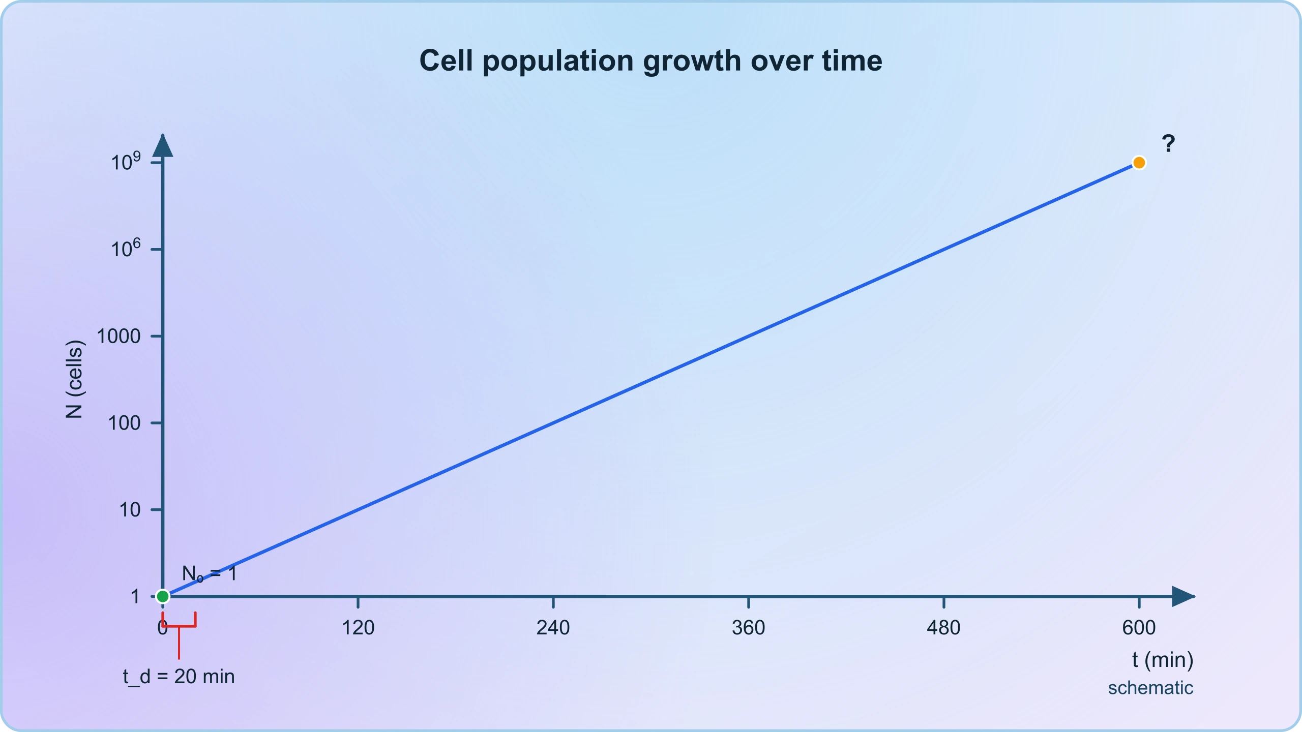
The mathematics of colony formation is instructive. If a single bacterium divides every twenty minutes under ideal conditions, the population follows exponential growth, ##N(t) = N_0 \cdot 2^{t/20}##, where ##t## is measured in minutes. After ten hours, or six hundred minutes, the exponent is thirty, giving roughly one billion cells from one. This is why a colony becomes visible within a day and why food spoilage can be rapid. It also explains why industrial fermentation requires careful control: the same exponential law that makes a starter culture efficient can make contamination catastrophic within hours if a foreign organism gains a foothold.
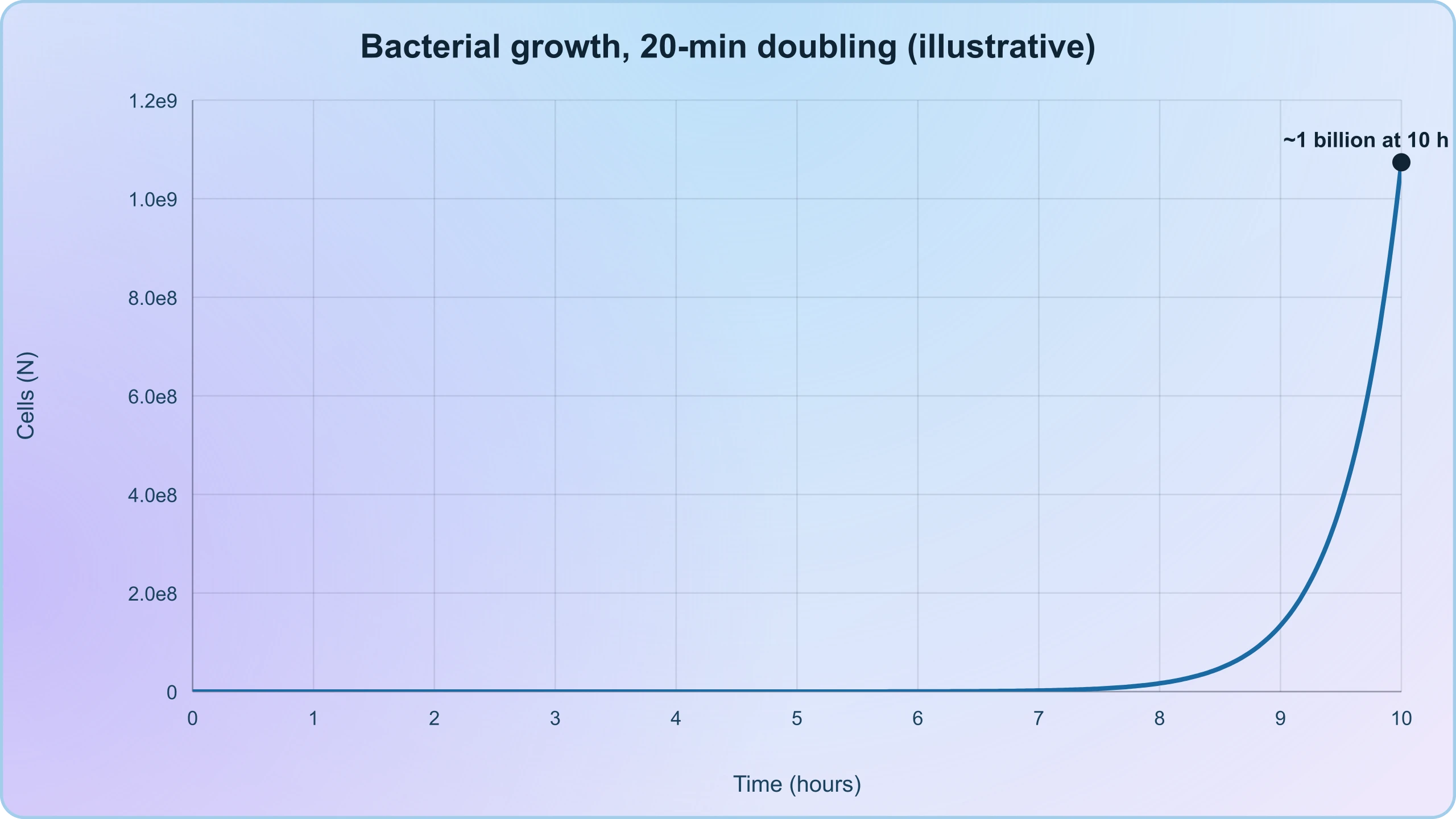
Practical work reinforces the point. A learner streaking a plate and incubating it overnight performs, in miniature, the same logic used in hospital diagnostics and in the dairy industry. The colony morphology — size, colour, edge, elevation — offers a first clue to identity, while biochemical tests and molecular methods confirm it. The reference material's figures showing rod-shaped and spherical bacteria at 1500X, a flagellated rod at 50,000X, and viruses at 1,00,000 to 1,50,000X illustrate the range of magnification required, and remind us that colony visibility and cellular visibility are two different thresholds entirely.
Koch's Postulates and the Germ Theory
The historical context for this lesson is the germ theory of disease, which displaced the older miasma explanation of illness during the nineteenth century. Robert Koch formalised the logic by which a specific microbe could be shown to cause a specific disease, and his criteria remain a model of rigorous causal reasoning. The four postulates state that the organism must be found in all cases of the disease, must be isolated and grown in pure culture, must reproduce the disease when introduced into a healthy host, and must be re-isolated from that host. Koch's identification of the tubercle bacillus and his work on anthrax and cholera established microbiology as a discipline.
These postulates are worth teaching not as dogma but as a template for causal inference, with acknowledged limits. Some pathogens cannot be cultured easily, some diseases require cofactors, and ethical constraints prevent deliberate infection of humans. Molecular equivalents, such as detecting pathogen nucleic acid consistently in diseased tissue, have extended the logic. The essential lesson for learners is that the germ theory was a triumph of evidence over assumption, and that the same discipline of controlled observation should be applied to claims about beneficial microbes. Not every microbe is a pathogen, and not every correlation is causation.
Harmful and Beneficial: Correcting the Great Misconception
The single most important correction this lesson must deliver is that the equation of microbes with disease is false. The reference material states it directly: microbes cause a large number of diseases in human beings, and they also cause diseases in animals and plants, but this should not make you think that all microbes are harmful, since several microbes are useful to man in diverse ways. Pathogens are a minority, and even that minority is context-dependent. An organism harmless in the gut can be dangerous in the bloodstream; a soil bacterium benign to a healthy adult may threaten an immunocompromised patient. Pathogenicity is a property of the interaction, not a fixed label attached to a species.
Why the Misconception Persists
The misconception has understandable roots. Medicine historically encountered microbes through disease, and the dramatic successes of sanitation, vaccination and antibiotics were framed as victories over germs. Public messaging about hygiene reinforced a binary. Yet the same century that produced germ theory also produced the fermentation industries, the dairy sciences and, later, antibiotics themselves, all of which depend on microbes. The corrective is not to deny pathogenesis but to restore proportion. Most microbes are neutral or beneficial to humans, many are essential to ecosystems, and a small number are dangerous under specific conditions. Learners who internalise this proportion will read the rest of the course correctly.
A useful exercise is to classify familiar examples. Curd formation by lactic acid bacteria, bread rising through baker's yeast, the antibiotic penicillin from a mould, and the decomposition of leaf litter are all microbial benefits. Tuberculosis, cholera and tobacco mosaic disease are microbial harms. The same genus can appear on both sides: some Lactobacillus strains are probiotics, while some Escherichia coli strains are harmless commensals and others cause severe illness. This strain-level nuance is a more accurate mental model than a species-level verdict, and it recurs throughout applied microbiology.
Beneficial Roles Across Household, Industry and Environment
The course's later lessons will elaborate, but the preview belongs here. In households, lactic acid bacteria convert milk to curd, improving nutritional quality by increasing vitamin B12, while yeasts leaven bread and batters. In industry, microbes synthesise beverages and antibiotics in large vessels called fermentors. In environmental management, they treat sewage, generate biogas, act as biocontrol agents against pests, and serve as biofertilisers that enrich soil nitrogen and phosphorus availability. Each of these is a deliberate harnessing of microbial metabolism, and each rests on the principles of diversity, growth and selection established in this lesson.
What unites these applications is control. Beneficial use of microbes requires selecting the right organism, providing the right substrate, excluding contaminants, and managing temperature and pH. The difference between a successful fermentation and a spoiled batch is often a few degrees or a few hours. This is why the lesson's emphasis on growth conditions and colony formation is not academic: it is the practical foundation of every industrial and household application that follows. Learners should leave this lesson able to explain not only that microbes are useful, but why their usefulness depends on understanding how they grow.
Microbial Diversity in Numbers: Scale, Growth and Measurement
Appreciating microbes requires a sense of scale, and the numbers are genuinely startling. A single gram of fertile soil can contain billions of bacterial cells and thousands of species, while a millilitre of seawater holds millions. The human body hosts tens of trillions of microbial cells. These figures are not trivia; they explain why microbes drive global biogeochemical cycles of carbon, nitrogen and sulphur, and why a handful of soil can contain the genetic diversity of a rainforest. For learners, the quantitative perspective transforms microbes from an abstract category into the dominant biomass of the biosphere.
Exponential Growth and Its Consequences
Microbial growth is exponential, and the mathematics is worth working through explicitly. Starting from ##N_0## cells with a doubling time ##t_d##, the population after time ##t## is ##N = N_0 \cdot 2^{t/t_d}##. Taking logarithms gives ##\log_{10} N = \log_{10} N_0 + \dfrac{t}{t_d}\log_{10} 2##, a straight line against time. This linearity on a semi-log plot is the basis of growth curves used in every microbiology laboratory. It also explains why a seemingly clean food can become unsafe overnight, and why industrial fermentations are monitored continuously rather than sampled occasionally.
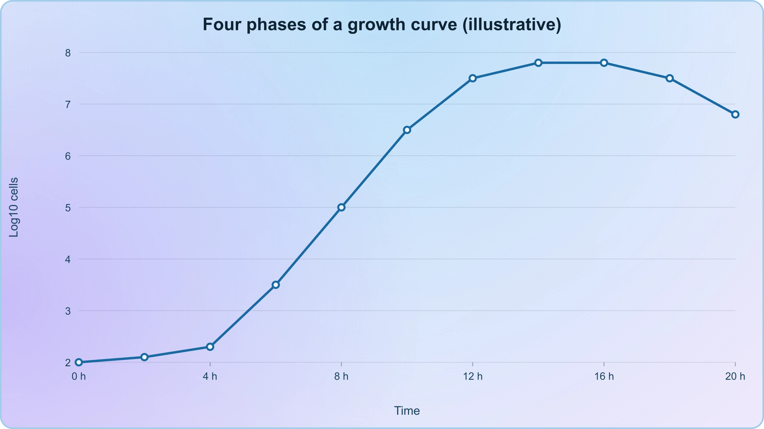
Growth curves in practice show four phases: lag, exponential, stationary and death. The lag phase represents adaptation, the exponential phase rapid division, the stationary phase nutrient exhaustion and waste accumulation, and the death phase decline. Understanding these phases is essential for the applications ahead. A fermentation halted at stationary phase may have maximum product, while one allowed to enter death phase may be spoiled. The reference material's observation that LAB multiply at suitable temperatures to convert milk to curd is a practical instance of exponential growth under controlled conditions.
Counting the Uncountable
Measurement is a recurring theme in microbiology because most microbes cannot be seen. Viable plate counts estimate living cells by counting colonies and multiplying by the dilution factor, giving results in colony-forming units per millilitre. Direct microscopic counts, turbidity measurements and molecular methods such as quantitative PCR offer alternatives with different trade-offs. A simple calculation illustrates the principle: if a ##10^{-5}## dilution yields 50 colonies on a plate inoculated with 0.1 mL, the original concentration is ##50 \times 10^{5} / 0.1 = 5 \times 10^{7}## CFU per mL. Such arithmetic is routine in dairy, water and pharmaceutical quality control.
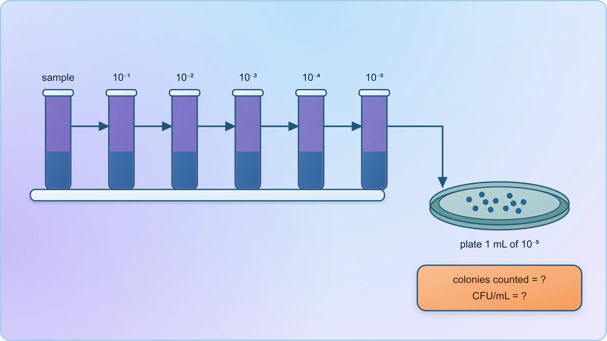
These methods matter for human welfare directly. Drinking water standards, food safety limits and pharmaceutical sterility tests all rest on microbial enumeration. When a municipal water authority reports that treated water meets safety criteria, it is reporting the outcome of exactly these counting techniques. The lesson's emphasis on colonies visible to the naked eye thus connects to a chain of reasoning that runs from a petri dish to public health policy. Learners should see that the humble colony count is a instrument of governance as much as of science.
From Diversity to Application: Preparing for the Lessons Ahead
This lesson has established four pillars that the remainder of the course will build upon. First, microbes are diverse across every domain of life and beyond it, including acellular agents such as viruses, viroids and prions. Second, they are ubiquitous, occupying habitats from thermal vents to the human gut. Third, they can be studied through culture and colony formation, and their growth follows predictable exponential mathematics. Fourth, and most importantly, the harmful minority must not obscure the beneficial majority whose contributions to human welfare are the subject of the course. Each pillar will be revisited with increasing specificity.
What the Next Lesson Will Build Upon
The next lesson turns to microbes in household products, beginning with curd, dough and cheese. It will assume that learners understand what lactic acid bacteria are, that they are prokaryotic, that they grow exponentially at suitable temperatures, and that their metabolic products acidify milk and coagulate proteins. It will also assume familiarity with the idea that different microbes produce different flavours and textures, which is why cheese varieties are specific to their microbial starters. The reference material's examples of Lactobacillus converting milk to curd, of carbon dioxide puffing dough, and of Propionibacterium sharmanii creating the holes in Swiss cheese will be developed in detail there.
Learners preparing for that lesson should consolidate three things from this one: the distinction between prokaryotic and eukaryotic microbes, the concept of a starter culture as an inoculum containing millions of cells, and the principle that fermentation outcomes depend on controlled conditions. A short self-check is useful. Can you name the kingdom-level groups that contain microbes? Can you explain why archaea are not bacteria? Can you state Koch's postulates and one limitation? Can you give three beneficial and three harmful microbial examples? If these are secure, the household products lesson will read as an extension rather than a new beginning.
Common Mistakes to Avoid
Several errors recur in examinations and in public discussion. The first is treating "microbe" as synonymous with "pathogen," which this lesson has corrected. The second is calling archaea a type of bacteria, when they form a separate domain. The third is assuming viruses are living cells; they are acellular obligate parasites, and viroids and prions are stranger still. The fourth is confusing colony counts with total cell counts, since plate methods detect only viable, culturable organisms. The fifth is imagining that beneficial microbes are always safe, when immunocompromised individuals can be vulnerable even to ordinarily harmless organisms.
A sixth error concerns the microbiome: it is not a fixed possession but a dynamic community shaped by diet, antibiotics and environment. A seventh is the assumption that fermentation is a single process, when in fact different organisms follow different metabolic routes, some producing acids, others alcohols, others gases. An eighth is neglecting the role of temperature and pH in determining which organism dominates a culture. Each of these errors will resurface in later lessons, and each is best corrected now, while the conceptual foundation is being laid and before the applications become more detailed.
To consolidate the lesson, a short programming exercise can model microbial growth and demonstrate why exponential behaviour matters in practice. The script below simulates population growth over time and prints the population at intervals, mirroring the mathematics developed earlier. Learners with an interest in computational biology can extend it to include a lag phase or a carrying capacity, which introduces the logistic model used in more advanced ecology.
import math

def microbial_growth(n0, doubling_time_min, total_time_min):
    """Return population after total_time_min under exponential growth."""
    generations = total_time_min / doubling_time_min
    return n0 * (2 ** generations)

n0 = 1
doubling = 20  # minutes, typical for fast-growing bacteria
for hours in range(0, 11, 2):
    t = hours * 60
    n = microbial_growth(n0, doubling, t)
    print(f"After {hours:2d} h: {n:,.0f} cells")
The output illustrates the central quantitative lesson of this section: from a single cell, ten hours of division at a twenty-minute doubling time yields roughly one billion cells. This is why a colony becomes visible overnight, why milk sours quickly in warm weather, and why industrial fermentations require rigorous contamination control. The same equation governs the spoilage of food and the success of a starter culture, and learners who can manipulate it will find the applied lessons far more intuitive.
Also Read
- No links available.
From our network :
- 10 Physics Numerical Problems with Solutions for IIT JEEPrepare for IIT JEE with 10 physics numerical problems, complete with detailed solutions and conceptual explanations. Ideal for mastering kinematics, thermodynamics, electromagnetism, and optics.
- 98% of Global MBA Programs Now Prefer GRE Over GMAT Focus EditionExplore why 98% of global MBA programs now prefer the GRE over GMAT Focus. Learn about the shorter GRE format, verbal logic importance, and strategic versatility for students.
- Analyzing Trump Deportation Numbers Insights Into The 2026 Immigration Crackdown
- How to Print Cheaply: Cutting the Cost per PageA practical, measured guide to lowering what each printed page costs — duplex and draft modes, third-party ink, yield mathematics, and knowing when a new printer beats refills.
- Do You Need a New Router or Is Your Internet Plan the Problem?A practical comparison of the signs that point to router limits versus signs your broadband speed or provider is the bottleneck, so you can decide what to replace before spending money.
- EV 2.0: The Solid-State Battery Breakthrough and Global Factory ExpansionDiscover the impact of the Solid-State Battery Breakthrough on the EV market. Learn about CATL and BYD stock growth, global factory expansion in Europe, and the future of EV 2.0 technology.
- AI-Powered 'Precision Diagnostic' Replaces Standard GRE Score ReportsDiscover the new GRE Precision Diagnostic. Learn how AI-powered behavioral analytics and time-to-solve tracking are replacing standard GRE score reports to empower students.
- Trump Political Strategy How Geopolitical Stunts Serve As Media Diversions
- Where to Put Your Wireless Router for Better CoverageA practical, physics-grounded guide to router placement: why the centre of the home beats the corner, how walls and metal sap signal, and how to run a simple survey that proves the improvement.
RESOURCES
- Resources will be updated soon.