The eukaryotic cell cycle is best understood not as a single continuous event but as a rhythm with two contrasting movements: a long, quiet period of preparation and a brief, dramatic period of division. In cultured human cells, this entire rhythm repeats roughly once every twenty-four hours, yet the actual act of division consumes only about one hour of that span. The remaining time, exceeding ninety-five percent of the cycle, belongs to interphase — a phase historically mislabelled as "resting" even though it is among the most metabolically demanding periods in a cell's life. Recognising this asymmetry is the first step toward reading the cell cycle correctly.
This lesson examines the two basic phases that structure the cycle: interphase and M phase. It establishes how long each phase lasts in human cells and in yeast, clarifies what happens at the boundary between them, and then maps the three sub-phases of interphase — G1, S and G2 — onto the work the cell performs before it divides. Particular attention falls on the arithmetic of DNA content during S phase, where the amount of DNA per cell doubles from 2C to 4C while the chromosome number remains fixed at 2n. That distinction between DNA quantity and chromosome count is a recurring source of confusion and deserves careful treatment.
The material is organised to build directly on the preceding discussion of the cell cycle as a coordinated sequence of division, replication and growth, and it prepares the ground for a closer inspection of each interphase sub-phase in the lesson that follows. Terminology, numerical values and the order of ideas are preserved throughout, with a short "Going further" block reserved for well-established background that aids comprehension without altering the core content.
On This Page
The Two Basic Phases of the Cell Cycle
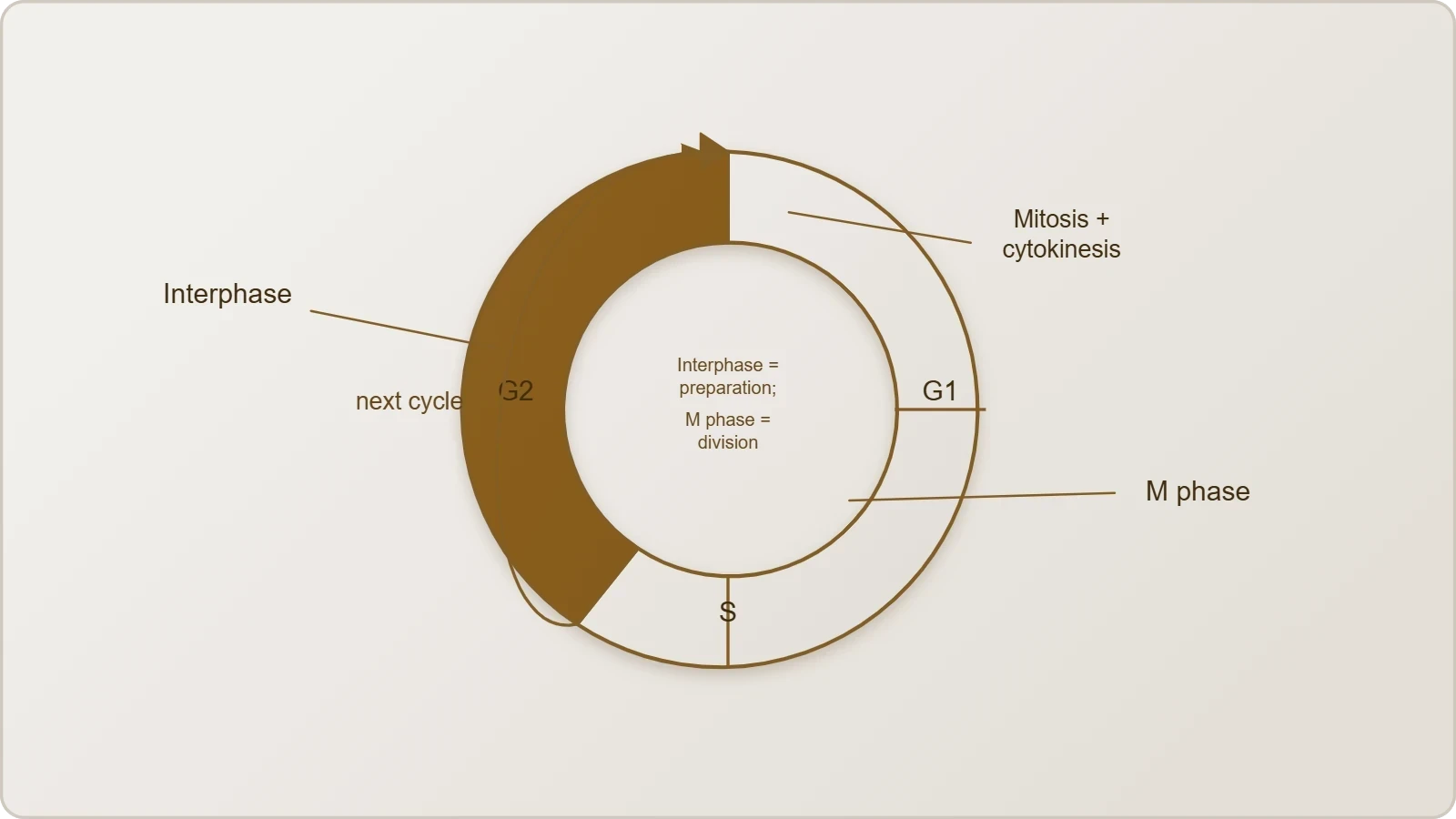
A typical eukaryotic cell cycle is divided into two basic phases: interphase and M phase, the latter also called the mitosis phase. The M phase represents the period when the actual cell division, or mitosis, occurs, while interphase represents the interval between two successive M phases. This division of labour is fundamental: one phase executes the separation of the cell into daughters, and the other assembles everything required for that separation to succeed. Every discussion of cycle timing, checkpoint control or DNA content ultimately refers back to this simple two-part architecture.
Defining Interphase and M Phase
Interphase is the phase between two successive M phases, and although it is conventionally called the resting phase, that label is misleading. It is the time during which the cell prepares for division by undergoing both cell growth and DNA replication in an orderly manner. The M phase, by contrast, begins with nuclear division, corresponding to the separation of daughter chromosomes, a process termed karyokinesis, and usually ends with division of the cytoplasm, known as cytokinesis. The word "usually" matters, because cytoplasmic division can occasionally be deferred or omitted while nuclear division proceeds.
The two phases are therefore complementary rather than sequential in a trivial sense. Interphase accumulates material and duplicates the genetic instructions; M phase distributes those instructions and partitions the cell. Neither phase can be shortened arbitrarily without consequences for the other, and the boundary between them is a controlled transition rather than a passive lapse. Keeping this complementarity in view prevents the common error of treating interphase as dead time and mitosis as the only event of consequence in the cycle.
Why the Division of the Cycle Matters
Separating the cycle into interphase and M phase gives biologists a workable framework for timing, measurement and comparison. When a culture is observed under a microscope, cells in M phase are visibly distinctive because chromosomes condense and the nucleus reorganises, whereas interphase cells present a relatively uniform nucleus. This visibility allows the proportion of cells in each phase to be estimated and compared across treatments. The framework also clarifies where errors in replication or chromosome segregation originate, since most of the preparatory work happens well before any chromosome becomes visible.
The division further supports the study of regulation. Because interphase occupies the overwhelming majority of the cycle, any factor that lengthens or shortens the cycle as a whole almost certainly acts on interphase rather than on mitosis. Growth conditions, nutrient availability and cell type all influence cycle duration primarily by stretching or compressing the interphase interval. Recognising this helps explain why two cells of the same organism can display markedly different cycle lengths despite executing an essentially identical M phase.
Duration of the Cell Cycle Across Organisms and Cell Types
The duration of the cell cycle is not a fixed constant of life. A typical eukaryotic cell cycle is illustrated by human cells in culture, which divide once in approximately every twenty-four hours. However, this duration can vary from organism to organism and also from cell type to cell type within the same organism. Yeast, for example, can progress through the cell cycle in only about ninety minutes. These two figures, twenty-four hours and ninety minutes, frame the entire discussion of cycle timing and demonstrate how profoundly the pace of division can differ.
Human Cells in Culture: The Twenty-Four Hour Reference
Human cells dividing once every twenty-four hours provide the standard reference against which other cycles are compared. Within that twenty-four hour average, cell division proper lasts for only about an hour, while interphase accounts for more than ninety-five percent of the total duration. The arithmetic is straightforward: if the cycle is roughly twenty-four hours and mitosis occupies about one hour, then interphase consumes the remaining twenty-three hours or so. This proportion is the single most instructive fact about cycle timing, because it reframes the cycle as a long preparation punctuated by a short execution.
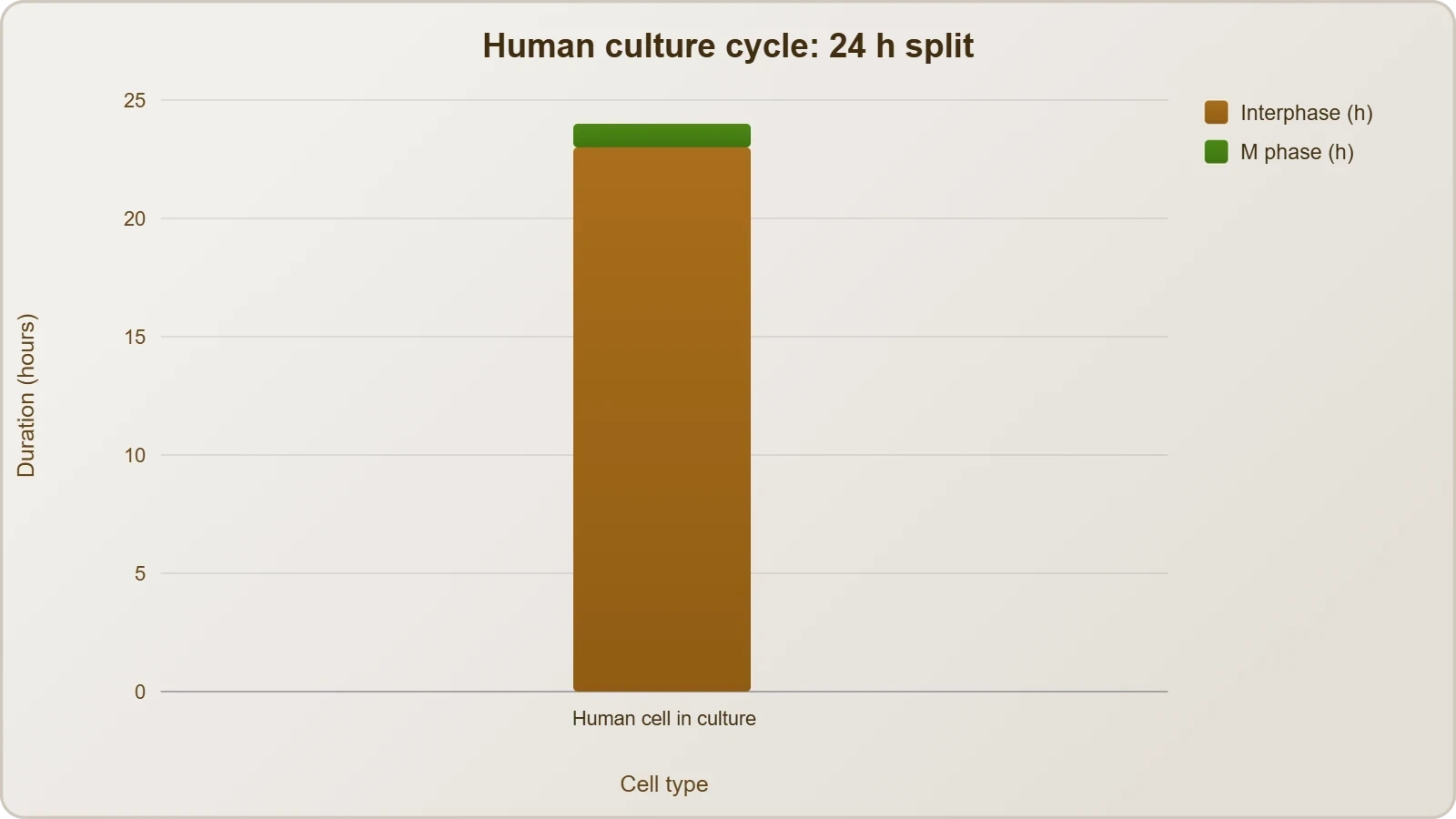
It is worth emphasising that the twenty-four hour figure describes cells in culture under favourable conditions, not every cell in the human body. The material presents it as an illustration of a typical eukaryotic cycle, and that framing should be retained. Cultured cells are a convenient model precisely because their cycles are relatively uniform and observable, yet the underlying principle — a long interphase and a brief M phase — holds broadly across eukaryotic systems even where absolute durations differ.
Yeast and the Range of Cycle Lengths
Yeast compresses the entire cycle into about ninety minutes, roughly one-sixteenth of the human culture figure. This comparison is not a curiosity but a demonstration that the same two-phase architecture can operate at radically different tempos. The phases themselves remain recognisable — interphase with its growth and replication, followed by M phase with nuclear and cytoplasmic division — but the intervals are scaled down dramatically. Such variation cautions against treating any single organism's timing as universal.
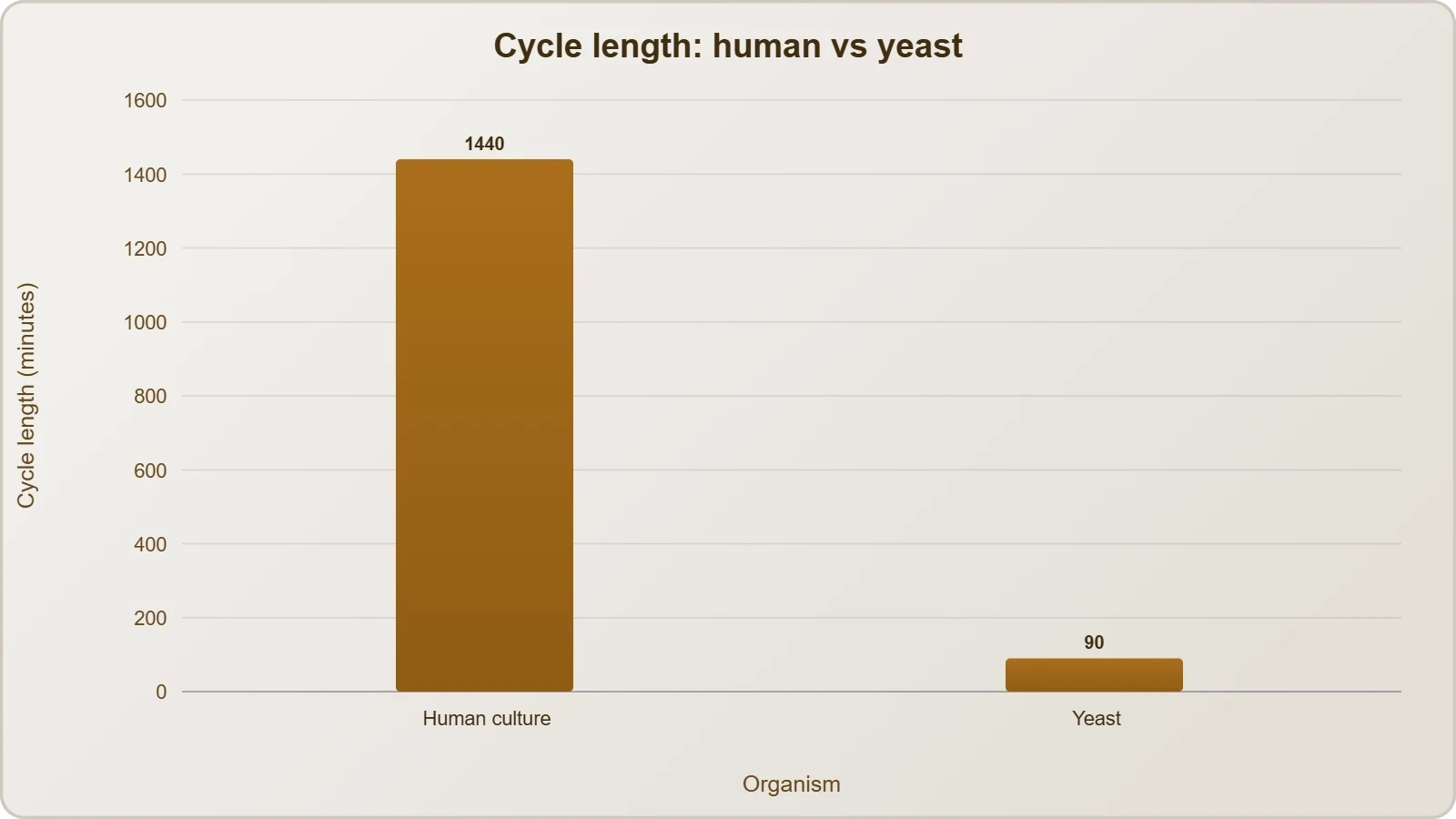
The variation extends beyond comparisons between species. Even within one organism, different cell types can cycle at different rates, and some cells exit the cycle altogether for extended periods. The material's statement that duration varies from cell type to cell type opens the door to this broader picture without developing it fully. For present purposes, the essential point is that the two-phase division of the cycle is universal, while the clock that governs it is adjustable and context-dependent.
Inside the M Phase: Karyokinesis and Cytokinesis
The M phase starts with the nuclear division, corresponding to the separation of daughter chromosomes, and this nuclear division is called karyokinesis. It usually ends with division of the cytoplasm, termed cytokinesis. The sequence is therefore ordered: chromosomes are first separated within the nuclear context, and the cytoplasm is then partitioned between the two emerging daughter cells. This ordering reflects the logic of division, since distributing genetic material correctly is the primary obligation, while dividing the surrounding cytoplasm is the concluding act that completes the formation of two cells from one.
Nuclear Division as the Opening Act
Karyokinesis is the defining event of the M phase. It corresponds to the separation of daughter chromosomes, ensuring that each prospective daughter cell receives a complete set of genetic instructions. Because this separation is the point at which genetic continuity is physically secured, it is unsurprising that the M phase begins here rather than with the cytoplasm. The material's phrasing places nuclear division first and treats it as the anchor of the phase, with everything else arranged around it. Any account of mitosis that begins with cytoplasmic events inverts the material's emphasis.
The visible changes that make M phase cells distinctive under a microscope stem from this nuclear reorganisation. Chromosomes condense and become individually discernible, the nuclear envelope undergoes profound rearrangement, and the machinery that moves chromosomes into position becomes active. These events are the substance of the detailed stages of mitosis treated elsewhere in the course; here they need only be located correctly within the two-phase framework. The essential point is that karyokinesis marks the transition from preparation to execution.
Cytoplasmic Division as the Usual Conclusion
Cytokinesis, the division of the cytoplasm, usually concludes the M phase. The qualifier "usually" is deliberate and important: the material does not claim that cytoplasmic division invariably follows nuclear division. When cytokinesis does occur, it partitions the cytoplasm so that two separate daughter cells result from the single parent cell. The diagrammatic view of the cell cycle, indicating the formation of two cells from one cell, captures this endpoint. Without cytokinesis, nuclear division alone would leave multiple nuclei within a shared cytoplasm rather than distinct cells.
The relationship between the two events can be summarised compactly. Nuclear division secures the genetic partition; cytoplasmic division completes the physical partition. Together they convert one cell into two, which is the outcome the entire cycle has been preparing for throughout interphase. Because interphase occupies more than ninety-five percent of the cycle, the M phase's brevity should not be mistaken for unimportance. It is the short, decisive conclusion to a long and carefully managed preparation, and its two components, karyokinesis and cytokinesis, are best understood in that light.
The Three Sub-Phases of Interphase
Interphase is divided into three further phases: G1 phase, also called Gap 1; S phase, also called Synthesis; and G2 phase, also called Gap 2. These three sub-phases partition the long preparatory interval into stages with distinct obligations. G1 corresponds to the interval between mitosis and the initiation of DNA replication. S marks the period during which DNA synthesis or replication takes place. G2 is the interval in which proteins are synthesised in preparation for mitosis while cell growth continues. Together they account for the more than ninety-five percent of the cycle that interphase occupies.
G1: Growth Without Replication
During G1 phase, the cell is metabolically active and continuously grows but does not replicate its DNA. This combination of activity and restraint defines the sub-phase: the cell is busy, synthesising materials and increasing in size, yet it holds back from copying its genetic instructions. G1 therefore corresponds to the interval between mitosis and the initiation of DNA replication, a period of assessment and accumulation. The cell emerges from division, resumes its ordinary metabolic work, and grows, but the decisive step of replication has not yet been taken.
The restraint in G1 is significant because it separates growth from replication in time. A cell that has just divided must rebuild its contents before it can consider duplicating its DNA, and G1 provides that interval. The material's emphasis on continuous growth without DNA replication captures the essential character of the phase. It is not a pause in activity but a specific kind of activity, directed toward increasing cell mass and metabolic capacity rather than toward copying the genome.
S: DNA Doubles While Chromosome Number Holds
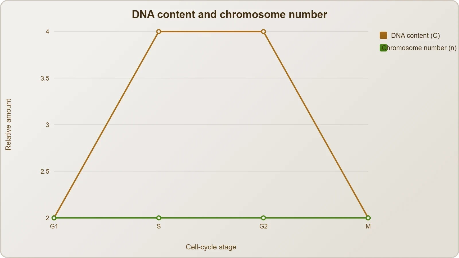
S phase marks the period during which DNA synthesis or replication takes place, and during this time the amount of DNA per cell doubles. If the initial amount of DNA is denoted as 2C, then it increases to 4C. Crucially, however, there is no increase in chromosome number: if the cell had a diploid or 2n number of chromosomes at G1, even after S phase the number of chromosomes remains the same, that is, 2n. This distinction between DNA content and chromosome count is the conceptual heart of the sub-phase.
The arithmetic can be stated compactly. Before S phase, DNA content is 2C and chromosome number is 2n; after S phase, DNA content is 4C while chromosome number remains 2n. The doubling of DNA reflects the creation of a second copy of each chromosome, but the two copies remain associated as sister chromatids and are not yet counted as separate chromosomes. In animal cells, during S phase, DNA replication begins in the nucleus, and the centriole duplicates in the cytoplasm, so replication proceeds in two compartments simultaneously.
G2: Protein Synthesis and Continued Growth
During G2 phase, proteins are synthesised in preparation for mitosis while cell growth continues. This sub-phase bridges replication and division: the genome has been duplicated in S phase, and G2 supplies the protein machinery that mitosis will require. Growth does not stop during G2, so the cell continues to increase in mass even as it assembles the components needed for chromosome separation and cytoplasmic division. G2 thus completes the preparation that G1 began, closing the interphase interval before the M phase opens.
Placing G2 after S rather than before it is logically necessary, since the proteins synthesised in G2 serve a genome that has already been duplicated. The sequence G1, then S, then G2 reflects a coherent progression: grow, replicate, then prepare the machinery for division. Each sub-phase depends on the completion of the one before it, and the order is not interchangeable. This ordered progression is what the material means when it describes the cell as preparing for division by undergoing growth and DNA replication in an orderly manner.
DNA Content and Chromosome Number Through the Cycle
The relationship between DNA content and chromosome number is the most frequently misread aspect of the cell cycle, and it repays careful attention. The material states the position precisely: during S phase the amount of DNA per cell doubles, rising from 2C to 4C, but there is no increase in chromosome number, which remains at 2n. The reason is that the newly synthesised DNA forms a second copy of each existing chromosome, and the two copies remain joined as sister chromatids. They are not yet independent chromosomes, so the count does not change.
Reading the 2C to 4C Transition
Denoting the initial amount of DNA as 2C and the post-replication amount as 4C gives a compact way to track the cycle's progress. A cell in G1 has 2C of DNA and 2n chromosomes. After S phase it has 4C of DNA but still 2n chromosomes. The doubling of C measures the duplication of genetic material; the constancy of n measures the number of centromere-bearing units. Confusing the two leads to the mistaken conclusion that replication doubles the chromosome number, which it does not.
This notation also clarifies what happens later. When the M phase separates daughter chromosomes, the sister chromatids part and each becomes an independent chromosome in a daughter cell, restoring the 2n count in each daughter while the DNA content per cell returns to 2C. The cycle therefore oscillates between 2C and 4C in DNA content while the chromosome number per cell remains 2n at the start and end of each cycle. The intermediate 4C state is a temporary doubling, not a permanent change in ploidy.
Why the Distinction Matters
The distinction matters because it underpins any quantitative analysis of the cycle. Measuring DNA content per cell, for instance by staining and flow analysis, reveals the 2C and 4C populations and the intermediate values characteristic of cells partway through S phase. Measuring chromosome number requires counting centromeres or visible chromosomes, which gives 2n throughout interphase. Two different measurements, two different pictures, and only a clear grasp of the underlying biology reconciles them.
The distinction also prepares the ground for understanding the mechanics of division. Because sister chromatids remain associated after replication, the M phase has something definite to separate: paired copies that must be pulled apart. If replication had simply doubled the chromosome number, division would be a different problem altogether. The 2C to 4C transition with constant 2n is thus not a technicality but the structural precondition for faithful segregation of genetic material into two daughter cells.
Going Further: Background That Aids Understanding
The letters used for the interphase sub-phases are abbreviations worth knowing explicitly. G1 stands for Gap 1, S stands for Synthesis, and G2 stands for Gap 2. The term "gap" reflects the historical observation that these intervals appeared as gaps between the two events that were visible under early microscopes, namely replication and division. The name has persisted even though the intervals are now known to be filled with intense biochemical activity. Recognising the expansion of the abbreviations helps students connect the labels to the processes they denote.
What the M Phase Includes
The M phase includes both nuclear division and, usually, cytoplasmic division. Nuclear division, or karyokinesis, corresponds to the separation of daughter chromosomes; cytoplasmic division, or cytokinesis, partitions the cytoplasm and completes the formation of two cells from one. Keeping both components in mind prevents the common shorthand of equating the M phase with mitosis alone. The material's careful phrasing, which places karyokinesis at the start and cytokinesis as the usual conclusion, is a precise description of what the phase encompasses.
This background does not alter the core content but sharpens it. Knowing that G1, S and G2 expand to Gap 1, Synthesis and Gap 2 makes the sequence easier to remember and easier to explain. Knowing that the M phase spans both nuclear and cytoplasmic division clarifies why the phase is described as beginning with one event and usually ending with another. These small clarifications support the material rather than extending beyond it, and they prepare the reader for the detailed treatment of each interphase sub-phase that follows.
Also Read
- No links available.
From our network :
- Vite 6/7 'Cold Start' Regression in Massive Module GraphsIn-depth analysis of Vite 6/7 cold start regressions in massive module graphs. Learn about barrel file issues, pre-bundling optimizations, and how to improve Vite dev server performance.
- Trump Political Strategy How Geopolitical Stunts Serve As Media Diversions
- EV 2.0: The Solid-State Battery Breakthrough and Global Factory ExpansionDiscover the impact of the Solid-State Battery Breakthrough on the EV market. Learn about CATL and BYD stock growth, global factory expansion in Europe, and the future of EV 2.0 technology.
- 10 Physics Numerical Problems with Solutions for IIT JEEPrepare for IIT JEE with 10 physics numerical problems, complete with detailed solutions and conceptual explanations. Ideal for mastering kinematics, thermodynamics, electromagnetism, and optics.
- Analyzing Trump Deportation Numbers Insights Into The 2026 Immigration Crackdown
- Mastering DB2 12.1 Instance Design: A Technical Deep Dive into Modern Database ArchitectureComprehensive guide to DB2 12.1 Instance Design. Explore architectural components, HADR setup, buffer pool tuning, and security policies for IBM DB2 12.1. Includes 10 functional SQL and Shell samples.
- Mastering DB2 LUW v12 Tables: A Comprehensive Technical GuideComprehensive guide on DB2 LUW v12 tables. Explore CREATE TABLE syntax, BLU Acceleration, range partitioning, data types, and security features for IBM DB2 LUW v12.
- AI-Powered 'Precision Diagnostic' Replaces Standard GRE Score ReportsDiscover the new GRE Precision Diagnostic. Learn how AI-powered behavioral analytics and time-to-solve tracking are replacing standard GRE score reports to empower students.
- 98% of Global MBA Programs Now Prefer GRE Over GMAT Focus EditionExplore why 98% of global MBA programs now prefer the GRE over GMAT Focus. Learn about the shorter GRE format, verbal logic importance, and strategic versatility for students.
RESOURCES
- Learn: Phases of the cell cycle (article) | Khan Academykhanacademy.orgDuring the mitotic (M) phase, the cell separates its DNA into two sets and divides its cytoplasm, forming two new cells. Interphase. Let's enter…
- Cell Cycle - National Human Genome Research Institute (NHGRI)genome.govThe next stage is M. M stands for mitosis. This is where the cell actually partitions the two copies of the genetic material into…
- Biology: The Cell Cycle Flashcards - Quizletquizlet.comThe two phases of the cell cycle are interphase and the M phase. The interphase includes G1, S, and G2. The M phase includes…
- Cell Cycle Assay Solution Deep Red - dojindodojindo.com... cell cycle phase. The cell cycle is classified into two major phases: interphase and the mitotic (M) phase. Interphase is composed of G1…
- The Eukaryotic Cell Cycle - The Cell - NCBI Bookshelf - NIHncbi.nlm.nih.govThe division cycle of most eukaryotic cells is divided into four discrete phases: M, G1, S, and G2. M phase (mitosis) is usually followed…
- The retinoblastoma protein is phosphorylated during specific phases ...pubmed.ncbi.nlm.nih.govSep 22, 1989 ... ... cycle, but is phosphorylated in a phase-specific manner. In the G0 and G1 phases of the cell cycle ...…
- An Overview of the Cell Cycle - Molecular Biology of the Cell - NCBIncbi.nlm.nih.govThe cell grows continuously in interphase, which consists of three phases: DNA replication is confined to S phase; G1 is the gap between M…
- Regulation of Cell Cycle Progression by Growth Factor-Induced Cell ...pmc.ncbi.nlm.nih.govNov 26, 2021 ... ... M phase than in interphase [168]. A further study shows that Cdc2 ... phases of cell cycle including S,…
- Yeast cells can enter a quiescent state through G1, S, G2, or M ...pubmed.ncbi.nlm.nih.gov... M phase of the cell cycle. We monitored entry to a quiescent state by ... M phases of the cell cycle. These findings…
- Phases Of Cell Cycle G0 G1 S G2 M - Mitosis - MCAT Contentjackwestin.comThe cell cycle has two major phases: interphase (G0, G1, S, G2) and the mitotic phase (M). The cell cycle is an ordered series…
- Cell cycle - Wikipediaen.wikipedia.org... cells, the cell cycle is divided into two main stages: interphase, and the M phase that includes mitosis and cytokinesis. During interphase, the…
- 3.2 The cell cycle | Cell division - Siyavulasiyavula.comThe first stage is interphase during which the cell grows and replicates its DNA. The second phase is the mitotic phase (M-Phase) during which…
- Cell Cycle Probability - LabXchangelabxchange.orgInterphase is divided into 3 subphases: the gap 1 (G1) phase, the synthesis ... The second phase of the cell cycle is the M…
- Reversible and effective cell cycle synchronization method for ...life-science-alliance.orgMar 4, 2025 ... The cell division cycle includes four major stages: G1, S, G2, and M phases, each with distinct functions. During the…
- The Cell Cycle | Listen to the Podcast on Spotifyopen.spotify.comMay 12, 2026 ... In this episode, we explore the different phases of the cell cycle: Interphase (G1, S, G2) and the M phase…