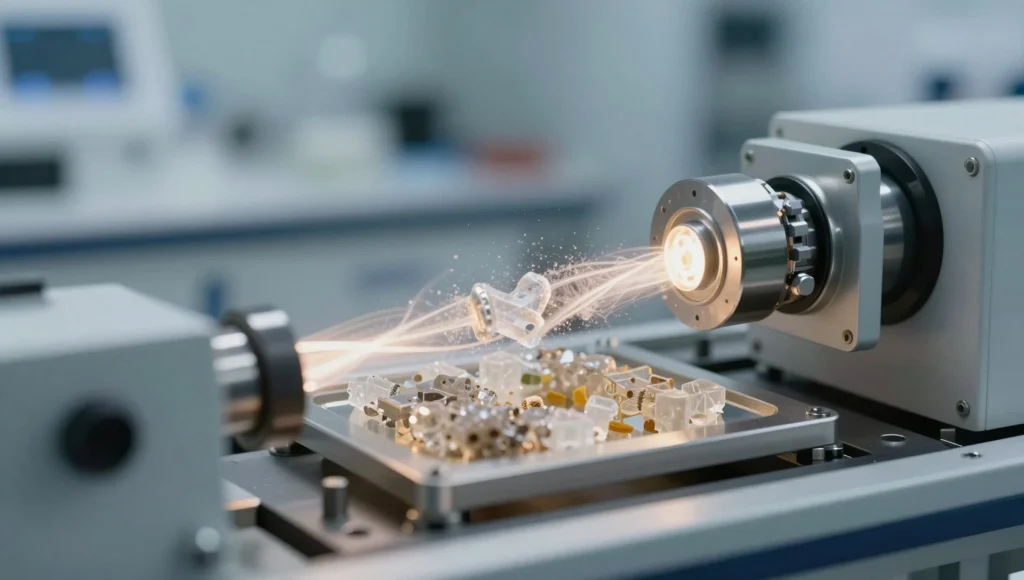
Neutron scattering represents one of the most powerful yet underappreciated windows into the atomic architecture of matter. Unlike X-rays, which interact with electron clouds and therefore struggle to locate light elements, neutrons strike atomic nuclei directly and reveal hydrogen, lithium, and oxygen positions with extraordinary clarity. This technique has quietly reshaped materials science, condensed-matter physics, and structural biology over the past seven decades.
The method exploits the wave nature of thermal neutrons produced in research reactors or spallation sources. When a beam of neutrons strikes a sample, the pattern of scattered intensity encodes both the positions of atoms and their dynamic motions. Researchers reconstruct these patterns mathematically to obtain pair-distribution functions, phonon dispersion curves, and magnetic structures that no other probe can deliver with comparable fidelity.
This analysis unpacks the physical principles behind neutron scattering, contrasts it with X-ray and electron techniques, and surveys its transformative applications across magnetism, polymer science, battery chemistry, and quantum materials. We also work through ten quantitative problems that illustrate how scattering cross-sections, momentum transfer, and energy resolution are computed in practice.
On This Page
- The Physical Foundations of Neutron Scattering
- Why Neutrons Reveal What X-Rays Cannot
- Instrumentation: Reactors, Spallation Sources, and Spectrometers
- Quantitative Analysis: Ten Worked Problems in Neutron Scattering
- Applications Across Magnetism, Soft Matter, and Energy Materials
- Challenges, Limitations, and the Future of Neutron Science
The Physical Foundations of Neutron Scattering
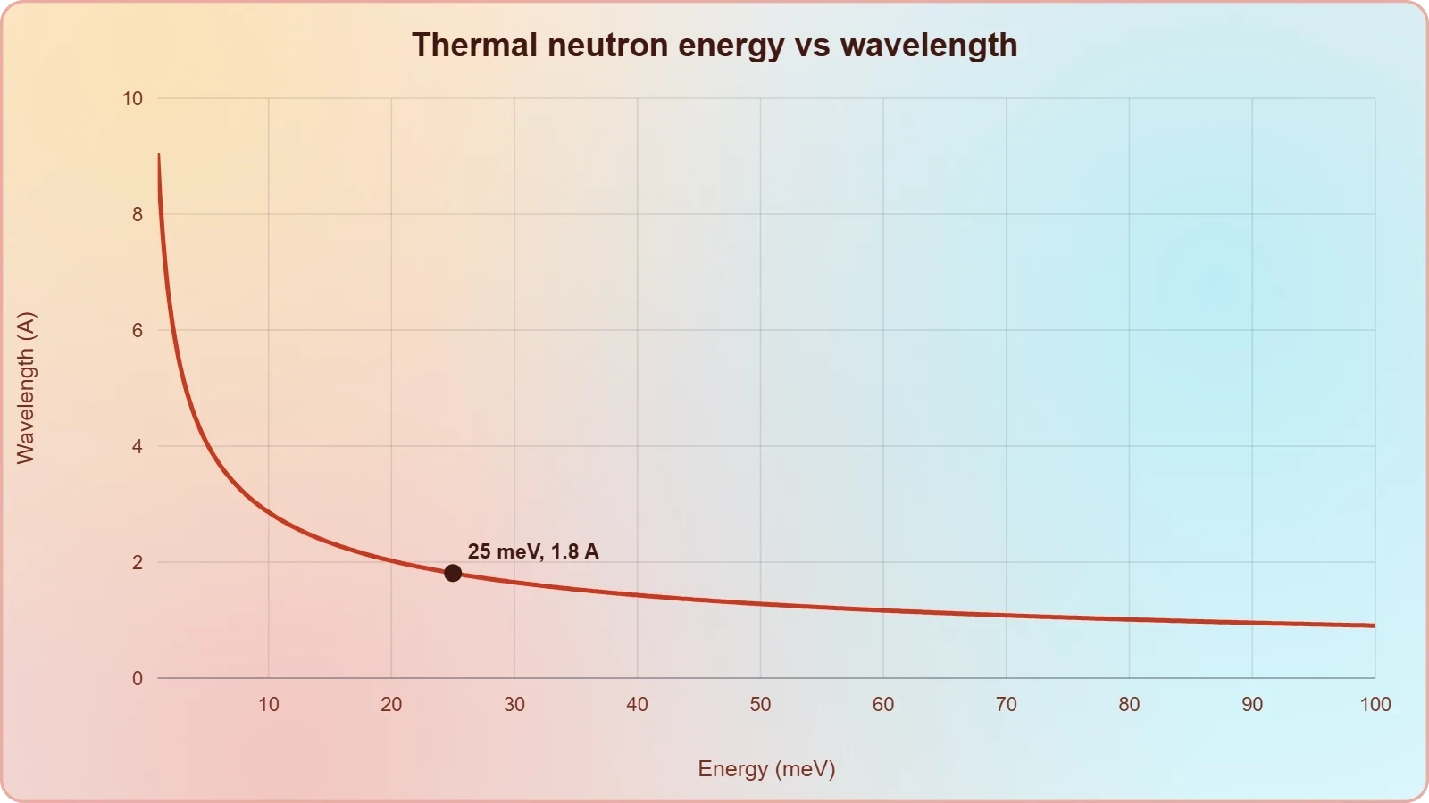
Neutron scattering rests on the quantum-mechanical description of a slow neutron as a matter wave whose de Broglie wavelength matches interatomic spacings. Thermal neutrons carry energies near ##[E \approx k_B T]## at room temperature, roughly 25 meV, which corresponds to wavelengths around 1.8 Å. This fortunate coincidence makes them ideal probes of crystalline lattices and molecular vibrations simultaneously.
Wave-Particle Duality and the Scattering Vector
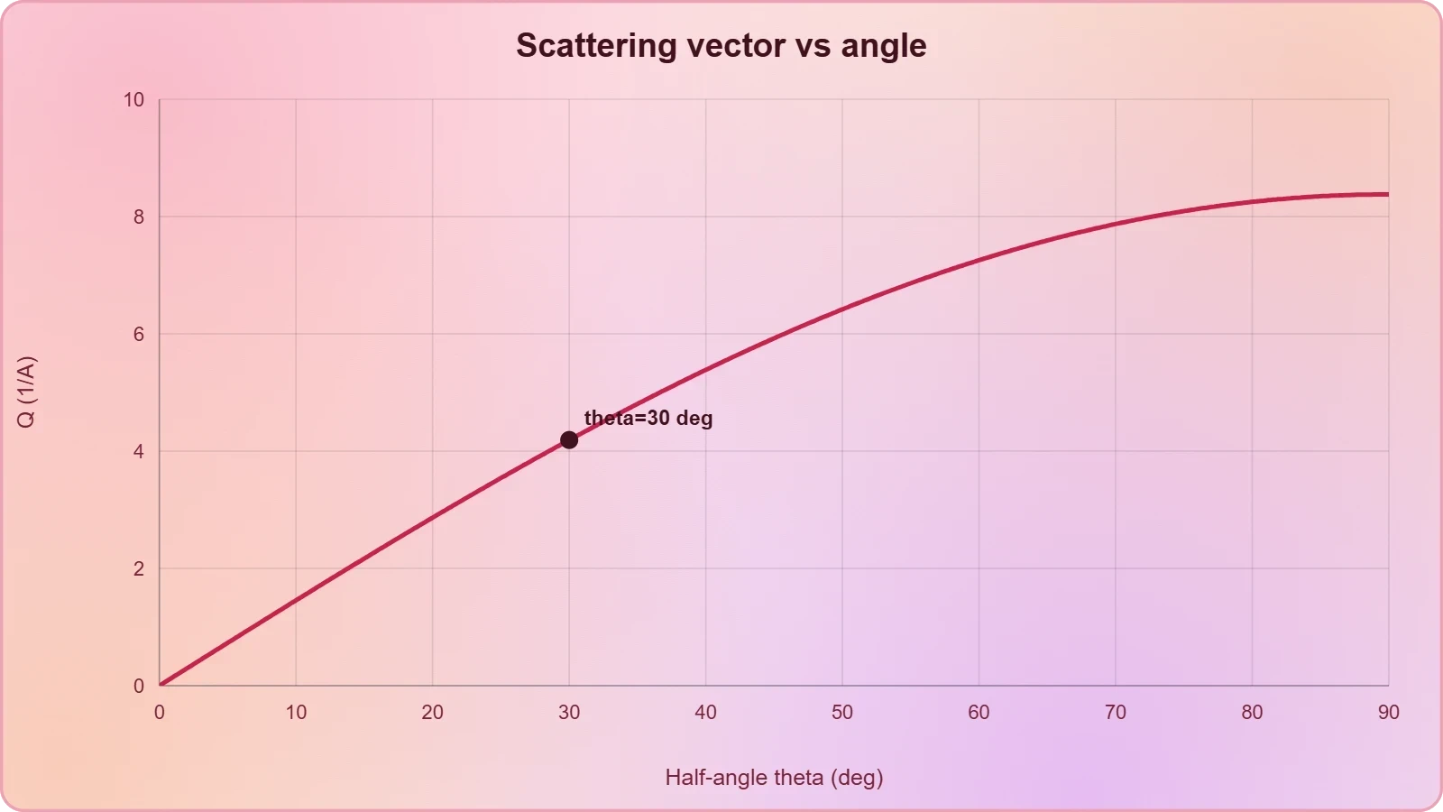
The scattering process is governed by conservation of momentum and energy between the incoming neutron and the sample. The momentum transfer, or scattering vector ##[\mathbf{Q}]##, is defined as the difference between outgoing and incoming wavevectors. Its magnitude determines the length scale probed, with large ##[Q]## resolving short interatomic distances and small ##[Q]## revealing mesoscopic structures.
The energy transfer ##[\hbar\omega]## captures inelastic events where neutrons gain or lose energy to excitations such as phonons, magnons, or molecular rotations. Elastic scattering, where ##[\omega = 0]##, yields diffraction patterns and static structure factors. Inelastic scattering maps the dynamical response function ##[S(\mathbf{Q},\omega)]##, which encodes the full spectrum of collective motions within the material.
Coherent and Incoherent Cross-Sections
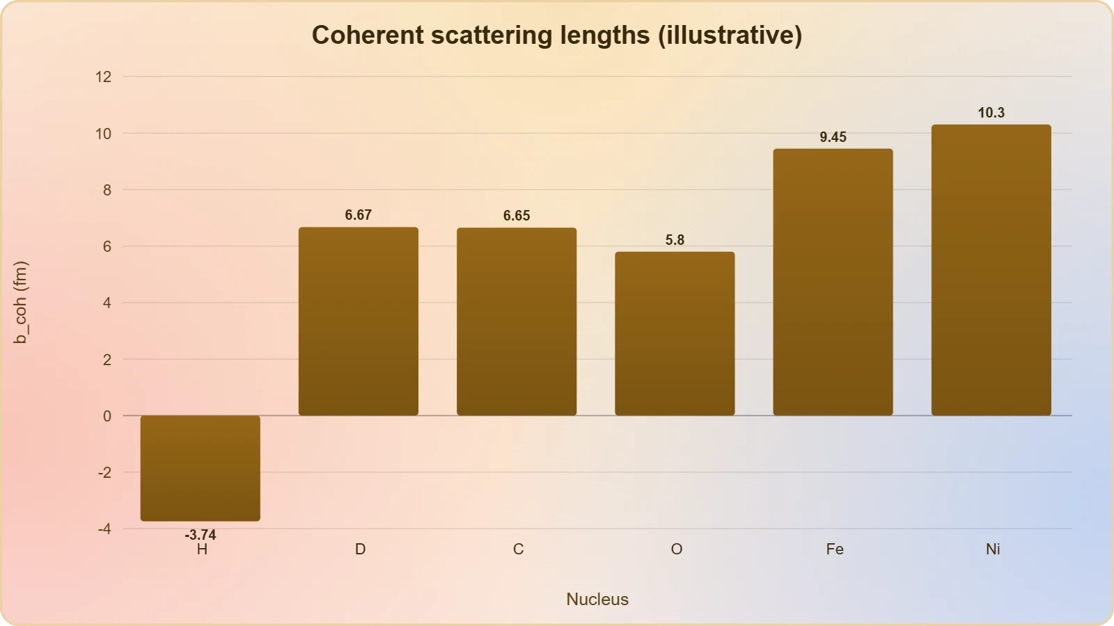
Each isotope possesses a coherent scattering length ##[b_{coh}]## that produces interference and structural information, plus an incoherent component arising from spin and isotope disorder. Hydrogen, notoriously, has an enormous incoherent cross-section, which makes it a superb probe of molecular diffusion but a nuisance for crystallography. Deuterium, by contrast, scatters coherently and is routinely substituted to suppress background.
The differential cross-section for scattering into a solid angle ##[d\Omega]## integrates over all final states and connects microscopic potentials to measurable intensities. For a collection of ##[N]## nuclei, the coherent cross-section scales with ##[|b_{coh}|^2]## and the static structure factor ##[S(\mathbf{Q})]##, which is the Fourier transform of the pair-correlation function ##[g(r)]##.
Why Neutrons Reveal What X-Rays Cannot
X-ray crystallography remains the workhorse of structural science, yet its interaction with electron density creates blind spots that neutron techniques elegantly fill. Because neutrons scatter from nuclei rather than electrons, their scattering power does not scale with atomic number. This property allows researchers to locate hydrogen atoms, distinguish neighboring transition metals, and probe magnetic moments with unmatched sensitivity.
Contrast with X-Ray and Electron Probes
X-rays are strongly absorbed by heavy elements and barely scattered by hydrogen, making light-atom positions inferable only indirectly. Electrons, meanwhile, suffer severe multiple scattering in thick specimens and require ultra-thin samples under vacuum. Neutrons penetrate centimeters of most materials, enabling bulk measurements inside pressure cells, cryostats, and operating batteries without destructive sample preparation.
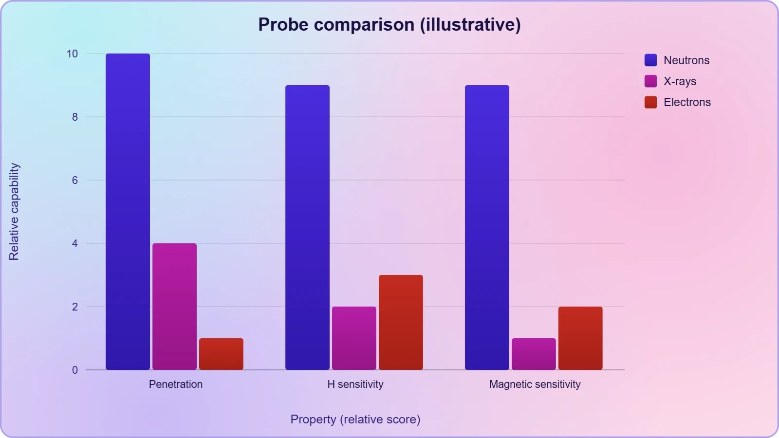
Magnetic neutron scattering exploits the neutron's intrinsic spin, which couples to unpaired electron moments through the dipole-dipole interaction. This coupling produces magnetic Bragg peaks and spin-wave dispersion that directly map ordered magnetic structures. No X-ray technique can access this magnetic channel with equivalent momentum and energy resolution across such broad ranges.
Penetration Depth and Isotope Sensitivity
The weak interaction between neutrons and matter translates into exceptional penetration, often tens of centimeters through aluminum, steel, or titanium. This penetration permits operando experiments where the sample remains inside realistic environments, from high-pressure diamond anvil cells to functioning electrochemical cells. Researchers observe structural evolution in real time under conditions that mimic industrial operation.
Isotope sensitivity provides another decisive advantage, since scattering lengths vary erratically across the periodic table and even between isotopes of the same element. Replacing hydrogen with deuterium shifts scattering contrast dramatically without altering chemistry, enabling selective labeling of specific molecular segments. This trick underpins small-angle neutron scattering studies of polymers, micelles, and biological membranes.
We Also Published
Instrumentation: Reactors, Spallation Sources, and Spectrometers
Producing usable neutron beams demands either nuclear fission reactors or spallation sources where high-energy protons strike heavy-metal targets. Reactors deliver continuous, steady flux ideal for monochromatic instruments, while spallation sources generate pulsed beams whose time-of-flight structure enables broad bandwidth measurements. Both facility classes host dozens of specialized spectrometers serving thousands of annual experiments.
Continuous Versus Pulsed Neutron Sources
Research reactors such as those at Grenoble and Oak Ridge provide stable thermal and cold neutron fluxes through moderator systems. Their continuous operation suits triple-axis spectrometers that scan energy and momentum point by point with exceptional resolution. Spallation sources like ISIS and the Spallation Neutron Source instead produce intense bursts, allowing time-of-flight diffraction and total-scattering measurements across wide ##[Q]##-ranges in single exposures.
Cold neutron sources, moderated by liquid hydrogen or deuterium, shift the spectrum toward longer wavelengths suited to soft matter and magnetic studies. Guide halls transport these cold neutrons tens of meters with minimal loss, feeding small-angle scattering instruments and reflectometers. The choice of source and moderator fundamentally shapes which science questions become experimentally tractable.
Diffractometers, Spectrometers, and Reflectometers
Diffractometers measure elastic scattering to determine crystal and magnetic structures, often using Rietveld refinement against powder patterns. Triple-axis spectrometers isolate specific ##[(\mathbf{Q},\omega)]## points to map phonon and magnon dispersion relations with meV precision. Time-of-flight chopper spectrometers capture entire dynamical spectra simultaneously, trading resolution for throughput in studies of complex excitations.
Small-angle neutron scattering instruments probe structures from one to hundreds of nanometers, revealing polymer coils, nanoparticle assemblies, and protein complexes in solution. Neutron reflectometry resolves layered thin films with sub-nanometer depth resolution, critical for studying interfaces in organic electronics and biological membranes. Each instrument class addresses a distinct region of the vast length-and-energy landscape.
Quantitative Analysis: Ten Worked Problems in Neutron Scattering
Mastering neutron scattering requires fluency with the quantitative relationships linking beam parameters, sample properties, and measured intensities. The following ten problems progress from basic wavelength calculations to structure-factor analysis and inelastic kinematics. Each solution demonstrates the algebraic reasoning that underpins real instrument planning and data interpretation.
Wavelength, Energy, and Momentum Calculations
Problem 1. Compute the de Broglie wavelength of a neutron with kinetic energy ##[E = 25]## meV. Using ##[\lambda = h/\sqrt{2m_nE}]##, we substitute ##[h = 6.626\times10^{-34}]## J·s and ##[m_n = 1.675\times10^{-27}]## kg.
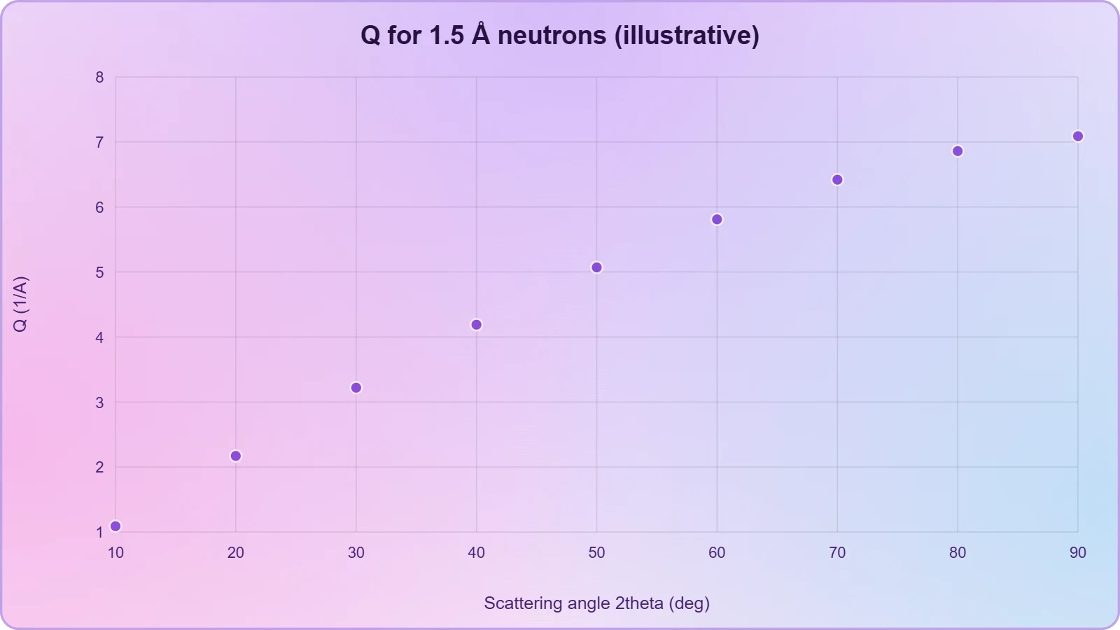
Problem 2. A neutron of wavelength 1.5 Å scatters elastically at ##[2\theta = 60°]##. Find the magnitude of the scattering vector ##[Q]##. The relation is ##[Q = \dfrac{4\pi}{\lambda}\sin\theta]##, with ##[\theta = 30°]##.
Problem 3. Determine the Bragg angle for the (111) reflection of copper (##[d = 2.087]## Å) using 1.8 Å neutrons. Bragg's law gives ##[\sin\theta = \lambda/(2d)]##, so ##[\sin\theta = 1.8/(2\times2.087) = 0.431]##, yielding ##[\theta \approx 25.5°]## and ##[2\theta \approx 51°]##.
Problem 4. Calculate the energy of a phonon detected at an energy transfer of ##[\hbar\omega = 12]## meV, expressed in wavenumbers. Since ##[1\,\text{meV} = 8.066\,\text{cm}^{-1}]##, the phonon wavenumber is ##[12 \times 8.066 \approx 96.8\,\text{cm}^{-1}]##, corresponding to a frequency near 2.9 THz.
Cross-Sections, Structure Factors, and Resolution
Problem 5. Estimate the coherent scattering intensity from ##[10^{20}]## carbon nuclei given ##[b_{coh} = 6.65]## fm and ##[S(Q) = 2.5]##. The cross-section is ##[N|b|^2 S(Q)]##, so ##[10^{20}\times(6.65\times10^{-15})^2\times2.5 \approx 1.1\times10^{-8}]## m², a substantial signal for a bulk sample.
Problem 6. A sample contains equal hydrogen and deuterium. Compute the average coherent scattering length given ##[b_H = -3.74]## fm and ##[b_D = 6.67]## fm. The mean is ##[(-3.74 + 6.67)/2 = 1.465]## fm, but the variance contributes strongly to incoherent scattering.
Problem 7. For a time-of-flight spectrometer with 100 m flight path, find the neutron velocity corresponding to 4 Å wavelength. Using ##[v = h/(m_n\lambda)]##, we obtain ##[v = 6.626\times10^{-34}/(1.675\times10^{-27}\times4\times10^{-10}) \approx 989]## m/s, giving a flight time near 101 ms.
Problem 8. Compute the magnetic scattering amplitude for a spin-only moment of ##[S = 5/2]## using ##[p = g\sqrt{S(S+1)}]## with ##[g = 2]##. This gives ##[p = 2\sqrt{35/4} = \sqrt{35} \approx 5.92]## Bohr magnetons, which sets the scale of magnetic Bragg intensities.
Problem 9. A powder pattern shows the first reflection at ##[2\theta = 28°]## for ##[\lambda = 1.5]## Å. Determine the d-spacing via ##[d = \lambda/(2\sin\theta)]##, giving ##[d = 1.5/(2\sin 14°) \approx 3.10]## Å, consistent with a layered structure.
Problem 10. Estimate the energy resolution required to resolve a phonon at 5 meV with 5% precision. The resolution must satisfy ##[\Delta E/E \leq 0.05]##, so ##[\Delta E \leq 0.25]## meV, demanding a chopper spectrometer with fine time channels and long flight paths.
Applications Across Magnetism, Soft Matter, and Energy Materials
Neutron scattering's unique capabilities translate into decisive contributions across an unusually broad scientific frontier. In magnetism, it resolves spin structures and excitations inaccessible to any other probe. In soft matter, deuterium labeling reveals polymer conformations and self-assembly. In energy science, operando measurements track lithium transport and hydrogen storage inside functioning devices.
Magnetic Structures and Quantum Materials
Magnetic neutron diffraction determines the arrangement of spins in antiferromagnets, multiferroics, and frustrated magnets where competing interactions suppress conventional order. Inelastic scattering maps spin-wave dispersion, revealing exchange constants and anisotropy gaps that govern quantum behavior. These measurements guide the design of spintronic devices and topological quantum materials with tailored magnetic properties.
Frustrated magnets such as spin ices and kagome lattices host exotic excitations including magnetic monopoles and Dirac spinons. Neutron scattering provides the definitive experimental signature of these phenomena, distinguishing genuine quantum spin liquids from conventional ordered states. Such studies inform the broader quest for robust quantum information platforms operating at elevated temperatures.
Polymers, Biomolecules, and Battery Materials
Small-angle neutron scattering with deuterium contrast resolves polymer chain conformations in melts and blends, testing reptation and scaling theories directly. In structural biology, neutron crystallography locates hydrogen atoms in enzyme active sites, revealing protonation states essential for catalytic mechanism. Membrane protein structures benefit enormously from neutron reflectometry and diffraction under near-physiological conditions.
Lithium-ion battery research exploits neutron diffraction and quasielastic scattering to track lithium occupancy and ionic diffusion during charge cycling. Operando cells allow researchers to observe phase transitions and degradation pathways inside functioning electrodes. Hydrogen storage materials, fuel-cell membranes, and thermoelectric compounds similarly benefit from neutron probes that see light elements and bulk behavior simultaneously.
Challenges, Limitations, and the Future of Neutron Science
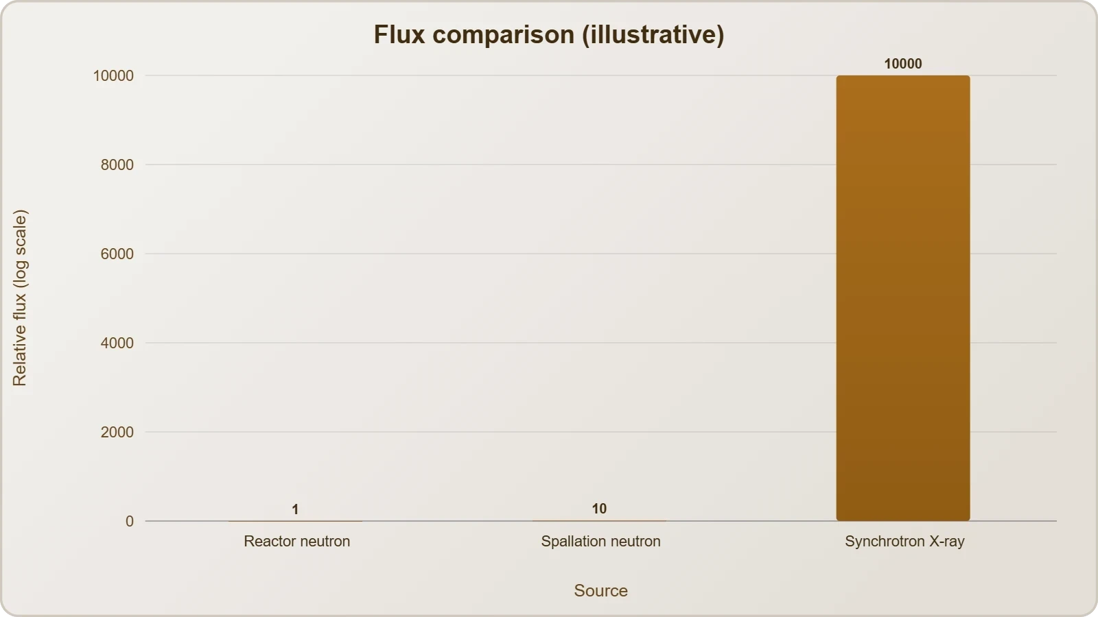
Despite its extraordinary capabilities, neutron scattering faces persistent constraints that shape its scientific reach. Neutron sources are large, expensive facilities with limited beam time, and the flux available at samples remains orders of magnitude below synchrotron X-ray sources. These realities demand careful experiment design, efficient detectors, and increasingly sophisticated data analysis to extract maximum information from precious beam hours.
Flux Limitations and Sample Requirements
Because neutron production is inherently inefficient, samples must often be larger than those used in X-ray work, typically milligrams to grams for diffraction. This requirement complicates studies of rare materials, thin films, and biological crystals that grow only in microscopic quantities. Instrument scientists continually push detector efficiency and focusing optics to reduce sample demands while preserving resolution.
Background from incoherent scattering, especially from hydrogen, can overwhelm weak signals and requires isotopic substitution or careful sample environment design. Data reduction pipelines must correct for detector efficiency, multiple scattering, and absorption with high precision. These corrections, while routine, consume substantial expertise and computational resources in every experiment.
Emerging Sources, Machine Learning, and Operando Methods
The European Spallation Source and upgrades to existing facilities promise order-of-magnitude gains in brightness, opening new frontiers in quantum materials and biological systems. Machine learning accelerates data reduction, phase identification, and structure refinement, reducing the burden on scarce beam time. These computational advances increasingly complement rather than replace traditional analysis.
Operando and in-situ experiments represent a growing fraction of neutron science, capturing materials under realistic operating conditions rather than idealized laboratory states. Combined with complementary X-ray and electron probes, neutron scattering anchors a multimodal characterization ecosystem. This integration promises deeper mechanistic understanding of catalysts, batteries, and functional materials under real-world stress.
From our network :
- AI-Powered 'Precision Diagnostic' Replaces Standard GRE Score ReportsDiscover the new GRE Precision Diagnostic. Learn how AI-powered behavioral analytics and time-to-solve tracking are replacing standard GRE score reports to empower students.
- Analyzing Trump Deportation Numbers Insights Into The 2026 Immigration Crackdown
- 10 Physics Numerical Problems with Solutions for IIT JEEPrepare for IIT JEE with 10 physics numerical problems, complete with detailed solutions and conceptual explanations. Ideal for mastering kinematics, thermodynamics, electromagnetism, and optics.
- Mastering DB2 12.1 Instance Design: A Technical Deep Dive into Modern Database ArchitectureComprehensive guide to DB2 12.1 Instance Design. Explore architectural components, HADR setup, buffer pool tuning, and security policies for IBM DB2 12.1. Includes 10 functional SQL and Shell samples.
- 98% of Global MBA Programs Now Prefer GRE Over GMAT Focus EditionExplore why 98% of global MBA programs now prefer the GRE over GMAT Focus. Learn about the shorter GRE format, verbal logic importance, and strategic versatility for students.
- Vite 6/7 'Cold Start' Regression in Massive Module GraphsIn-depth analysis of Vite 6/7 cold start regressions in massive module graphs. Learn about barrel file issues, pre-bundling optimizations, and how to improve Vite dev server performance.
- Trump Political Strategy How Geopolitical Stunts Serve As Media Diversions
- Mastering DB2 LUW v12 Tables: A Comprehensive Technical GuideComprehensive guide on DB2 LUW v12 tables. Explore CREATE TABLE syntax, BLU Acceleration, range partitioning, data types, and security features for IBM DB2 LUW v12.
- EV 2.0: The Solid-State Battery Breakthrough and Global Factory ExpansionDiscover the impact of the Solid-State Battery Breakthrough on the EV market. Learn about CATL and BYD stock growth, global factory expansion in Europe, and the future of EV 2.0 technology.
RESOURCES
- MSE Neutron Scattering | U.S. DOE Office of Science (SC)science.osti.govThe neutron scattering core research area supports innovative applications of neutron scattering to achieve transformative understanding of atomic and ...
- Naval Research Laboratory Study Uses Neutron Scattering to Clarify ...nrl.navy.milScientists at the U.S. Naval Research Laboratory (NRL) have identified the true source of a magnetic effect seen in the material ruthenium dioxide (RuO₂), ...
- Materials Science - MU Research Reactormurr.missouri.eduInteraction with nucleus: Neutrons interact via the strong nuclear force with the nucleus of an atom, making neutron scattering dependent on the isotope. This ...
- 2023 Neutron Scattering Conference GRCgrc.orgThis GRC will be held in conjunction with the "Neutron Scattering" Gordon Research Seminar (GRS). ... "Looking Into the Future: Energy and Materials Research ...
- Neutron Scattering - MU Research Reactormurr.missouri.eduA university-based neutron facility that provides world-class neutron measurement capabilities for addressing key scientific questions about materials at ...
- Neutron and X-Ray Scattering - Argonne National Laboratoryanl.govThe objective of our research is to obtain an understanding of the critical roles of local disorder and short-range correlations in driving materials properties ...
- Neutron Scattering Group - BNL - Brookhaven National Laboratorybnl.govCondensed Matter Physics & Materials Science. Home; Research Groups. Advanced Ultrafast Optical Probes · Chiral Materials and Unconventional Superconductivity ...
- Australian Centre for Neutron Scattering - ANSTOansto.gov.auNeutrons in Research. Research at the Australian Centre for Neutron Scattering been used to determine the internal structure of many types of materials ...
- Canadian Institute for Neutron Scattering – Discover materials ...cins.caOur Research Community. Materials researchers who use neutron beam facilities in Canada are spread out throughout Canada and the world, forming a global network ...
- What makes neutron scattering uniqueornl.govApr 1, 2020 ... The penetrating power of neutrons also allows researchers to look within materials ... material,” ORNL neutron scattering scientist Clarina ...
- The Center for High Resolution Neutron Scattering (CHRNS ... - NISTnist.govThe instruments are used by university, government and industrial researchers in materials science, chemistry, biology and condensed matter physics to ...
- Dr. Gu Receives Prestigious National Neutron Scattering Science ...usm.eduSep 14, 2026 ... Gu's research uses neutron scattering to study how polymer ... science, neutron scattering and advanced functional materials,” Gu said.
- Neutron scattering for quantum materials research - CIFARcifar.caJan 13, 2021 ... One of the key techniques for materials characterization in quantum materials research is neutron scattering. Because neutrons are electrically ...
- Neutron Scattering Studies of Materials for Hydrogen Storage - NISTnist.govAug 28, 2021 ... Neutron scattering techniques—including diffraction, inelastic, and quasielastic scattering—are integral in investigating these materials. Here, ...
- Applications of Neutron Scattering to Inorganic Materials - Booksbooks.rsc.orgSep 23, 2026 ... ... neutron scattering techniques that underpin modern inorganic materials research. Edited by Dr Lucy Clark (University of Birmingham), this ...