Every living cell maintains an internal architecture that is far more deliberate than its fluid appearance suggests. Beneath the surface of the cytoplasm lies a filamentous scaffolding that gives the cell its shape, anchors its organelles, and drives the movements that sustain life. Alongside this framework sit the hair-like appendages that propel cells through their surroundings, the organising centres that govern cell division, the command nucleus that safeguards genetic information, and a family of small oxidative compartments known as microbodies. Together these structures complete the picture of the eukaryotic cell as an integrated, self-regulating system.
This closing lesson of the course examines the cytoskeleton, the elegant 9+2 architecture of cilia and flagella, the centrosome and its perpendicular centrioles, the nucleus with its envelope, nucleolus, chromatin and chromosomes, the centromere-based classification of chromosomes, and the microbodies. It builds directly on the membrane-bound organelles discussed earlier, particularly mitochondria, plastids and ribosomes, and shows how the cell coordinates structure with function at every scale. The terminology, definitions and numerical details follow the established order of the chapter so that learners can consolidate the full sequence of ideas.
By the end of this lesson, a learner should be able to describe the three filament systems of the cytoskeleton, explain the axonemal arrangement of microtubules in a cilium or flagellum, distinguish a centrosome from its centrioles, identify the parts of the nucleus and the types of chromosomes, and state the role of microbodies. Each section pairs the descriptive content with a structured reference table so that the facts remain easy to revise before examinations.
On This Page
The Cytoskeleton as the Cell's Internal Framework
The cytoplasm of a eukaryotic cell is not a formless solution. It contains an elaborate network of filamentous proteinaceous structures that collectively form the cytoskeleton. This network is built from three distinct classes of filaments: microtubules, microfilaments and intermediate filaments. Each class has its own diameter, protein composition and mechanical character, yet all three cooperate to give the cytoplasm its organisation. The cytoskeleton is involved in many functions, including mechanical support, motility and the maintenance of the shape of the cell. Without it, the cell would collapse into an unstructured droplet and lose the spatial order on which organelle function depends.
Composition and Mechanical Roles
Microtubules are the thickest of the three filament types and act as rigid tracks along which organelles and vesicles are transported. Microfilaments, the thinnest, are contractile elements that generate force at the cell periphery and help reshape the plasma membrane during movement. Intermediate filaments occupy a middle position in diameter and provide tensile strength, resisting stretching and mechanical stress. The three systems overlap and interconnect, so that a pull on one filament is distributed across the network. This cooperative arrangement allows the cell to remain both stable and adaptable, responding to internal demands and external forces without losing structural integrity.
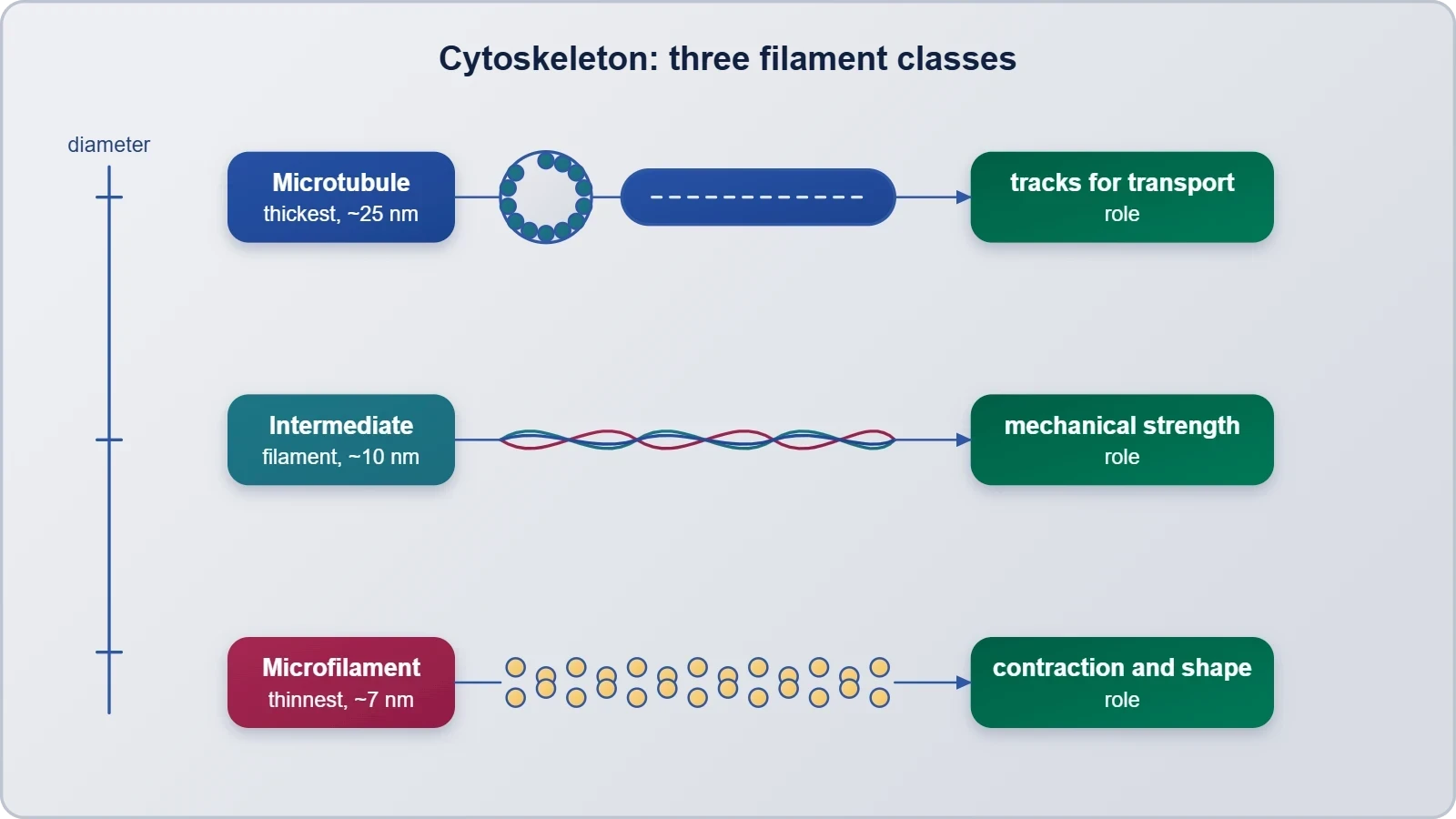
The functional importance of the cytoskeleton extends well beyond passive support. It participates in motility at the cellular level, contributes to the maintenance of cell shape, and provides the internal scaffolding that positions organelles in their correct locations. Because the filaments are dynamic, they can assemble and disassemble in response to signals, allowing the cell to change shape during division, migration or differentiation. This dynamism is what distinguishes the cytoskeleton from a static skeleton: it is a living framework that is continuously remodelled. The same filament systems also serve as the anchoring points for the cilia and flagella described in the next section.
How the Framework Connects to Other Organelles
The cytoskeleton does not operate in isolation. It links the plasma membrane to internal membranes and to the nucleus, creating a continuous mechanical pathway through the cell. Microtubules radiate from organising centres and guide the movement of vesicles between the endoplasmic reticulum, the Golgi apparatus and the plasma membrane. Microfilaments concentrate beneath the membrane, where they support the cell cortex and drive changes in surface shape. Intermediate filaments form a resilient meshwork around the nucleus and across the cytoplasm. This interconnected design means that a change in one part of the framework can be transmitted rapidly to distant regions of the cell.
Understanding the cytoskeleton also clarifies why certain cellular processes fail when the framework is disrupted. Transport of materials, positioning of organelles and the mechanics of cell division all depend on intact filaments. The framework therefore represents a unifying principle: structure and function are inseparable in the living cell. The table below summarises the three filament classes and their principal contributions.
Cilia and Flagella and the 9+2 Microtubule Array
Cilia and flagella are hair-like outgrowths of the cell membrane. Cilia are small structures that work like oars, causing the movement of either the cell or the surrounding fluid. Flagella are comparatively longer and are responsible for cell movement. The prokaryotic bacteria also possess flagella, but these are structurally different from eukaryotic flagella. This distinction matters because the two structures share a name but not an architecture. In eukaryotic cells, electron microscopy reveals that both cilia and flagella are covered with plasma membrane, and their core, called the axoneme, contains a number of microtubules running parallel to the long axis.
The Axoneme and Its Nine Radial Spokes
The axoneme usually has nine doublets of radially arranged peripheral microtubules and a pair of centrally located microtubules. This arrangement of axonemal microtubules is referred to as the 9+2 array. The central tubules are connected by bridges and are also enclosed by a central sheath, which is connected to one of the tubules of each peripheral doublet by a radial spoke. There are therefore nine radial spokes. The peripheral doublets are also interconnected by linkers. This precise geometry converts sliding of microtubules into the bending motion that propels a cell or sweeps fluid across a surface.
The structural logic of the 9+2 array can be expressed simply as a count of microtubules. Each peripheral doublet contributes two tubules, and the central pair contributes two more, giving a total of twenty microtubules per axoneme:
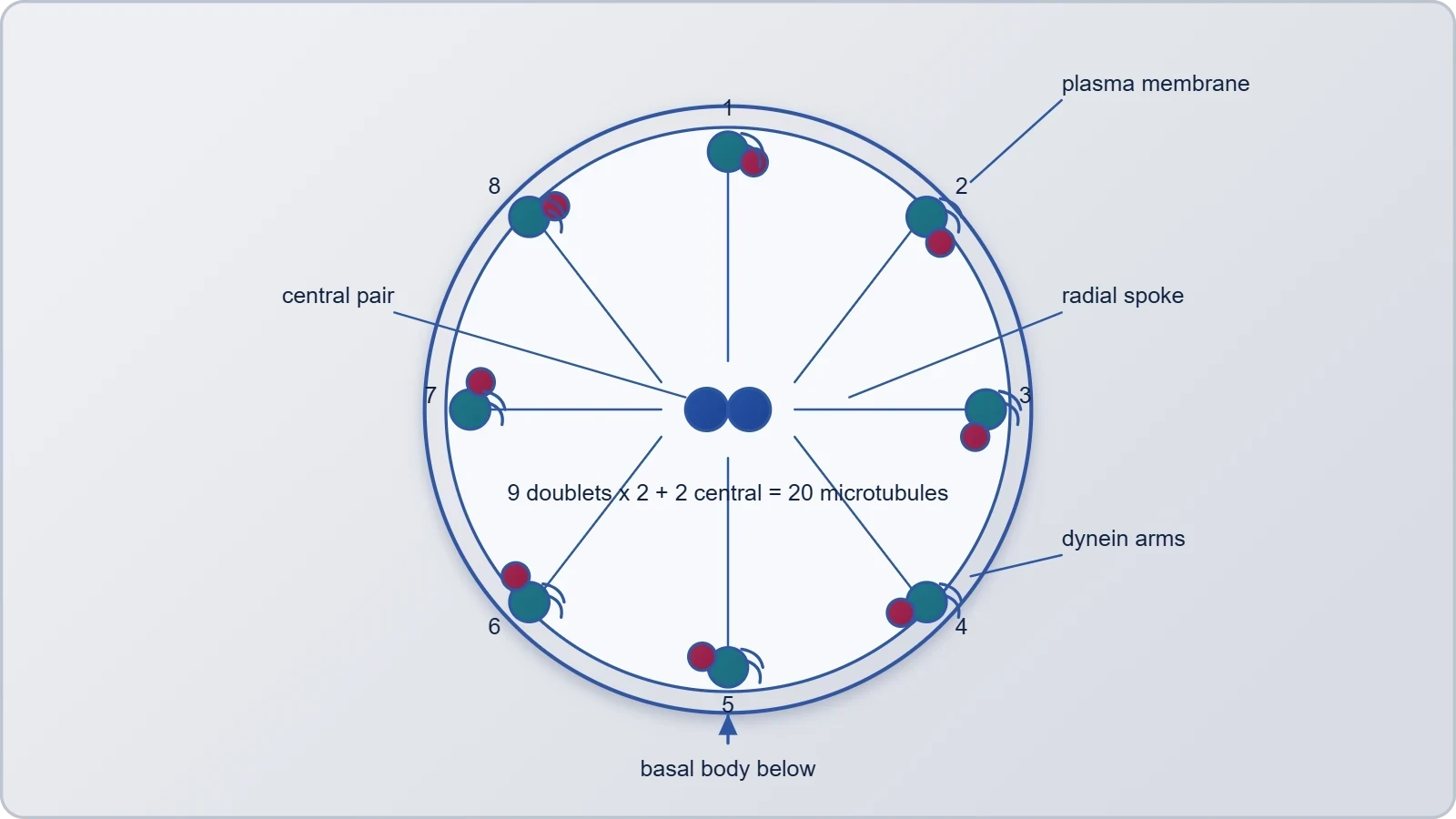
Both the cilium and the flagellum emerge from a centriole-like structure called the basal body. The basal body anchors the axoneme in the cytoplasm and serves as the template from which the microtubule arrangement is organised. Because the axoneme is enclosed by the plasma membrane, the entire appendage is effectively an extension of the cell surface rather than a separate organelle. This continuity explains why cilia and flagella can respond to signals at the membrane while still generating mechanical force deep within their core.
Movement, Length and Functional Contrast
The functional contrast between cilia and flagella is largely one of scale and motion pattern. Cilia are short and numerous, beating in coordinated waves that move fluid or propel the cell. Flagella are longer and typically fewer, producing a undulating motion that drives the cell forward. Both depend on the same 9+2 core, so the difference lies in dimensions and beat dynamics rather than in fundamental design. Recognising this shared architecture helps learners see that nature reuses a single molecular solution across a wide range of cellular tasks.
The basal body deserves particular attention because it links the appendage to the rest of the cell. It is centriole-like in organisation, which foreshadows the next section on the centrosome. In many cells, the basal body and the centriole share structural features, reflecting a common evolutionary and developmental logic. The table below sets out the principal features of cilia and flagella for quick reference.
Centrosome and Centrioles
The centrosome is an organelle usually containing two cylindrical structures called centrioles. They are surrounded by amorphous pericentriolar materials. Both centrioles in a centrosome lie perpendicular to each other, and each has an organisation like the cartwheel. This perpendicular arrangement is a defining feature and is easy to recognise in electron micrographs. The centrosome functions as a microtubule-organising centre, and its centrioles provide the template for the assembly of the cytoskeletal tracks that radiate through the cytoplasm. The basal bodies of cilia and flagella are centriole-like, which connects this section directly to the previous one.
Organisation of the Centriole Pair
Each centriole is a cylindrical structure built from microtubules arranged in a characteristic pattern, and the cartwheel organisation refers to the spoke-like arrangement seen in cross-section. The two centrioles sit at right angles, a geometry that is thought to support their role during cell division. The pericentriolar material around them is amorphous, meaning it lacks a fixed crystalline form, yet it is functionally essential because it nucleates microtubules. Together, the centrioles and the surrounding material form a single organelle that coordinates the spatial arrangement of the entire microtubule network.
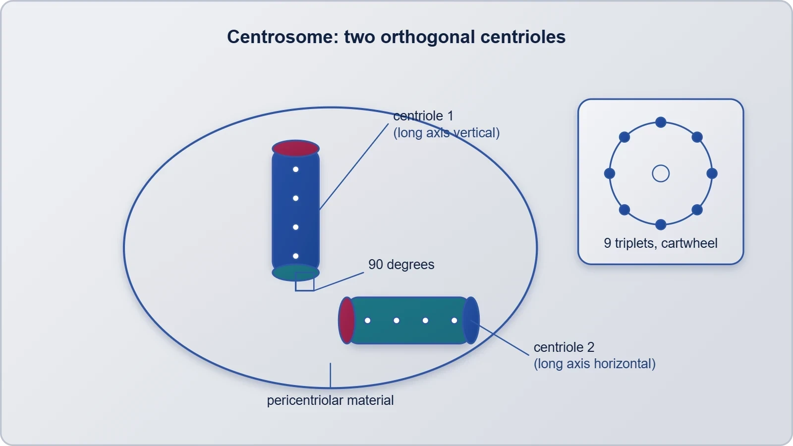
The perpendicular relationship between the two centrioles can be described geometrically. If the long axes of the two cylinders are represented as vectors, their mutual orientation is approximately orthogonal:
This orthogonality is not a trivial detail. It reflects a highly ordered internal geometry that is reproduced each time the centrosome is duplicated. For learners, the key point is that the centrosome is not a random cluster of tubules but a structured organelle with a defined number, arrangement and orientation of centrioles. That order is what allows it to perform its organising role reliably across successive cell generations.
Relationship to Basal Bodies and Cell Division
The structural resemblance between centrioles and basal bodies is one of the most instructive connections in cell biology. A basal body anchors a cilium or flagellum, while a centriole sits within the centrosome. Both share a cylindrical, microtubule-based organisation, and both serve as nucleation sites. Recognising this shared design helps learners appreciate how the cell reuses a single structural motif for different purposes. The centrosome, through its centrioles, thus stands at the intersection of cytoskeletal organisation, appendage formation and cell division.
During division, the centrosome duplicates and the two centrosomes move to opposite poles of the cell, where they help organise the microtubules that separate chromosomes. This function ties the centrosome to the nucleus, which is the subject of the next section. The table below summarises the defining features of the centrosome and its centrioles.
The Nucleus: Envelope, Nucleolus, Chromatin and Chromosomes
The nucleus is the largest and most conspicuous organelle of the eukaryotic cell, and it houses the genetic material that directs cellular activity. Its boundary is the nuclear envelope, a double membrane that separates the nucleoplasm from the cytoplasm. Within the nucleus, the nucleolus is a distinct region associated with the assembly of ribosomal components, while chromatin represents the diffuse form of the genetic material. When the cell prepares to divide, chromatin condenses into discrete chromosomes. This progression from diffuse chromatin to condensed chromosome is one of the central themes of nuclear biology and explains why the same genetic content can appear in such different forms.
Nuclear Envelope and Nucleolus
The nuclear envelope encloses the nucleus and regulates the exchange of materials between the nucleoplasm and the cytoplasm. Its double-membrane construction mirrors the pattern seen in mitochondria and chloroplasts, reinforcing the idea that membrane organisation is a recurring theme in eukaryotic cells. The nucleolus lies within the nucleus and is associated with the production of ribosomal components, linking nuclear activity directly to the ribosomes described in the previous lesson. Together, the envelope and the nucleolus make the nucleus both a protective container and an active site of synthesis and assembly.
The relationship between chromatin and chromosomes can be understood as a change of state rather than a change of substance. Chromatin is the extended, diffuse form of the genetic material, while chromosomes are the condensed structures visible during division. The same material is simply packaged differently. This distinction is important because it explains how genetic information can be both accessible for routine cellular work and compact enough to be moved accurately during division. The nucleus therefore combines storage, expression and transmission of genetic information within a single bounded compartment.
Centromere-Based Chromosome Types
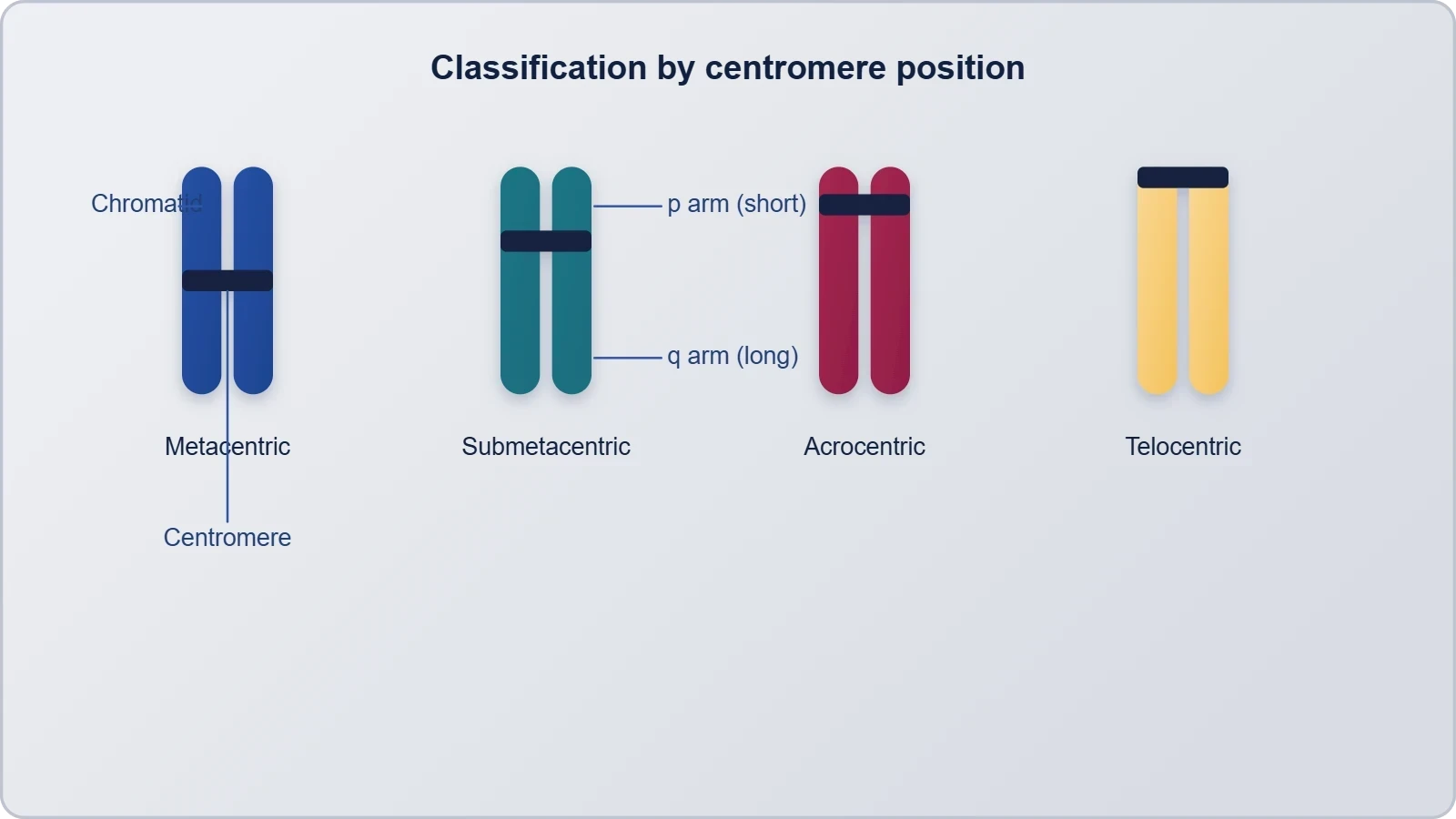
Chromosomes are classified according to the position of the centromere, the region that connects the two chromatids and serves as the attachment point for the spindle fibres during division. The centromere-based classification recognises several types depending on where the centromere lies along the chromosome. This positional criterion is a standard part of chromosome description and provides a simple way to compare chromosomes within a cell. The table below presents the centromere-based chromosome types as a structured reference for revision.
Microbodies and the Closing View of the Cell
Microbodies are small, membrane-bound compartments that complete the inventory of eukaryotic organelles. They are distinct from the larger organelles discussed earlier and are characterised by their compact size and their association with oxidative reactions. Although small, they contribute to the cell's metabolic versatility and demonstrate that even the least conspicuous compartments have defined roles. Their presence alongside mitochondria and plastids reinforces the principle that the eukaryotic cell is a community of specialised membrane-bound structures, each with its own internal chemistry and its own contribution to the whole.
Position Within the Organelle Community
Placing microbodies within the wider organelle community helps learners see the cell as an integrated system rather than a list of parts. Mitochondria and plastids were described in the previous lesson as double-membrane organelles with their own DNA and ribosomes. Microbodies, by contrast, are smaller and single-membrane bound, yet they participate in the same metabolic network. The cytoskeleton positions them, the nucleus directs their abundance, and the endomembrane system supplies their membranes. This interdependence is the central lesson of the course: no organelle functions in isolation.
The closing view of the cell therefore emphasises coordination. The cytoskeleton provides mechanical order, cilia and flagella provide motility, the centrosome organises microtubules, the nucleus safeguards and expresses genetic information, and microbodies contribute specialised chemistry. Each structure examined in this lesson connects to the others, forming a coherent whole. The table below summarises the organelles covered in this final lesson and their principal roles.
Going Further
The 9+2 array is a widely conserved arrangement of microtubules found in eukaryotic cilia and flagella. Its persistence across diverse organisms suggests that the geometry is tightly constrained by function: the nine peripheral doublets and the central pair must be positioned precisely for coordinated sliding to produce useful bending. Learners who encounter cilia and flagella in later studies will find that the same 9+2 description recurs, which makes it a reliable anchor for revision. The conservation of this pattern is one of the clearest examples of structural unity in eukaryotic biology.
Human erythrocytes and sieve tube cells of vascular plants lack a nucleus in their mature form. This observation is a useful counterpoint to the general rule that eukaryotic cells contain a nucleus. It shows that the nucleus is essential during development but can be dispensed with once a cell has specialised for a particular function. Mature erythrocytes, for instance, are optimised for oxygen transport, while sieve tube cells are optimised for conduction. These exceptions remind learners that biological generalisations describe the typical case rather than an invariable law.
Both observations reinforce the lesson's central message: cellular structures are adapted to function, and their presence or absence reflects the demands of the cell's role. The 9+2 array illustrates conservation of a mechanical design, while the nucleus-free mature cells illustrate specialisation through loss. Together they show that structure and function are two sides of the same biological principle, and that exceptions often illuminate the rule more clearly than the rule itself.
Consolidating the Final Lesson
This lesson has traced the structures that complete the eukaryotic cell: the cytoskeleton with its three filament systems, the cilia and flagella built on the 9+2 axoneme, the centrosome with its perpendicular centrioles, the nucleus with its envelope, nucleolus, chromatin and chromosomes, and the microbodies. Each structure was presented in the order established by the course, with its terminology and numerical details preserved. The aim has been to show how these components cooperate rather than to treat them as isolated facts. A learner who can connect the cytoskeleton to the basal body, the centrosome to the spindle, and the nucleus to the ribosomes has grasped the integrative logic of the cell.
Key Points to Retain
The cytoskeleton consists of microtubules, microfilaments and intermediate filaments and provides mechanical support, motility and shape maintenance. Cilia and flagella are membrane-covered outgrowths whose axoneme has nine peripheral doublets and a central pair, the 9+2 array, with nine radial spokes and interdoublet linkers. The centrosome usually contains two perpendicular centrioles with cartwheel organisation, surrounded by amorphous pericentriolar material. The nucleus contains the nuclear envelope, nucleolus, chromatin and chromosomes, and chromosomes are classified by centromere position. Microbodies are small membrane-bound compartments with specialised chemistry.
These points form a compact revision set. Learners preparing for examinations should be able to reproduce the 9+2 description, state the number of radial spokes, describe the perpendicular centrioles, and distinguish chromatin from chromosomes. Each of these facts is anchored in a structural diagram or table within the lesson, so revision can proceed by recalling the visual arrangement rather than memorising isolated sentences. The final table below offers a last consolidated check across the whole lesson.
Also Read
- No links available.
From our network :
- Vite 6/7 'Cold Start' Regression in Massive Module GraphsIn-depth analysis of Vite 6/7 cold start regressions in massive module graphs. Learn about barrel file issues, pre-bundling optimizations, and how to improve Vite dev server performance.
- EV 2.0: The Solid-State Battery Breakthrough and Global Factory ExpansionDiscover the impact of the Solid-State Battery Breakthrough on the EV market. Learn about CATL and BYD stock growth, global factory expansion in Europe, and the future of EV 2.0 technology.
- 98% of Global MBA Programs Now Prefer GRE Over GMAT Focus EditionExplore why 98% of global MBA programs now prefer the GRE over GMAT Focus. Learn about the shorter GRE format, verbal logic importance, and strategic versatility for students.
- AI-Powered 'Precision Diagnostic' Replaces Standard GRE Score ReportsDiscover the new GRE Precision Diagnostic. Learn how AI-powered behavioral analytics and time-to-solve tracking are replacing standard GRE score reports to empower students.
- Mastering DB2 LUW v12 Tables: A Comprehensive Technical GuideComprehensive guide on DB2 LUW v12 tables. Explore CREATE TABLE syntax, BLU Acceleration, range partitioning, data types, and security features for IBM DB2 LUW v12.
- 10 Physics Numerical Problems with Solutions for IIT JEEPrepare for IIT JEE with 10 physics numerical problems, complete with detailed solutions and conceptual explanations. Ideal for mastering kinematics, thermodynamics, electromagnetism, and optics.
- Analyzing Trump Deportation Numbers Insights Into The 2026 Immigration Crackdown
- Mastering DB2 12.1 Instance Design: A Technical Deep Dive into Modern Database ArchitectureComprehensive guide to DB2 12.1 Instance Design. Explore architectural components, HADR setup, buffer pool tuning, and security policies for IBM DB2 12.1. Includes 10 functional SQL and Shell samples.
- Trump Political Strategy How Geopolitical Stunts Serve As Media Diversions
RESOURCES
- Resources will be updated soon.