Every living cell operates as a highly organised economy of compartments, and few compartments illustrate that principle more vividly than the mitochondrion, the plastid and the ribosome. These three structures carry out tasks that no other organelle can substitute: mitochondria convert the chemical energy of food into ATP through aerobic respiration, plastids capture light and store nutrients in plant cells, and ribosomes translate genetic instructions into the proteins on which every cellular process depends. Understanding how each is built explains, in turn, why each performs its task so efficiently.
This lesson examines these organelles in the order in which cell biology traditionally presents them, beginning with the double-membrane architecture of the mitochondrion and its cristae, moving through the classification of plastids into chloroplasts, chromoplasts and leucoplasts, and closing with the ribosome and its subunits. The treatment builds directly on the endomembrane system covered previously, where the endoplasmic reticulum and Golgi apparatus were shown to package and modify proteins, and it prepares the ground for the cytoskeleton, cilia, flagella, centrosome, nucleus and microbodies that follow.
Throughout, the emphasis falls on structure as the explanation of function. A membrane folded into cristae, a stack of thylakoids arranged like coins, or a ribosome split into two subunits of unequal size are not decorative details; each arrangement is the physical basis of a measurable biochemical capability. Learners who grasp this connection will find that the numbers, dimensions and sedimentation coefficients discussed here become memorable rather than merely listable, and that the vocabulary of the chapter begins to organise itself around a small number of governing principles.
On This Page
- Mitochondria: Double-Membrane Power Houses of the Cell
- Plastids: Chloroplasts, Chromoplasts and Leucoplasts
- Chloroplast Dimensions and the Architecture of the Thylakoid
- Ribosomes: Granular Structures Without a Membrane
- Reading the Numbers: Sedimentation, Size and Surface Area
- Going Further
- Consolidating the Lesson: A Structured Revision Path
Mitochondria: Double-Membrane Power Houses of the Cell
Mitochondria, written in the singular as mitochondrion, are not easily visible under the microscope unless they are specifically stained, a practical point that reflects how thinly they are distributed through the cytoplasm. Their number per cell varies with the physiological activity of the cell, so a metabolically demanding tissue may contain far more of them than a quiescent one. Shape and size also show considerable variability, though the typical form is sausage-shaped or cylindrical, with a diameter of 0.2–1.0 μm (average 0.5 μm) and a length of 1.0–4.1 μm.
Outer Membrane, Inner Membrane and the Two Compartments
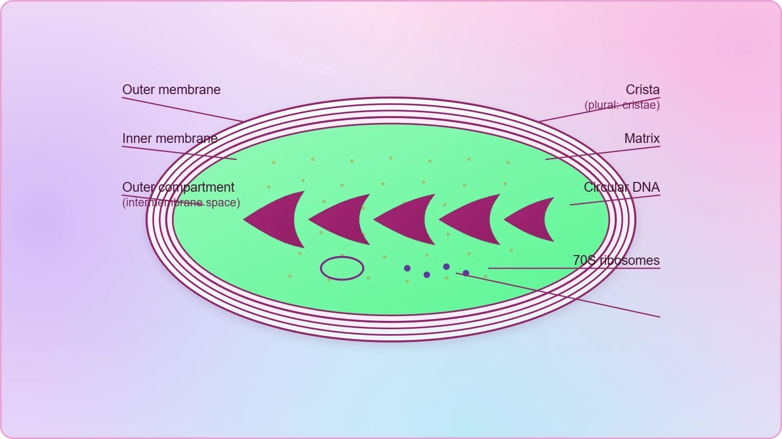
Each mitochondrion is a double membrane-bound structure. The outer membrane and the inner membrane divide its lumen distinctly into two aqueous compartments, the outer compartment and the inner compartment. The outer membrane forms the continuous limiting boundary of the organelle, while the inner membrane folds inward to create the cristae. The two membranes each carry their own specific enzymes associated with mitochondrial function, so the boundary is not a passive wrapper but an enzymatically active interface between the organelle and the rest of the cell.
The inner compartment is filled with a dense, homogeneous substance called the matrix. The cristae, singular crista, project toward this matrix and thereby increase the surface area available for the membrane-associated reactions of respiration. This geometric arrangement matters: a larger membrane surface accommodates more of the enzyme complexes that drive oxidative phosphorylation, so the folded inner membrane is directly tied to the organelle's capacity to generate energy rather than being an incidental feature of its shape.
Matrix Contents, Energy Production and Fission
Mitochondria are the sites of aerobic respiration, and they produce cellular energy in the form of ATP, which is why they are called the power houses of the cell. The matrix itself possesses a single circular DNA molecule, a few RNA molecules, ribosomes of the 70S type, and the components required for the synthesis of proteins. This internal machinery means the organelle carries a measure of genetic autonomy, and the mitochondria divide by fission, a simple splitting process rather than a complex mitotic apparatus.
The presence of 70S ribosomes inside the matrix is a detail worth pausing over, because the cytoplasmic ribosomes of a eukaryotic cell are of the 80S type. The mitochondrion therefore contains a protein-synthesis system that differs in sedimentation behaviour from the one operating in the surrounding cytosol. Learners preparing for examinations should be able to state both facts together: the mitochondrion is double membrane-bound, its inner membrane forms cristae, its matrix holds circular DNA and 70S ribosomes, and it divides by fission.
Plastids: Chloroplasts, Chromoplasts and Leucoplasts
Plastids are found in all plant cells and in euglenoides, and because they are large they are easily observed under the microscope. They bear specific pigments that impart particular colours to plants, and on the basis of pigment type they are classified into chloroplasts, chromoplasts and leucoplasts. This threefold division is the organising principle of the topic: each class is defined by what it contains and, consequently, by the colour and function it confers on the tissue where it resides.
Pigments and the Three Classes of Plastids
Chloroplasts contain chlorophyll and carotenoid pigments, which are responsible for trapping light energy essential for photosynthesis. Chromoplasts contain fat-soluble carotenoid pigments such as carotene, xanthophylls and others, giving parts of the plant a yellow, orange or red colour. Leucoplasts are the colourless plastids, of varied shapes and sizes, with stored nutrients. The distinction is functional as well as visual: one class harvests light, one supplies colour, and one serves as a storehouse for reserves.
The leucoplasts are further subdivided by the nutrient they store. Amyloplasts store carbohydrates in the form of starch, as in the potato; elaioplasts store oils and fats; and aleuroplasts store proteins. This internal classification rewards careful reading, because the three names differ by only a few letters yet refer to entirely different storage roles. A learner who can pair each name with its stored material has mastered the part of the topic that examiners most often test through matching or one-word-answer questions.
Chloroplast Structure: Stroma, Thylakoids and Grana
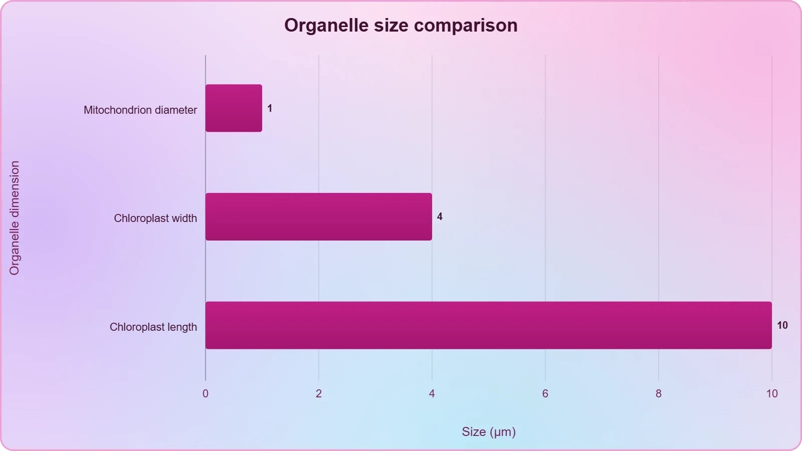
The majority of chloroplasts in green plants are found in the mesophyll cells of the leaves. They are lens-shaped, oval, spherical, discoid or even ribbon-like organelles, with variable length of 5–10 μm and width of 2–4 μm. Their number varies from one per cell in Chlamydomonas, a green alga, to 20–40 per cell in the mesophyll. Like mitochondria, chloroplasts are double membrane-bound, and of the two membranes the inner one is relatively less permeable.
The space limited by the inner membrane is called the stroma. Within the stroma are organised, flattened membranous sacs called thylakoids, arranged in stacks like piles of coins called grana, singular granum, or as intergranal thylakoids. Flat membranous tubules called stroma lamellae connect the thylakoids of different grana. The membrane of the thylakoids encloses a space called the lumen. Chlorophyll pigments are present in the thylakoids, so the light-trapping apparatus is localised precisely where the membrane stacks are densest.
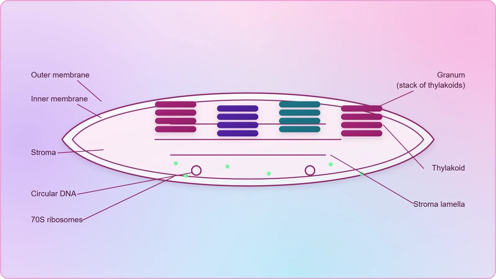
The stroma contains enzymes required for the synthesis of carbohydrates and proteins, together with small, double-stranded circular DNA molecules and ribosomes. The ribosomes of the chloroplasts are smaller, at 70S, than the cytoplasmic ribosomes at 80S. This parallel with the mitochondrion is deliberate and instructive: both organelles are double membrane-bound, both carry circular DNA, and both use 70S ribosomes, which is why the two are frequently compared in examination questions and in discussions of organelle autonomy.
Chloroplast Dimensions and the Architecture of the Thylakoid
The chloroplast repays close attention to its measurements because its scale explains its visibility. With a length of 5–10 μm and a width of 2–4 μm, it is large enough to be seen under the light microscope, unlike the mitochondrion at 0.2–1.0 μm in diameter. The number per cell ranges from a single chloroplast in Chlamydomonas to 20–40 in a mesophyll cell, a range that reflects the differing photosynthetic demands of a unicellular alga and a differentiated leaf tissue.
Stroma, Thylakoids, Grana and Stroma Lamellae
The inner chloroplast membrane is relatively less permeable than the outer, and the space it limits is the stroma. Within the stroma lie the thylakoids, flattened membranous sacs that may be stacked into grana or left as intergranal thylakoids. Stroma lamellae, flat membranous tubules, connect the thylakoids of different grana, creating a continuous membrane network across the organelle. The thylakoid membrane encloses the lumen, and chlorophyll pigments reside in this membrane rather than in the stroma.
This layered architecture separates two chemically distinct environments. The stroma holds the enzymes for carbohydrate and protein synthesis along with circular DNA and ribosomes, while the thylakoid membranes hold the pigment and enclose the lumen. The arrangement is functionally analogous to the mitochondrial division between matrix and cristae, and recognising the parallel helps learners retain both structures without confusing their contents. The chloroplast is, in effect, a double-membrane organelle with an internal membrane system specialised for light capture.
Comparing Mitochondria and Chloroplasts
Placing the two organelles side by side clarifies what is shared and what is distinctive. Both are double membrane-bound, both contain circular DNA and 70S ribosomes, and both are capable of protein synthesis within their own compartments. They differ in their internal folding: the mitochondrion folds its inner membrane into cristae, whereas the chloroplast builds separate thylakoid sacs that may stack into grana. They also differ in function, one releasing energy through aerobic respiration and the other trapping light energy for photosynthesis.
A useful way to consolidate this comparison is to express the sedimentation distinction explicitly. The cytoplasmic ribosome of a eukaryote is 80S, while the organellar ribosome is 70S, so the two systems can be distinguished by their sedimentation behaviour. The Svedberg unit is defined through the sedimentation coefficient, which depends on both mass and shape, so a 70S ribosome is not simply the arithmetic sum of its 50S and 30S subunits. Writing the relationship as ##80S \neq 60S + 40S## and ##70S \neq 50S + 30S## reinforces why the unit measures behaviour rather than simple addition.
Ribosomes: Granular Structures Without a Membrane
Ribosomes are the granular structures first observed under the electron microscope as dense particles by George Palade in 1953. They are composed of ribonucleic acid, RNA, and proteins, and they are not surrounded by any membrane. That absence of a membrane is a defining feature rather than a trivial omission: it distinguishes the ribosome from every membrane-bound organelle discussed so far and explains why the particle can be described purely in terms of its two subunits and their sedimentation behaviour.
80S and 70S Ribosomes and Their Subunits
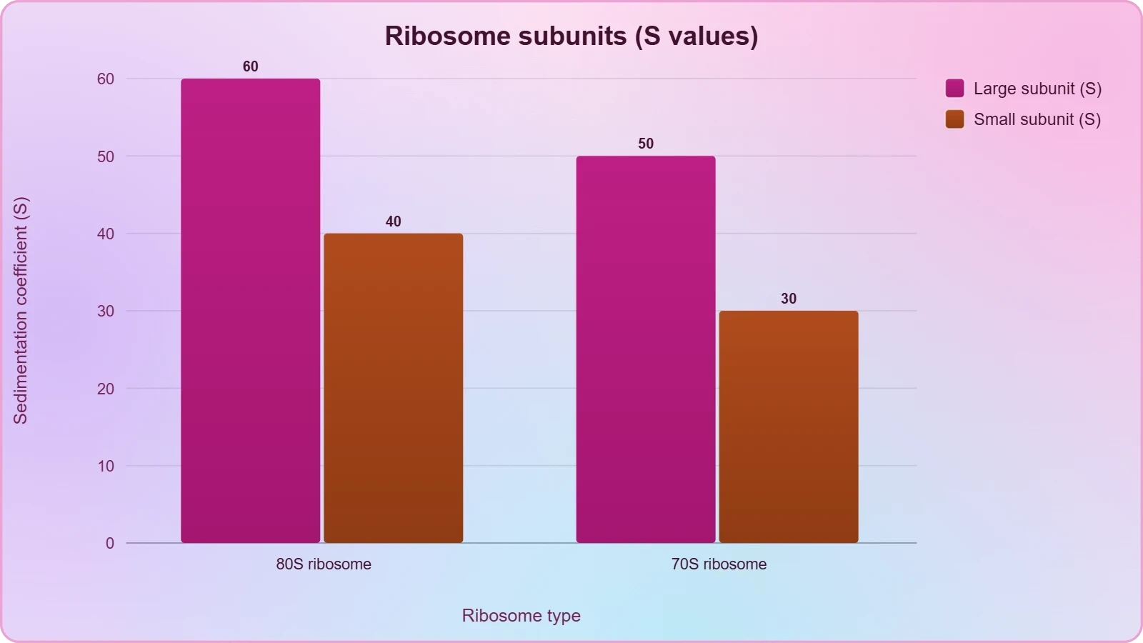
The eukaryotic ribosomes are 80S, while the prokaryotic ribosomes are 70S. Each ribosome has two subunits, a larger and a smaller one. The two subunits of the 80S ribosome are 60S and 40S, while those of the 70S ribosome are 50S and 30S. Both the 70S and the 80S ribosomes are composed of two subunits, so the two-subunit plan is universal even though the sedimentation values differ between the two cell types.
The letter S stands for Svedberg's unit, which denotes the sedimentation coefficient and is indirectly a measure of density and size. Because the coefficient reflects how a particle behaves in a centrifugal field, the values of the subunits do not simply add to the value of the whole ribosome. This is why the 80S ribosome is not the sum of 60 and 40 in any straightforward arithmetic sense, and why the 70S ribosome is not the sum of 50 and 30. The unit describes a physical property, not a count of parts.
Where Ribosomes Occur and Why the Distinction Matters
Ribosomes occur free in the cytoplasm and associated with the endoplasmic reticulum, where the proteins they synthesise enter the endomembrane system for modification and packaging. The 70S type also appears inside mitochondria and chloroplasts, as noted earlier, which links this section back to the two organelles already discussed. Recognising that the organellar ribosome is of the 70S type, while the cytoplasmic ribosome of a eukaryote is 80S, is one of the most frequently examined facts in the chapter.
The practical significance of the distinction extends beyond classification. Because the prokaryotic and organellar ribosome is 70S and the eukaryotic cytoplasmic ribosome is 80S, the two are structurally different targets, a difference that underlies the selective action of several well-known antibacterial agents. The lesson does not require the pharmacology, but the structural contrast is worth stating plainly: the sedimentation value is a shorthand for a genuine difference in composition and behaviour, not merely a label attached for convenience.
Reading the Numbers: Sedimentation, Size and Surface Area
The quantitative details in this lesson are not incidental decoration; they encode the logic of the organelles. A mitochondrion of average diameter 0.5 μm and length 1.0–4.1 μm is small enough to escape easy light-microscopic detection, while a chloroplast of length 5–10 μm and width 2–4 μm is large enough to be seen. The cristae increase the surface area of the inner mitochondrial membrane, and the grana stack thylakoids to concentrate the light-harvesting membrane. In each case, a measurement or a geometric arrangement explains a capability.
Why the Svedberg Unit Is Not Additive
The sedimentation coefficient depends on both the mass and the shape of a particle, so the values of the subunits cannot simply be summed. This is the conceptual heart of the ribosome topic, and it can be expressed compactly. For the eukaryotic ribosome, the whole particle sediments at 80S while its parts sediment at 60S and 40S; for the prokaryotic ribosome the corresponding values are 70S, 50S and 30S. The non-additivity is a consequence of how the coefficient is defined, not an error in the data.
In this expression, ##s## is the sedimentation coefficient, ##m## is the particle mass, ##\bar{v}## is its partial specific volume, ##\rho## is the density of the medium, and ##f## is the frictional coefficient, which depends on shape. Because ##f## changes when subunits associate into a whole ribosome, the coefficient of the assembled particle is not the sum of the coefficients of its separated parts. The formula therefore justifies the statement that S is indirectly a measure of density and size, and it explains why 60S plus 40S does not yield 80S.
Connecting Structure to Function Across the Three Organelles
A final synthesis ties the three subjects of this lesson together. The mitochondrion converts energy, the plastid captures or stores it, and the ribosome builds the proteins that make both possible. Each structure achieves its function through a specific arrangement: cristae for membrane surface, thylakoid stacks for light capture, and two unequal subunits for translation. When these arrangements are understood as solutions to physical problems, the vocabulary of the chapter becomes a connected system rather than a list of isolated definitions.
This prepares the ground for the next lesson, which turns to the cytoskeleton, cilia and flagella, the centrosome and centrioles, the nucleus and the microbodies. The cytoskeleton, an elaborate network of filamentous proteinaceous structures consisting of microtubules, microfilaments and intermediate filaments, provides mechanical support, motility and maintenance of cell shape, and it will connect naturally to the organelles discussed here. The nucleus, in turn, will complete the picture of how the instructions translated by ribosomes are stored and dispatched.
Going Further
The three leucoplast types reward a second look because their names encode their contents. Amyloplasts store carbohydrates in the form of starch, as in the potato; elaioplasts store oils and fats; and aleuroplasts store proteins. Keeping these three pairings distinct is the surest way to answer matching questions correctly, and it also clarifies why a single colourless plastid class can serve such different storage roles in different tissues and at different stages of a plant's life.
Because mitochondria and chloroplasts contain their own circular DNA and 70S ribosomes, they are often described as having some bacterial-like features. This observation is well established in cell biology and is usually discussed in connection with the idea that these organelles share a distant evolutionary relationship with free-living prokaryotes. The lesson does not require that argument, but the structural facts already established, namely circular DNA and 70S ribosomes inside a double membrane, are the evidence on which the description rests.
Consolidating the Lesson: A Structured Revision Path
A reliable way to revise this material is to move from the general to the specific. Begin with the shared plan of the double-membrane organelles, then distinguish the mitochondrion from the chloroplast by their internal folding and their primary function, and finish with the ribosome, which is defined by the absence of a membrane and the presence of two subunits. This sequence mirrors the order in which the topics were presented and keeps related facts adjacent in memory rather than scattered across unrelated lists.
Common Points of Confusion and How to Avoid Them
Several errors recur in examination answers. The matrix is often confused with the stroma, although the first belongs to the mitochondrion and the second to the chloroplast. The cristae are sometimes described as part of the outer membrane, when they are infoldings of the inner membrane. The 70S and 80S values are occasionally swapped between prokaryotes and eukaryotes. Each of these mistakes disappears once the underlying structure is visualised rather than memorised as a string of words.
A second cluster of confusions concerns the plastids. Chromoplasts are sometimes treated as a synonym for chloroplasts, when the former contain fat-soluble carotenoids and the latter contain chlorophyll and carotenoids for light trapping. The three leucoplast types are frequently mixed up, with amyloplasts, elaioplasts and aleuroplasts assigned to the wrong reserves. Careful pairing of name with stored material, and of pigment with plastid class, prevents these errors and makes the classification genuinely useful rather than merely recitable.
Preparing for the Next Lesson
The organelles studied here do not operate in isolation. The proteins made by ribosomes must be moved, supported and positioned, and that is the work of the cytoskeleton, an elaborate network of filamentous proteinaceous structures consisting of microtubules, microfilaments and intermediate filaments. The cytoskeleton is involved in mechanical support, motility and maintenance of the shape of the cell, and it will connect the energy-producing and protein-building functions described here to the structural framework of the whole cell.
The next lesson also takes up cilia and flagella, the centrosome and centrioles, the nucleus and the microbodies. The nucleus will complete the account of how the instructions translated by ribosomes are stored, while the centrosome and centrioles will extend the discussion of microtubules into cell division. Learners who have understood why cristae increase surface area and why thylakoids stack into grana will find the same logic of structure serving function waiting for them in the cytoskeleton and the flagellum.
Also Read
- No links available.
From our network :
- Analyzing Trump Deportation Numbers Insights Into The 2026 Immigration Crackdown
- Vite 6/7 'Cold Start' Regression in Massive Module GraphsIn-depth analysis of Vite 6/7 cold start regressions in massive module graphs. Learn about barrel file issues, pre-bundling optimizations, and how to improve Vite dev server performance.
- Mastering DB2 12.1 Instance Design: A Technical Deep Dive into Modern Database ArchitectureComprehensive guide to DB2 12.1 Instance Design. Explore architectural components, HADR setup, buffer pool tuning, and security policies for IBM DB2 12.1. Includes 10 functional SQL and Shell samples.
- 10 Physics Numerical Problems with Solutions for IIT JEEPrepare for IIT JEE with 10 physics numerical problems, complete with detailed solutions and conceptual explanations. Ideal for mastering kinematics, thermodynamics, electromagnetism, and optics.
- Mastering DB2 LUW v12 Tables: A Comprehensive Technical GuideComprehensive guide on DB2 LUW v12 tables. Explore CREATE TABLE syntax, BLU Acceleration, range partitioning, data types, and security features for IBM DB2 LUW v12.
- AI-Powered 'Precision Diagnostic' Replaces Standard GRE Score ReportsDiscover the new GRE Precision Diagnostic. Learn how AI-powered behavioral analytics and time-to-solve tracking are replacing standard GRE score reports to empower students.
- EV 2.0: The Solid-State Battery Breakthrough and Global Factory ExpansionDiscover the impact of the Solid-State Battery Breakthrough on the EV market. Learn about CATL and BYD stock growth, global factory expansion in Europe, and the future of EV 2.0 technology.
- Trump Political Strategy How Geopolitical Stunts Serve As Media Diversions
- 98% of Global MBA Programs Now Prefer GRE Over GMAT Focus EditionExplore why 98% of global MBA programs now prefer the GRE over GMAT Focus. Learn about the shorter GRE format, verbal logic importance, and strategic versatility for students.
RESOURCES
- Resources will be updated soon.