Every bacterial cell lives inside a boundary that must be strong enough to resist internal pressure, selective enough to admit nutrients, and flexible enough to permit growth and division. That boundary is not a single sheet but a layered assembly, and biologists call it the cell envelope. Understanding the envelope is the key to understanding why bacteria behave differently under a microscope, why some species resist certain dyes and antibiotics, and how a prokaryote interacts with the world beyond its cytoplasm. This lesson examines that architecture in detail, moving from the outermost glycocalyx inward through the wall and plasma membrane, and then outward again to the appendages that project from the surface.
The material that follows builds directly on the previous lesson on prokaryotic cells, where the basic prokaryotic plan was introduced. Here the focus narrows to the envelope itself: its three-layered organisation, the Gram positive and Gram negative classification that arises from it, the forms the glycocalyx can take, the fluid mosaic structure of the plasma membrane, and the internal membrane modifications known as mesosomes and chromatophores. The lesson closes with the surface structures — flagella, pili and fimbriae — that extend beyond the envelope and give the cell motility and the ability to attach. Each concept is presented in the order the source develops it, with its terminology and figures preserved.
Because the envelope sits at the interface between cell and environment, its details carry practical weight. The Gram stain, one of the oldest and most useful tools in microbiology, is essentially a readout of envelope chemistry. The proteins embedded in the plasma membrane determine what enters and leaves. The glycocalyx decides whether a bacterium can cling to a surface or form a biofilm. By the end of this lesson, you should be able to describe each layer, explain how the layers differ between the two major bacterial groups, and account for the function of every surface appendage discussed.
On This Page
The Three-Layered Cell Envelope and Its Gram Classification
The prokaryotic cell envelope is best understood as three concentric layers, each with a distinct chemical identity and function. The outermost layer is the glycocalyx, a covering of carbohydrate-rich material. Beneath it lies the cell wall, a rigid structure that gives the cell its shape and protects it from osmotic rupture. Innermost is the plasma membrane, the selectively permeable barrier that separates cytoplasm from the exterior. Together these three layers form a functional unit, and the differences in their arrangement between bacterial groups produce the Gram positive and Gram negative categories that microbiologists rely on daily.
Composition and Arrangement of the Layers
The glycocalyx may appear as a loose slime layer or as a more organised capsule, and its composition varies with species. The cell wall provides mechanical strength, and in bacteria it is built from peptidoglycan, a meshwork of sugar chains cross-linked by short peptides. The plasma membrane lies immediately inside the wall and consists of a phospholipid bilayer studded with proteins. The relative thickness of the wall and the presence or absence of an additional outer membrane are what separate the two Gram classes, and these structural facts explain the staining behaviour described below.
In Gram positive bacteria the wall is thick and retains the primary dye complex after decolourisation, so the cell appears purple under the microscope. In Gram negative bacteria the wall is thinner and is overlaid by an outer membrane, and the dye complex is washed out during decolourisation, leaving the cell to take up the pink counterstain. This is not a trivial laboratory curiosity: the outer membrane of Gram negative cells acts as an additional permeability barrier, which is one reason such organisms often show greater resistance to certain antibacterial agents.

Why the Classification Matters
The Gram classification is more than a staining artefact; it reflects real structural differences that influence how a bacterium responds to its surroundings. Because the Gram negative envelope includes an outer membrane, many substances that readily reach the plasma membrane of a Gram positive cell are slowed or excluded. This has direct consequences for the action of dyes, detergents and antibacterial compounds, and it explains why the two groups are treated as distinct categories in both laboratory practice and clinical microbiology. Recognising the envelope type is therefore a first step in predicting how an organism will behave.
It is worth noting that the classification is not absolute. Some bacteria, such as the mycobacteria, possess walls with unusual lipid content and do not stain reliably by either route, and are described as acid-fast. Others may appear Gram variable depending on culture age. These exceptions do not undermine the scheme; they simply remind us that the envelope is a biological structure with natural variation, and that the two-class model is a useful simplification rather than a complete description of every prokaryote.
The Glycocalyx and the Forms It Takes
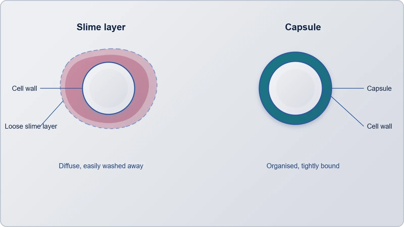
The glycocalyx is the outermost component of the envelope, a covering of carbohydrate-rich material that lies outside the cell wall. Its name reflects its composition: glyco for sugar and calyx for cup, since it forms a cup-like sheath around the cell. The glycocalyx is not a rigid structure like the wall; it is more variable in thickness and consistency, and its form differs between species and even between growth conditions. Two broad forms are recognised, the slime layer and the capsule, and the distinction between them rests on how tightly the material adheres to the cell surface.
Slime Layer and Capsule
A slime layer is a loose, diffuse covering that is easily washed away and does not adhere firmly to the wall. A capsule, by contrast, is a more organised and tightly bound structure that remains attached to the cell even when it is handled. Both are composed largely of polysaccharides, though some species incorporate polypeptide material. The capsule is often the more conspicuous of the two under appropriate staining, appearing as a halo around the cell, and it is the form most often associated with the protective functions attributed to the glycocalyx.
The functions of the glycocalyx follow from its position and chemistry. Because it is carbohydrate-rich and often hydrated, it can protect the cell against desiccation and help it adhere to surfaces, including the tissues of a host. In pathogenic species the capsule contributes to virulence by interfering with the host's ability to recognise and engulf the bacterium. The glycocalyx also participates in the formation of biofilms, communities of bacteria embedded in a shared matrix that adhere to surfaces and show increased resistance to adverse conditions.
Protective and Adhesive Roles
The glycocalyx illustrates a general principle of cell biology: structures at the surface are adapted to mediate interaction. A capsule that shields a bacterium from host defences is an advantage in a pathogenic lifestyle, while a slime layer that promotes attachment is an advantage on submerged surfaces where nutrients accumulate. Neither form is permanent; the cell can adjust its covering in response to conditions. This flexibility is part of why bacteria colonise so many environments, from soil particles to medical implants, and why the glycocalyx is studied alongside the wall and membrane as a functional component of the envelope.
From a practical standpoint, the glycocalyx also matters in the laboratory. Capsule staining is a distinct procedure because ordinary stains do not readily penetrate the hydrated matrix, and the capsule appears as an unstained halo against a stained background. Recognising this appearance helps in identifying species, and it reinforces the lesson that envelope structures are best studied with methods suited to their chemistry. The same principle applies to the membrane, whose detailed structure became accessible only with the electron microscope in the 1950s.
The Plasma Membrane and the Fluid Mosaic Model
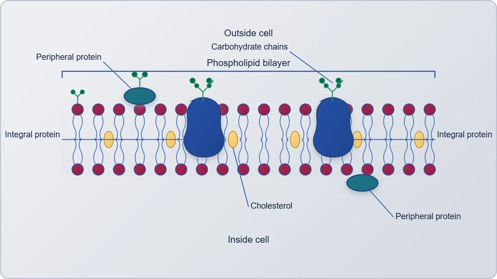
The plasma membrane is the innermost layer of the envelope and the one with the most demanding job: it must be selective. Chemical studies on the cell membrane, especially in human red blood cells, showed that the membrane is mainly composed of lipids and proteins. The major lipids are phospholipids arranged in a bilayer, with the polar head towards the outer sides and the hydrophobic tails towards the inner part. This arrangement ensures that the nonpolar tail of saturated hydrocarbons is protected from the aqueous environment, a point the source states explicitly and which is central to understanding membrane behaviour.
Lipid Bilayer, Cholesterol and Proteins
In addition to phospholipids, the membrane also contains cholesterol, which modulates the properties of the bilayer. Biochemical investigation clearly revealed that cell membranes also possess protein and carbohydrate. The ratio of protein to lipid varies considerably in different cell types; in human beings, the membrane of the erythrocyte has approximately 52 per cent protein and 40 per cent lipids. These figures are a useful reminder that the membrane is not a uniform material but a composite whose composition is tuned to the function of the particular cell.
Depending on the ease of extraction, membrane proteins can be classified as integral and peripheral. Peripheral proteins lie on the surface of the membrane, while integral proteins are partially or totally buried in the membrane. This distinction is operational as well as structural: peripheral proteins can be removed relatively gently, whereas integral proteins require disruption of the bilayer. The arrangement of lipids, proteins and carbohydrate is captured in the fluid mosaic model of the plasma membrane, which describes a dynamic structure in which components are free to move laterally within the plane of the membrane.
Selective Permeability and Membrane Behaviour
The bilayer arrangement has a direct functional consequence: the hydrophobic interior restricts the free passage of polar and charged substances, while small nonpolar molecules can diffuse across. This is the physical basis of selective permeability. Proteins embedded in the bilayer provide the routes by which ions, sugars and other polar solutes cross, and peripheral proteins participate in signalling and in linking the membrane to other structures. The fluid character of the membrane allows proteins to move within the plane of the bilayer, which is essential for processes such as the assembly of transport complexes and the redistribution of components during growth.
The fluid mosaic model, illustrated in the source as the structure of the plasma membrane, remains the standard description because it accounts for both the lipid bilayer and the dispersed proteins. It also explains why membrane composition matters: a membrane rich in protein, like that of the erythrocyte, is equipped for a particular set of transport and recognition tasks, while a membrane with a different ratio serves a different cell. The model is a framework, not a fixed picture, and later refinements have added detail about domains and asymmetry without displacing its central idea.
Mesosomes and Chromatophores: Internal Membrane Modifications
The plasma membrane of prokaryotes is not always a simple smooth sheet. In some bacteria it invaginates to form internal structures, and two such modifications are named in the source: the mesosome and the chromatophore. These are extensions of the plasma membrane itself, continuous with it, rather than separate organelles. Their existence shows that the prokaryotic envelope is not merely a boundary but a system that can be elaborated inward to increase surface area or to house specialised functions, a theme that anticipates the membrane-bound organelles of eukaryotic cells.
Mesosomes
A mesosome is an infolding of the plasma membrane, appearing as a coiled or vesicular structure within the cytoplasm. Because it is continuous with the membrane, it increases the membrane surface area available for processes that occur at the membrane. Mesosomes have been associated with a range of cellular activities, including the organisation of the membrane during cell division and the provision of membrane for secretion. Their prominence in some preparations has also made them a subject of careful interpretation, since the extent to which they exist in living cells rather than arising during fixation has been debated.
For the purposes of this lesson, the mesosome is best understood as a membrane modification: a region where the plasma membrane folds inward to create a larger working surface. This is a recurring solution in cell biology, and it reappears in the cristae of mitochondria and the thylakoids of chloroplasts, where infoldings of membrane serve to pack more functional area into a confined space. Recognising the mesosome as an infolding of the plasma membrane therefore connects the prokaryotic envelope to the broader theme of membrane specialisation.
Chromatophores
Chromatophores are another form of internal membrane modification, found in certain photosynthetic bacteria. They are extensions of the plasma membrane that carry the pigments and machinery needed to capture light energy. Like the mesosome, the chromatophore is not a separate organelle bounded by its own membrane; it is a specialised region of the plasma membrane itself, folded or arranged to accommodate the photosynthetic apparatus. This arrangement allows a prokaryote to perform photosynthesis without the membrane-bound chloroplasts found in eukaryotic cells.
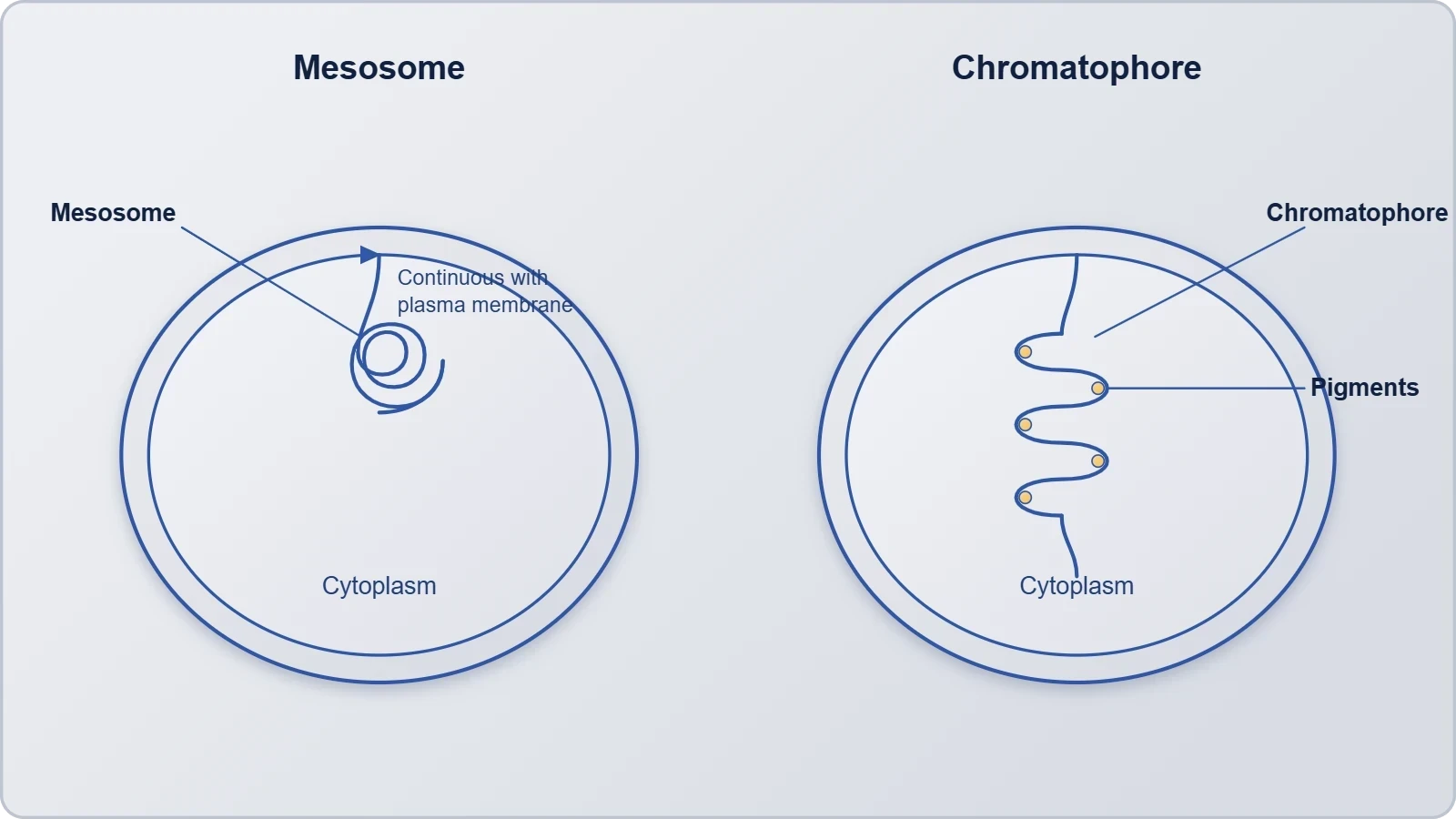
The contrast with eukaryotic organisation is instructive. In a plant cell, photosynthesis occurs in chloroplasts, which are distinct organelles with their own envelope. In a photosynthetic bacterium, the same function is housed in infoldings of the plasma membrane. The chromatophore thus illustrates how the prokaryotic envelope can be adapted to a specialised function while remaining a single continuous membrane system, and it prepares the ground for the later study of plastids in eukaryotic cells.
Flagella, Pili and Fimbriae: Surface Structures
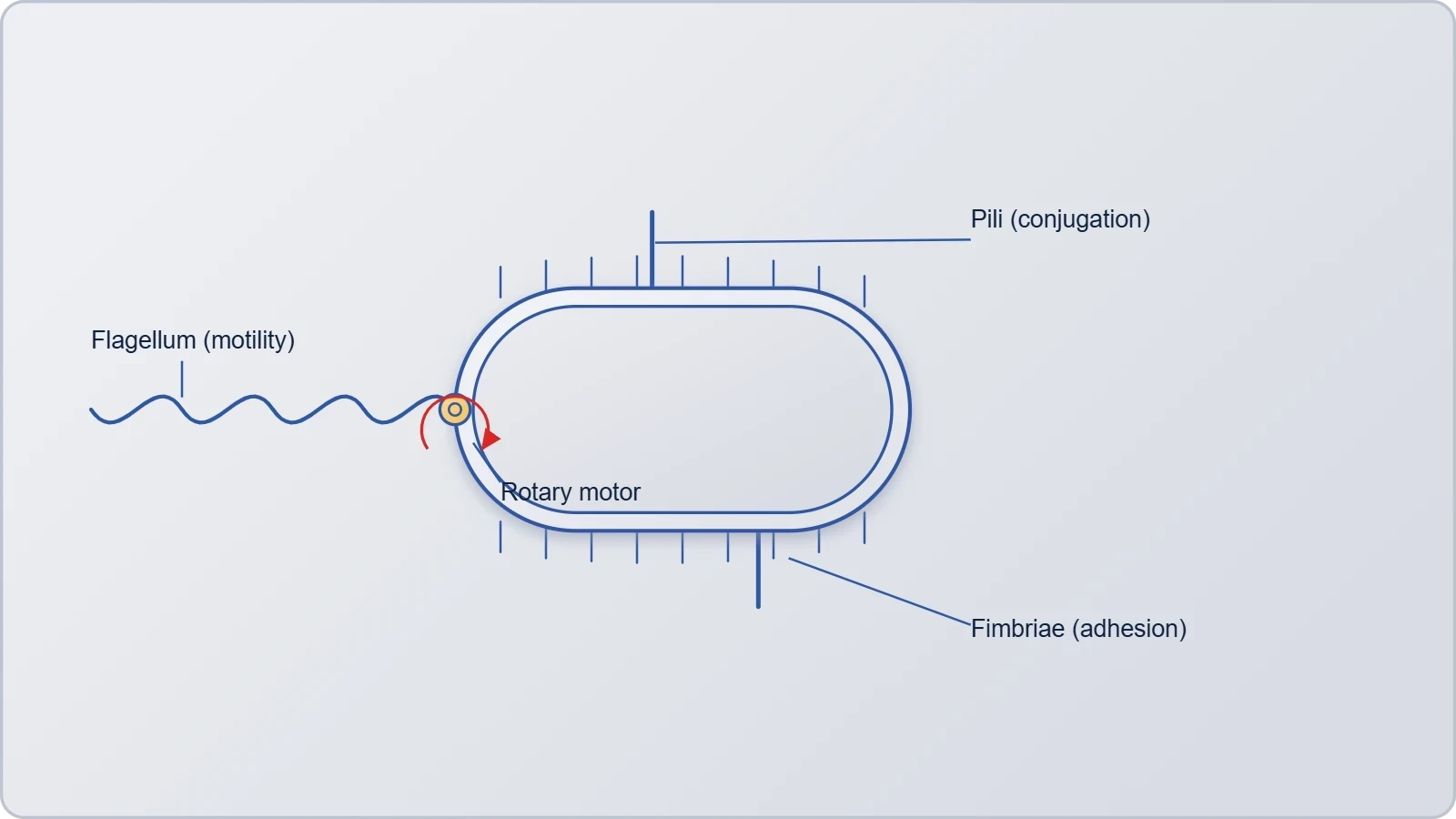
Beyond the envelope, many bacteria carry appendages that project from the cell surface. The source names three: flagella, pili and fimbriae. These structures are not part of the envelope proper, but they are anchored in it and extend outward, and they mediate the cell's interaction with its environment in ways the envelope alone cannot. Flagella provide motility, allowing a bacterium to swim toward favourable conditions or away from harmful ones. Pili and fimbriae are shorter and more numerous, and they serve primarily in attachment and, in some cases, in the exchange of genetic material between cells.
Flagella and Motility
A flagellum is a long, thread-like appendage that functions in locomotion. Its arrangement on the cell varies: a single flagellum at one pole, a tuft at one or both poles, or flagella distributed over the entire surface. The number and placement of flagella are characteristic of species and are used in classification. The flagellum is anchored in the envelope and extends through it, and its motion propels the cell through liquid. Because motility allows a bacterium to seek nutrients and escape adverse conditions, flagella are significant for survival and for colonisation of hosts.
The bacterial flagellum is structurally distinct from the eukaryotic flagellum, a point worth emphasising because the two are often confused. The prokaryotic structure is a relatively simple filament driven by a rotary mechanism at its base, whereas the eukaryotic flagellum is a complex extension of the cell bounded by the plasma membrane. The functional outcome, movement, is similar, but the underlying machinery is not. This is a clear example of analogy rather than homology, and it reminds us that similar functions can be achieved by different structures in different groups of organisms.
Pili and Fimbriae
Pili and fimbriae are short, hair-like structures that cover the surface of many bacteria. Fimbriae are numerous and fine, and they help the cell adhere to surfaces such as host tissues or the surfaces of other cells. Pili are similar in appearance but are typically fewer and longer, and certain specialised pili, often called sex pili, mediate the transfer of genetic material between bacteria during conjugation. Both structures are anchored in the envelope and project outward, and both contribute to the ability of bacteria to establish themselves in a particular niche.
The distinction between pili and fimbriae is not always sharp, and the terms are sometimes used interchangeably in the literature. What matters for this lesson is their shared role: they are surface structures that extend the reach of the cell and allow it to interact with surfaces and with other cells. Together with the flagella, they complete the picture of a prokaryotic cell whose envelope is not a passive boundary but an active interface, equipped with appendages that sense, attach and move.
Going Further
The Gram stain is named after the Danish bacteriologist Hans Christian Gram, who developed it in the 1880s. His method exploited the very differences in envelope structure described above, and it remains in use because it is rapid, inexpensive and informative. The fact that a technique devised more than a century ago still guides the first steps of bacterial identification is a testament to how directly the chemistry of the envelope reflects the biology of the organism.
The bacterial flagellum rotates like a propeller, which is why it can move the cell even though it is structurally different from the eukaryotic flagellum. This rotary motion is driven by a motor embedded in the envelope, and it allows the filament to spin rather than beat. The contrast with the bending motion of eukaryotic flagella is a useful reminder that motility has evolved more than once, and that the envelope is the platform on which these different solutions are mounted.
Also Read
- No links available.
From our network :
- 98% of Global MBA Programs Now Prefer GRE Over GMAT Focus EditionExplore why 98% of global MBA programs now prefer the GRE over GMAT Focus. Learn about the shorter GRE format, verbal logic importance, and strategic versatility for students.
- Trump Political Strategy How Geopolitical Stunts Serve As Media Diversions
- Analyzing Trump Deportation Numbers Insights Into The 2026 Immigration Crackdown
- Mastering DB2 12.1 Instance Design: A Technical Deep Dive into Modern Database ArchitectureComprehensive guide to DB2 12.1 Instance Design. Explore architectural components, HADR setup, buffer pool tuning, and security policies for IBM DB2 12.1. Includes 10 functional SQL and Shell samples.
- AI-Powered 'Precision Diagnostic' Replaces Standard GRE Score ReportsDiscover the new GRE Precision Diagnostic. Learn how AI-powered behavioral analytics and time-to-solve tracking are replacing standard GRE score reports to empower students.
- 10 Physics Numerical Problems with Solutions for IIT JEEPrepare for IIT JEE with 10 physics numerical problems, complete with detailed solutions and conceptual explanations. Ideal for mastering kinematics, thermodynamics, electromagnetism, and optics.
- EV 2.0: The Solid-State Battery Breakthrough and Global Factory ExpansionDiscover the impact of the Solid-State Battery Breakthrough on the EV market. Learn about CATL and BYD stock growth, global factory expansion in Europe, and the future of EV 2.0 technology.
- Mastering DB2 LUW v12 Tables: A Comprehensive Technical GuideComprehensive guide on DB2 LUW v12 tables. Explore CREATE TABLE syntax, BLU Acceleration, range partitioning, data types, and security features for IBM DB2 LUW v12.
- Vite 6/7 'Cold Start' Regression in Massive Module GraphsIn-depth analysis of Vite 6/7 cold start regressions in massive module graphs. Learn about barrel file issues, pre-bundling optimizations, and how to improve Vite dev server performance.
RESOURCES
- Resources will be updated soon.