Prokaryotic cells represent the oldest, simplest, and most abundant form of cellular life on Earth, and understanding their architecture is essential for anyone studying biology at the foundational level. Unlike the eukaryotic cells that make up plants, animals, fungi, and protists, prokaryotes lack a well-defined nucleus and the membrane-bound organelles that compartmentalise eukaryotic cytoplasm. Yet this apparent simplicity conceals remarkable biochemical sophistication. Bacteria and other prokaryotes carry out every essential life process—growth, reproduction, metabolism, and response to environment—within a single, compact cellular space, often measuring just one to two micrometres across.
This lesson examines prokaryotic cells as they are presented in the standard biology curriculum, focusing on the organisms they represent, their characteristic shapes, the nature of their genetic material, and the unique structures that distinguish them from eukaryotic cells. We will explore the naked genomic DNA and the small circular plasmids that confer special traits such as antibiotic resistance, the absence of membrane-bound organelles apart from ribosomes, and the specialised membrane infoldings known as mesosomes. Each concept builds directly on the overview of the cell established in the previous lesson and prepares the ground for the detailed study of the cell envelope that follows.
By the end of this lesson, you should be able to describe the defining features of prokaryotic cells with precision, explain the functional significance of plasmids and mesosomes, and articulate why prokaryotes are considered structurally distinct from eukaryotes despite sharing the same fundamental molecular machinery of life. The material follows a clear conceptual order: first the general organisation of the prokaryotic cell, then its genetic material, then its unique inclusions and membrane specialisations, and finally its ribosomes and storage bodies.
On This Page
- Defining Prokaryotic Cells and the Organisms They Represent
- Naked Genetic Material and the Role of Plasmids
- Absence of Membrane-Bound Organelles and the Mesosome
- Ribosomes and Inclusion Bodies in Prokaryotic Cells
- Surface Structures: Flagella, Pili, and Fimbriae
- Prokaryotic Size, Shapes, and the Range of Cellular Forms
- Consolidating the Prokaryotic Blueprint
Defining Prokaryotic Cells and the Organisms They Represent
Prokaryotic cells are the cellular form found in bacteria and archaea, and they constitute the vast majority of living organisms by both number and biomass. The term "prokaryote" itself signals the defining architectural feature: pro- meaning before, and karyon meaning nucleus, so a prokaryote is literally a cell that exists before the nucleus—or more precisely, without one. These cells are typically between one and two micrometres in diameter, placing them far below the resolution of the unaided human eye but comfortably within the range of light microscopy. Their small size confers a high surface-area-to-volume ratio, which supports rapid nutrient exchange and fast growth rates.
The Absence of a Well-Defined Nucleus
The most fundamental distinction between prokaryotic and eukaryotic cells lies in the organisation of the genetic material. In prokaryotes, there is no well-defined nucleus. The genetic material is basically naked, meaning it is not enveloped by a nuclear membrane. This naked DNA lies free in the cytoplasm, concentrated in a region sometimes called the nucleoid, though it is not bounded by any membrane. This arrangement stands in sharp contrast to eukaryotic cells, where the nuclear membrane separates transcription from translation and allows for extensive RNA processing before genetic messages reach the cytoplasm.
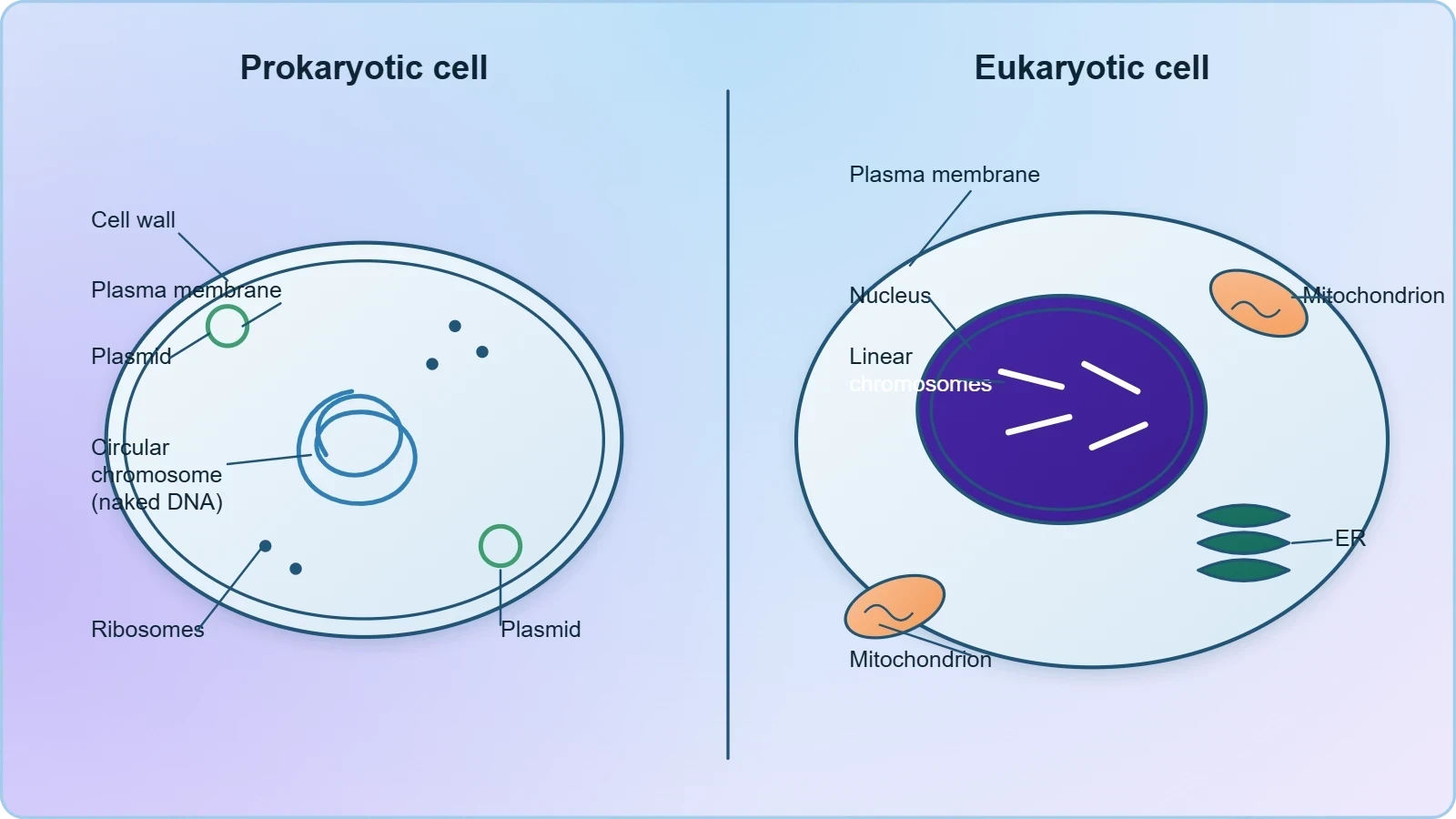
The absence of a nuclear envelope has profound consequences for gene expression. Because transcription and translation occur in the same compartment, prokaryotes can couple these processes directly, producing proteins from newly transcribed messenger RNA almost instantaneously. This efficiency contributes to the rapid responsiveness of bacteria to environmental change. It also means that prokaryotic gene regulation operates through mechanisms quite different from those in eukaryotes, relying heavily on operons and sigma factors rather than on chromatin remodelling and nuclear transport.
Cell Wall and Cytoplasm in Prokaryotes
Most prokaryotes possess a cell wall surrounding the cell membrane, with the notable exception of mycoplasma, which lacks a cell wall entirely. This exception is significant because it explains why mycoplasma are resistant to antibiotics that target cell wall synthesis, such as penicillin. The semi-fluid matrix filling the cell is the cytoplasm, which contains the genetic material, ribosomes, and various inclusions but lacks the membrane-bound compartments characteristic of eukaryotic cells. The cytoplasm of a prokaryote is therefore a single continuous compartment in which all metabolic reactions occur.
The cell wall itself performs essential functions: it determines cell shape, provides structural support, and prevents the bacterium from bursting under osmotic pressure. In the next lesson, we will examine the cell envelope in detail, including the glycocalyx, the cell wall, and the plasma membrane as a tightly bound three-layered structure. For now, it is enough to recognise that the wall is a defining feature of most prokaryotes and that its composition varies among bacterial groups, forming the basis for classifications such as Gram-positive and Gram-negative bacteria.
Naked Genetic Material and the Role of Plasmids
The genetic material of a prokaryotic cell is fundamentally different from that of a eukaryote in both organisation and packaging. In prokaryotes, the genetic material is basically naked, not enveloped by a nuclear membrane. The primary genome consists of a single chromosome, which is a circular DNA molecule. This circularity is a defining feature: unlike the linear chromosomes of eukaryotes, prokaryotic DNA forms a closed loop. The DNA is not associated with histone proteins in the same way eukaryotic DNA is, though some archaeal and bacterial species do possess histone-like proteins that help compact the genome.
Genomic DNA and the Single Circular Chromosome
The genomic DNA of a bacterium contains all the genes essential for its survival and reproduction. It is a single circular DNA molecule that replicates semi-conservatively before cell division. Because the genome is not enclosed in a nucleus, replication, transcription, and translation can occur simultaneously in the cytoplasm. This coupling is a hallmark of prokaryotic biology and underlies the rapid growth rates observed in many bacterial species. The circular chromosome is typically anchored to the plasma membrane, which helps ensure proper segregation of genetic material into daughter cells during binary fission.
The compactness of the prokaryotic genome is remarkable. Many bacteria package roughly a thousand times their own length of DNA into a cell just a micrometre or two across, achieving this through supercoiling and the action of nucleoid-associated proteins. This efficient packaging allows the cell to maintain a large genetic repertoire within a very small volume, supporting metabolic versatility and rapid adaptation to changing environments.
Plasmids and Antibiotic Resistance
In addition to the genomic DNA, many bacteria have small circular DNA outside the genomic DNA. These smaller DNA molecules are called plasmids. The plasmid DNA confers certain unique phenotypic characters to such bacteria, and one such character is resistance to antibiotics. Plasmids replicate independently of the main chromosome and can be transferred between bacteria, which explains how antibiotic resistance spreads so efficiently through bacterial populations. This plasmid DNA is also used in laboratories to monitor bacterial transformation with foreign DNA, a technique central to genetic engineering.
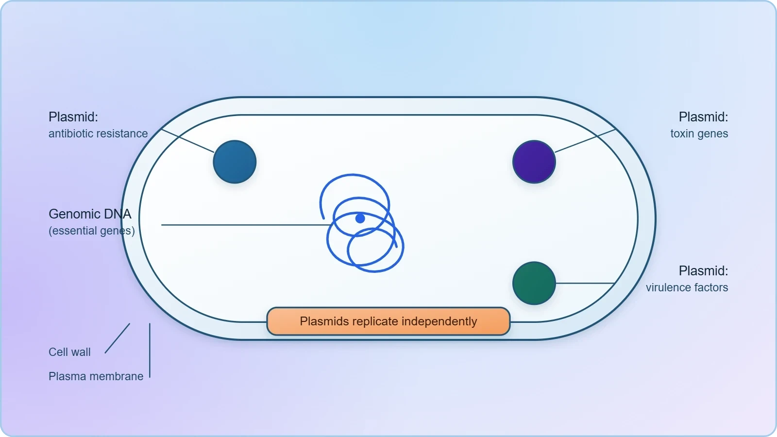
The phenotypic traits conferred by plasmids extend beyond antibiotic resistance. Some plasmids carry genes for the production of toxins, for metabolic pathways that allow the bacterium to use unusual carbon sources, or for proteins that enhance virulence. Because plasmids can be gained and lost, they represent a form of genetic flexibility that complements the more stable genomic chromosome. This dual-genome arrangement—one large circular chromosome plus one or more small plasmids—gives bacteria an extraordinary capacity to adapt to new challenges without altering their core genetic identity.
Absence of Membrane-Bound Organelles and the Mesosome
One of the most striking features of prokaryotic cells is the absence of the membrane-bound organelles found in eukaryotes. No organelles, like the ones in eukaryotes, are found in prokaryotic cells except for ribosomes. There are no mitochondria, no endoplasmic reticulum, no Golgi apparatus, no lysosomes, and no plastids. This absence does not mean prokaryotes are metabolically limited; rather, it means that the functions performed by these organelles in eukaryotes are carried out by the plasma membrane and the cytoplasm in prokaryotes. The plasma membrane itself hosts many of the enzymatic activities that eukaryotes localise to internal membranes.
Inclusions and the Mesosome
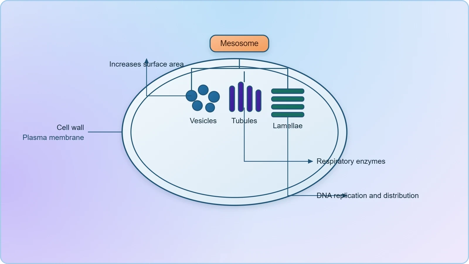
Prokaryotes have something unique in the form of inclusions. A specialised differentiated form of cell membrane called the mesosome is the characteristic of prokaryotes. These are essentially infoldings of the cell membrane. The mesosome increases the surface area of the plasma membrane and provides a larger area for enzymatic content. In some prokaryotes like cyanobacteria, there are other membranous extensions into the cytoplasm called chromatophores, which contain pigments. These structures allow the cell to carry out membrane-associated processes without the need for separate membrane-bound organelles.
The mesosome is formed by extensions of the plasma membrane into the cell, taking the form of vesicles, tubules, and lamellae. These extensions help in cell wall formation, DNA replication, and the distribution of genetic material to daughter cells. They also assist in respiration and secretion processes. By folding the membrane inward, the mesosome effectively creates internal compartments within a cell that otherwise lacks them, demonstrating how prokaryotes achieve functional compartmentalisation through membrane topology rather than through separate organelles.
Functional Significance of Membrane Infoldings
The functional logic of the mesosome becomes clear when we consider the constraints of prokaryotic cell architecture. Without mitochondria, the cell must carry out respiratory electron transport on its plasma membrane. Without a nuclear envelope, it must coordinate DNA replication and segregation using membrane-associated machinery. The mesosome provides additional membrane surface for these processes, effectively multiplying the cell's capacity for energy transduction and macromolecular synthesis. This is an elegant solution to the problem of achieving metabolic complexity within a single membrane-bound compartment.
Chromatophores in cyanobacteria serve a parallel function for photosynthesis. These membranous extensions contain the pigments required to capture light energy, allowing photosynthetic bacteria to convert solar energy into chemical energy without the chloroplasts found in eukaryotic plants. The presence of such specialised membrane systems in prokaryotes illustrates that membrane infolding is a general strategy for expanding functional surface area, one that appears in diverse forms across the bacterial world.
Ribosomes and Inclusion Bodies in Prokaryotic Cells
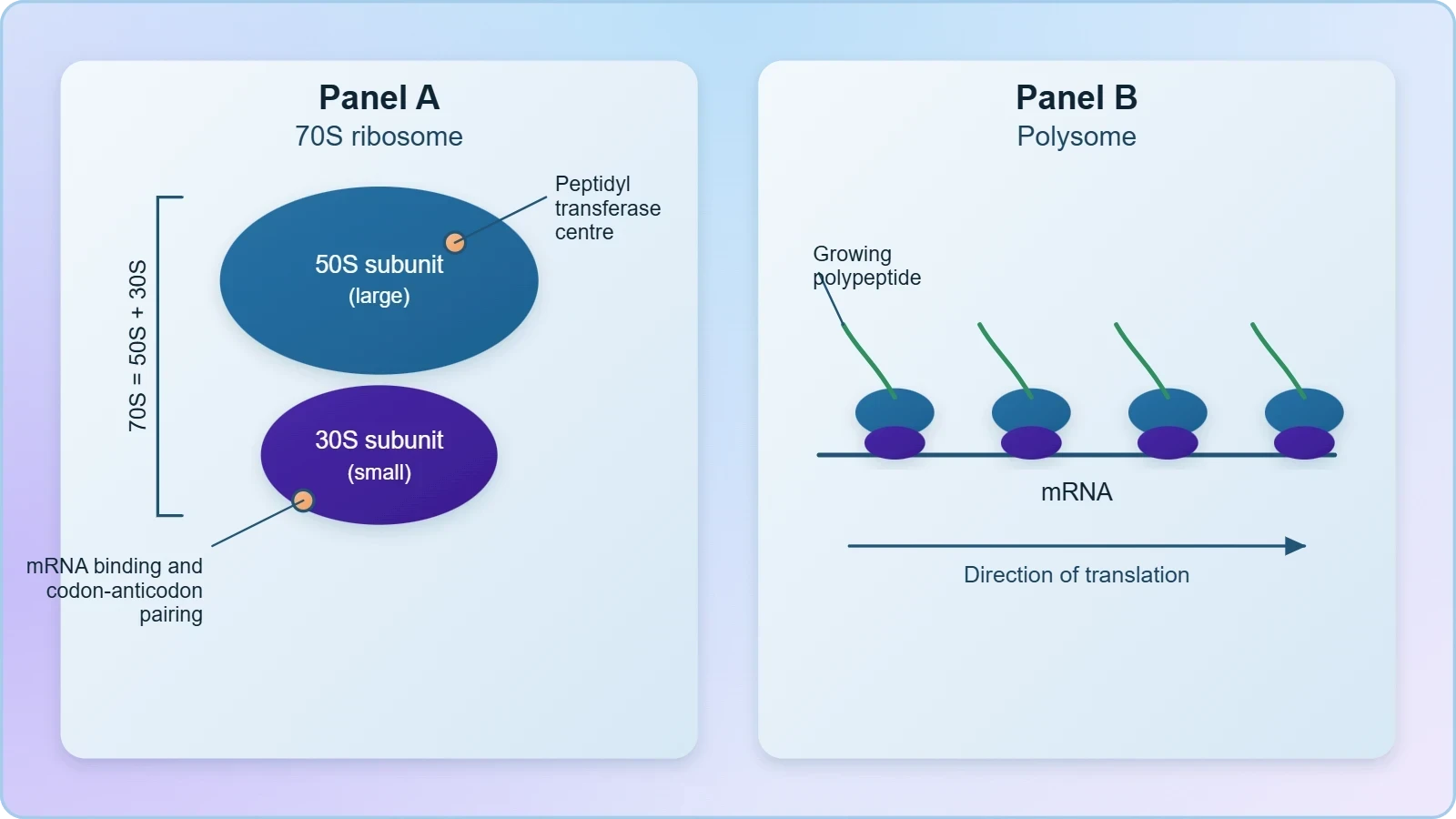
Although prokaryotes lack membrane-bound organelles, they do possess ribosomes, which are the site of protein synthesis. In prokaryotes, ribosomes are associated with the plasma membrane of the cell. They are about 15 nm by 20 nm in size and are made of two subunits: the 50S and 30S units, which when present together form the 70S prokaryotic ribosome. The S denotes Svedberg units, a measure of sedimentation rate during centrifugation, and the values are not additive because sedimentation depends on both mass and shape. The 70S ribosome is a defining feature of prokaryotes and a key target for many antibiotics.
Ribosome Structure and Polyribosomes
Ribosomes are the site of protein synthesis, translating messenger RNA into polypeptide chains. Several ribosomes may attach to a single mRNA and form a chain called polyribosomes or polysome. The ribosomes of a polysome translate the mRNA into proteins simultaneously, allowing the cell to produce multiple copies of a protein from a single transcript. This efficiency is particularly important for rapidly growing bacteria, which must synthesise proteins at high rates to support cell division. The association of ribosomes with the plasma membrane in prokaryotes further streamlines the coupling of transcription, translation, and protein secretion.
The 70S ribosome consists of a large 50S subunit and a small 30S subunit. The 50S subunit contains the peptidyl transferase centre, which catalyses peptide bond formation, while the 30S subunit binds messenger RNA and is the site of codon-anticodon recognition. This division of labour is structurally conserved across all domains of life, though the eukaryotic ribosome is larger (80S) and includes additional RNA and protein components. The differences between prokaryotic and eukaryotic ribosomes are clinically significant because they allow antibiotics to target bacterial protein synthesis without harming human cells.
Inclusion Bodies as Reserve Material
Reserve material in prokaryotic cells is stored in the cytoplasm in the form of inclusion bodies. These are not bound by any membrane system and lie free in the cytoplasm. Examples include phosphate granules, cyanophycean granules, and glycogen granules. Gas vacuoles are found in blue-green, purple, and green photosynthetic bacteria. Inclusion bodies allow the cell to store nutrients and energy in a concentrated form, releasing them when needed. Because they are not membrane-bound, they can be rapidly mobilised, giving prokaryotes a flexible strategy for managing resources in fluctuating environments.
The diversity of inclusion bodies reflects the metabolic versatility of prokaryotes. Phosphate granules store phosphorus, an essential nutrient that may be scarce in the environment. Glycogen granules store carbon and energy. Cyanophycean granules, found in cyanobacteria, store nitrogenous reserves. Gas vacuoles provide buoyancy, allowing photosynthetic bacteria to position themselves at optimal depths in the water column where light is available. Each type of inclusion body represents an adaptation to a specific ecological challenge, and together they illustrate how prokaryotes manage resources without the membrane-bound compartments used by eukaryotes.
Surface Structures: Flagella, Pili, and Fimbriae
Bacterial cells may be motile or non-motile. If motile, they have thin filamentous extensions from their cell wall called flagella. Bacteria show a range in the number and arrangement of flagella, and this variation is used in classification. The bacterial flagellum is composed of three parts: the filament, the hook, and the basal body. The filament is the longest portion and extends from the cell surface to the outside. The hook connects the filament to the basal body, which is embedded in the cell envelope and acts as the motor that drives rotation. This rotary mechanism is fundamentally different from the undulatory movement of eukaryotic flagella.
Flagellar Structure and Motility
The flagellum is a remarkable molecular machine. The basal body spans the cell envelope and contains a series of rings that anchor the structure and generate torque. The hook is a curved segment that transmits the rotational force to the filament. The filament itself is a hollow tube made of flagellin protein, assembled by transport through the hollow core. Rotation of the filament propels the bacterium through its environment, allowing it to move toward nutrients and away from harmful substances. The number and arrangement of flagella vary widely among bacteria, from a single polar flagellum to tufts at one or both poles to flagella distributed over the entire surface.
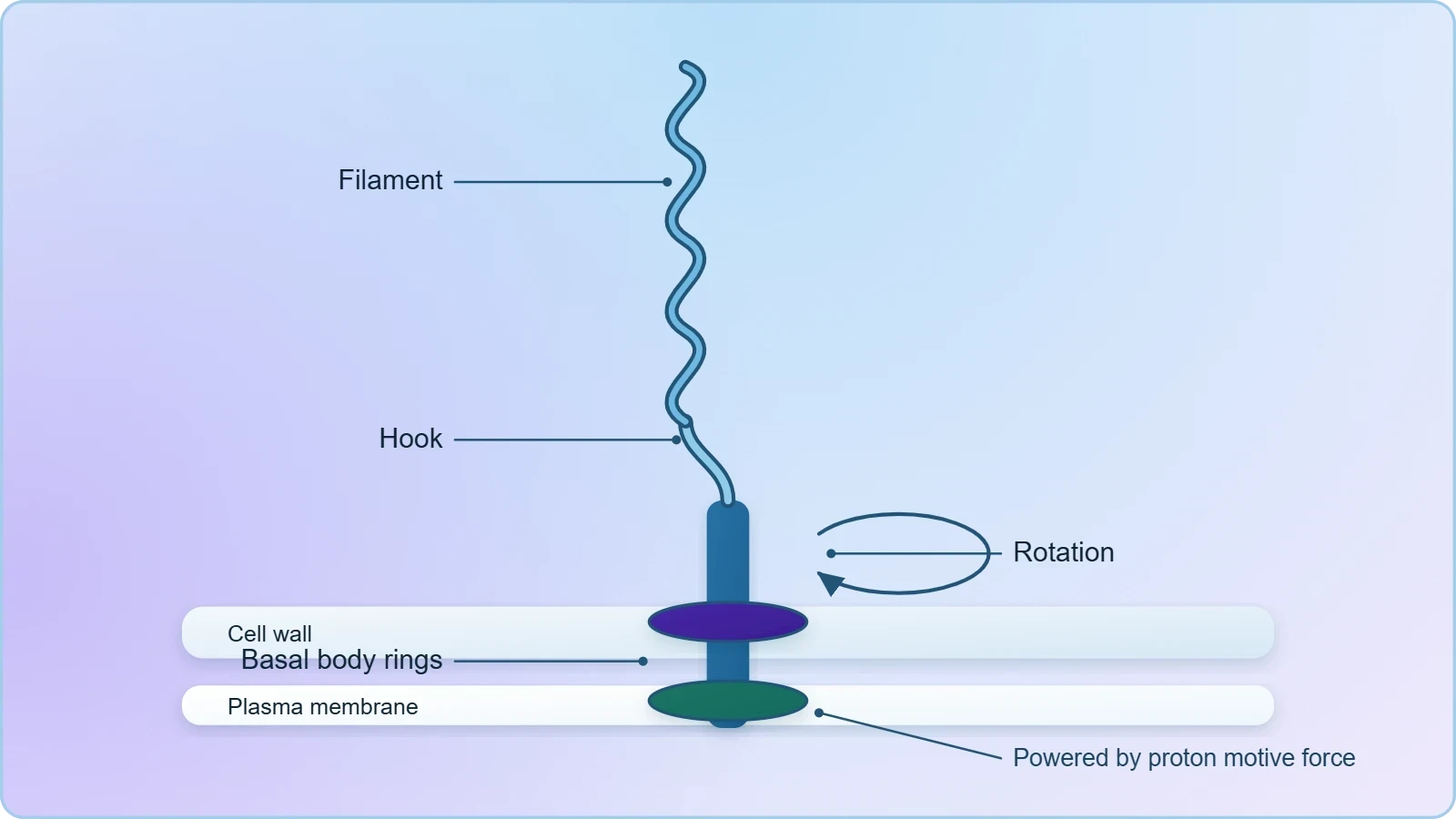
The energy for flagellar rotation comes from the proton motive force, the electrochemical gradient across the plasma membrane. This is a striking example of how prokaryotes use their plasma membrane for functions that eukaryotes assign to specialised organelles. The flagellar motor is one of the most efficient rotary engines known in biology, capable of spinning at hundreds of revolutions per second. Its structure and mechanism have been studied intensively not only for their biological significance but also for their potential applications in nanotechnology.
Pili and Fimbriae
Besides flagella, pili and fimbriae are also surface structures of the bacteria but do not play a role in motility. The pili are elongated tubular structures made of a special protein. The fimbriae are small bristle-like fibres sprouting out of the cell. In some bacteria, they are known to help attach the bacteria to rocks in streams and also to the host tissues. This attachment function is critical for colonisation and infection, allowing bacteria to establish themselves on surfaces where they can grow and multiply. Pili also play a role in conjugation, the transfer of genetic material between bacteria, which is one mechanism by which plasmid-borne traits such as antibiotic resistance spread.
The distinction between pili and fimbriae is subtle but important. Both are proteinaceous surface appendages that mediate attachment, but pili are typically longer and fewer in number, while fimbriae are shorter and more numerous. The adhesive properties of these structures are central to bacterial pathogenesis, as they allow bacteria to adhere to host tissues and resist being washed away by fluids. Understanding these surface structures is therefore essential for understanding how bacteria colonise their hosts and how infections can be prevented.
Prokaryotic Size, Shapes, and the Range of Cellular Forms
Prokaryotes exhibit a remarkable range of sizes and shapes, from the tiny PPLO at about 0.1 micrometres to typical bacteria at 1 to 2 micrometres. For comparison, viruses range from 0.02 to 0.2 micrometres, and a typical eukaryotic cell measures 10 to 20 micrometres. This size range places prokaryotes firmly in the microscopic domain, but their small size is not a limitation; rather, it is an adaptation that supports rapid nutrient uptake and fast reproduction. The high surface-area-to-volume ratio of small cells means that every part of the cytoplasm is close to the plasma membrane, facilitating efficient exchange with the environment.
PPLO and the Smallest Cells
PPLO stands for Pleuro Pneumonia Like Organisms, and these represent some of the smallest known cells, measuring about 0.1 micrometres. Their small size and lack of a cell wall make them structurally distinct from typical bacteria. Because they lack a cell wall, they are not affected by antibiotics that target cell wall synthesis, and they can adopt irregular shapes. The study of PPLO illustrates the lower size limit of cellular life and the minimal set of structures required for independent existence. Their parasitic lifestyle allows them to survive with a reduced genome, relying on their host for many essential metabolites.
The existence of PPLO also highlights the importance of the cell wall in maintaining cell shape and integrity. Without a wall, the cell membrane alone must contain the cytoplasm, which limits the size and shape the cell can achieve. This trade-off between size, shape, and structural support is a recurring theme in cell biology, and it helps explain why most bacteria have cell walls while a few specialised groups do not.
Shapes and Arrangements of Bacterial Cells
Bacteria display a variety of shapes, including spherical (cocci), rod-shaped (bacilli), and spiral forms. These shapes are determined by the cell wall and are maintained during growth and division. The arrangement of cells—whether single, in pairs, in chains, or in clusters—depends on the pattern of cell division and separation. Shape and arrangement are important for classification and identification, and they also influence how bacteria interact with their environment and with host tissues. The diversity of bacterial forms is a testament to the adaptability of prokaryotic life.
The relationship between cell shape and function is a rich area of study. Rod-shaped bacteria can swim more efficiently than spherical ones, while spiral shapes may aid in penetrating viscous environments. The cell wall, which determines shape, is therefore not just a static container but a dynamic structure that influences the cell's ecological strategy. Understanding the range of prokaryotic forms provides a foundation for understanding bacterial diversity and the many roles bacteria play in ecosystems, industry, and disease.
Going Further
Two points of context help consolidate this lesson. First, PPLO stands for Pleuro Pneumonia Like Organisms, a name that reflects their association with respiratory disease in livestock and their resemblance to the pneumonia-causing organisms studied in early microbiology. Second, because prokaryotes multiply rapidly, they can build up large populations quickly, which is one reason plasmid-borne antibiotic resistance can spread so efficiently through bacterial communities. A single resistant bacterium can, under favourable conditions, give rise to a vast population carrying the same plasmid, and the plasmid can also be transferred horizontally to other bacteria. This combination of rapid reproduction and horizontal gene transfer explains why antibiotic resistance is such a pressing public health concern and why understanding plasmid biology is essential for modern medicine.
Consolidating the Prokaryotic Blueprint
The prokaryotic cell, for all its apparent simplicity, is a highly efficient and adaptable biological machine. Its defining features—the absence of a well-defined nucleus, naked genetic material, a single circular chromosome supplemented by plasmids, the absence of membrane-bound organelles except ribosomes, and the presence of mesosomes and inclusion bodies—together constitute a coherent architectural plan. This plan allows prokaryotes to grow rapidly, adapt quickly to environmental change, and colonise virtually every habitat on Earth. The lessons learned from studying prokaryotes inform fields as diverse as medicine, ecology, biotechnology, and evolutionary biology.
As we move to the next lesson on the cell envelope and its modifications, keep in mind that the structures we have discussed here—the plasma membrane, the cell wall, and the mesosome—are not isolated features but parts of an integrated system. The cell envelope is the interface between the prokaryotic cell and its environment, and its modifications determine how the cell interacts with the world. Understanding the basic blueprint of the prokaryotic cell prepares you to appreciate the sophistication of the envelope and the many ways bacteria have adapted it to suit their diverse lifestyles.
Also Read
- No links available.
From our network :
- Mastering DB2 12.1 Instance Design: A Technical Deep Dive into Modern Database ArchitectureComprehensive guide to DB2 12.1 Instance Design. Explore architectural components, HADR setup, buffer pool tuning, and security policies for IBM DB2 12.1. Includes 10 functional SQL and Shell samples.
- 10 Physics Numerical Problems with Solutions for IIT JEEPrepare for IIT JEE with 10 physics numerical problems, complete with detailed solutions and conceptual explanations. Ideal for mastering kinematics, thermodynamics, electromagnetism, and optics.
- 98% of Global MBA Programs Now Prefer GRE Over GMAT Focus EditionExplore why 98% of global MBA programs now prefer the GRE over GMAT Focus. Learn about the shorter GRE format, verbal logic importance, and strategic versatility for students.
- AI-Powered 'Precision Diagnostic' Replaces Standard GRE Score ReportsDiscover the new GRE Precision Diagnostic. Learn how AI-powered behavioral analytics and time-to-solve tracking are replacing standard GRE score reports to empower students.
- Analyzing Trump Deportation Numbers Insights Into The 2026 Immigration Crackdown
- Vite 6/7 'Cold Start' Regression in Massive Module GraphsIn-depth analysis of Vite 6/7 cold start regressions in massive module graphs. Learn about barrel file issues, pre-bundling optimizations, and how to improve Vite dev server performance.
- Mastering DB2 LUW v12 Tables: A Comprehensive Technical GuideComprehensive guide on DB2 LUW v12 tables. Explore CREATE TABLE syntax, BLU Acceleration, range partitioning, data types, and security features for IBM DB2 LUW v12.
- Trump Political Strategy How Geopolitical Stunts Serve As Media Diversions
- EV 2.0: The Solid-State Battery Breakthrough and Global Factory ExpansionDiscover the impact of the Solid-State Battery Breakthrough on the EV market. Learn about CATL and BYD stock growth, global factory expansion in Europe, and the future of EV 2.0 technology.
RESOURCES
- Resources will be updated soon.