A leaf held up to the light reveals a working factory, and the vertical section of a dorsiventral leaf shows exactly how that factory is organised. This ninth lesson in the course moves from the stems examined earlier into the lamina of a dicotyledonous leaf, where three main parts — epidermis, mesophyll and vascular system — divide the labour of protection, photosynthesis and transport. The treatment stays close to the chapter's own terminology and order of ideas, so learners revising the topic meet the same definitions, the same structural sequence and the same emphasis on how form serves function.
The lesson begins with the two epidermal surfaces and their conspicuous cuticle, then descends into the mesophyll, where palisade parenchyma and spongy parenchyma are distinguished by shape, arrangement and position. From there it follows the vascular system through veins and midrib, noting how bundle size tracks vein size and how thick-walled bundle sheath cells enclose each bundle. Because the previous lesson dealt with the monocotyledonous stem, nothing here repeats that ground tissue or scattered-bundle arrangement; instead, the dicot leaf is treated on its own terms, preparing the ground for the isobilateral leaf and anatomical variation that follow.
Anatomy rewards careful looking, and this lesson is built around that habit. A vertical section through the lamina is a small window into a highly ordered structure, and every layer encountered has a reason for being where it is. Learners who can name each layer, describe its cells and explain its contribution to the leaf's life will find the remaining lessons of the course far easier to absorb, because the vocabulary and the logic of tissue systems carry across roots, stems and leaves alike.
On This Page
The Three Main Parts of the Leaf Lamina
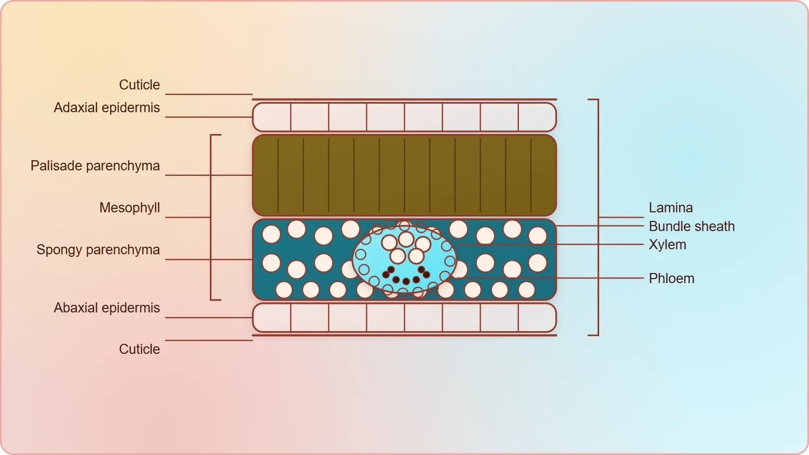
A vertical section of a dorsiventral leaf through the lamina shows three main parts, namely, epidermis, mesophyll and vascular system. This threefold division is the organising principle for everything that follows, and it mirrors the tissue-system thinking introduced at the start of the course. The epidermis forms the boundary, the mesophyll fills the space between the two surfaces, and the vascular system threads through the mesophyll as veins and midrib. Keeping these three parts distinct in the mind makes the detailed descriptions of cells and layers much easier to place and recall.
Epidermis: Adaxial and Abaxial Surfaces
The epidermis covers both the upper surface, called the adaxial epidermis, and the lower surface, called the abaxial epidermis, and it has a conspicuous cuticle. The cuticle is a thin covering layer that limits water loss, and the chapter notes that the epidermis may also bear trichomes and a few stomata. Because the leaf is dorsiventral, its two surfaces are not identical, and this asymmetry begins at the outermost layer. Recognising which surface faces the stem and which faces away is the first step in reading any leaf section correctly.
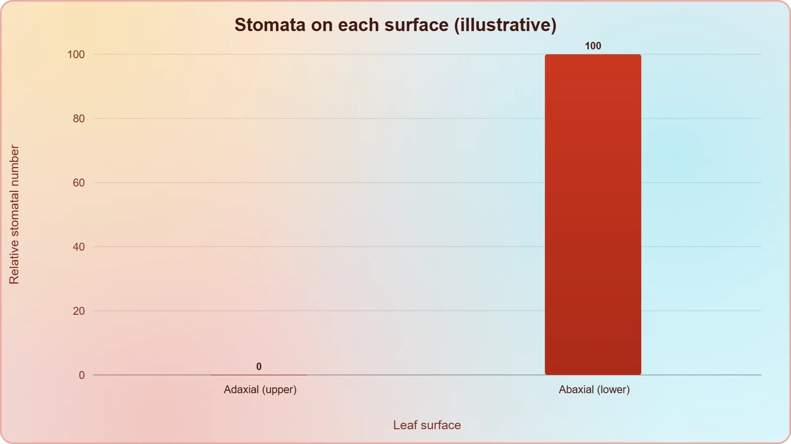
A striking feature of the dorsiventral leaf is the unequal distribution of stomata. The abaxial epidermis generally bears more stomata than the adaxial epidermis, and the latter may even lack stomata altogether. This arrangement places the majority of the gas-exchange pores on the shaded lower surface, which is consistent with the leaf's overall design. For the learner, the practical lesson is simple: when examining a peel or a section, count stomata on both surfaces rather than assuming they are evenly spread, and record which surface you sampled.
Mesophyll: The Photosynthetic Tissue
The tissue between the upper and the lower epidermis is called the mesophyll. It possesses chloroplasts and carries out photosynthesis, and it is made up of parenchyma. Within this single tissue the chapter distinguishes two types of cells — the palisade parenchyma and the spongy parenchyma — and this internal differentiation is precisely what makes the leaf dorsiventral. The mesophyll therefore illustrates a general principle of plant anatomy: identical cell lineage can produce structurally distinct populations when position and function demand it.
The adaxially placed palisade parenchyma is made up of elongated cells, which are arranged vertically and parallel to each other. The oval or round and loosely arranged spongy parenchyma is situated below the palisade cells and extends to the lower epidermis. Between the spongy cells there are numerous large spaces and air cavities. Reading the two descriptions side by side shows how shape and packing differ between the layers, and how the loose arrangement below complements the tight columnar arrangement above.
Reading the Vertical Section Layer by Layer
Once the three main parts are named, the next task is to move through the section in order, from the upper surface to the lower, and to describe what each layer contributes. This sequential reading is how anatomists actually work with a slide: they establish orientation, identify boundaries, then characterise the tissue in between. The table below summarises the layers of the dorsiventral leaf as the chapter presents them, and it can serve as a revision checklist before attempting a labelled diagram from memory.
Why the Two Surfaces Differ
The dorsiventral leaf is named for its two distinct faces, and the anatomy justifies the name at every level. The upper epidermis may be almost free of stomata, the palisade layer sits immediately beneath it, and the spongy tissue with its air cavities lies below. This vertical asymmetry is not incidental decoration; it reflects the leaf's habitual orientation to light. A leaf that presents one face to the sun and the other to the shaded, more humid air beneath can afford to specialise its surfaces, and the section shows that specialisation clearly.
For examination purposes, the contrast between surfaces is a favourite point of comparison. Candidates are often asked to explain why the abaxial epidermis bears more stomata, or to identify which surface a given peel came from. The answer lies in the arrangement just described: the adaxial surface is the light-facing one, while the abaxial surface, generally bearing more stomata, handles the bulk of gas exchange. Stating this relationship precisely, using the chapter's own terms, earns marks that vague references to "the underside" never will.
The Vascular System in Veins and Midrib
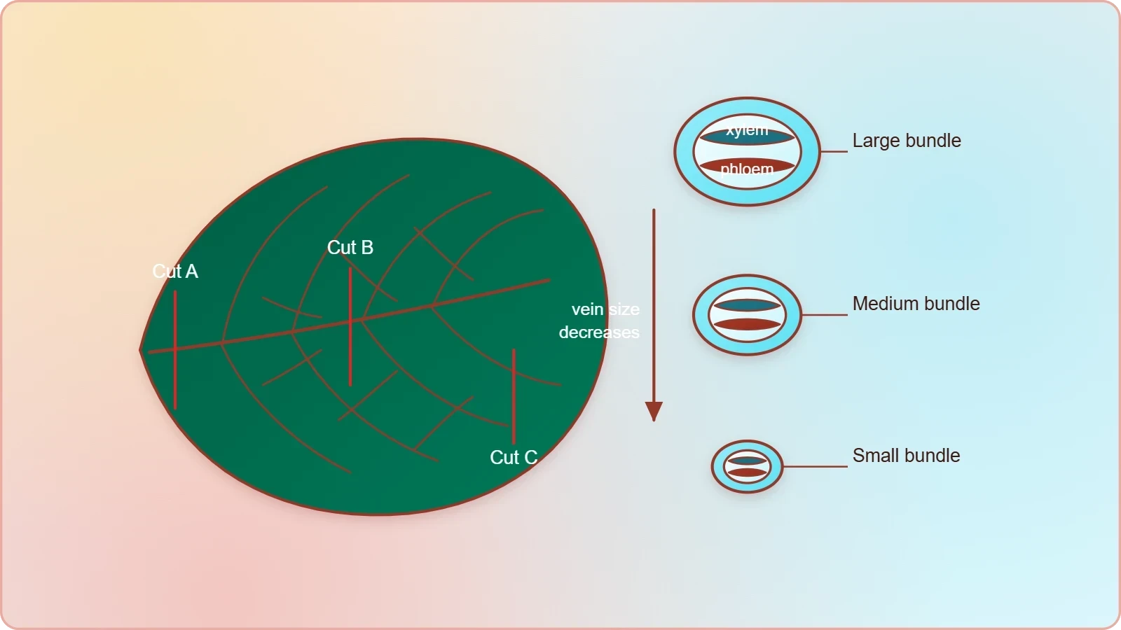
The vascular system includes vascular bundles, which can be seen in the veins and the midrib. The size of the vascular bundles is dependent on the size of the veins, so a section through a large vein reveals a correspondingly large bundle, while finer veins carry smaller ones. This proportionality is a useful diagnostic habit: when scanning a section, the bundle dimensions tell you something about the vein you have cut. The midrib, being the principal vein, naturally presents the most substantial vascular tissue in the lamina.
Bundle Sheath and Xylem Position
The vascular bundles are surrounded by a layer of thick-walled bundle sheath cells. This sheath is a defining feature of the leaf's vascular system as the chapter presents it, and it separates the conducting tissues from the surrounding mesophyll. Learners are directed to examine the position of xylem within the vascular bundle, a detail worth fixing firmly in memory. Being able to locate the xylem, and to state that the bundle lies within a sheath, demonstrates that the section has been read rather than merely glanced at.
Vein thickness varies across the lamina, and this variation is tied to the reticulate venation characteristic of dicot leaves. A branching, net-like pattern of veins means that sections cut at different points encounter bundles of different sizes, from the massive midrib to the finest veinlets. The anatomy therefore records the venation pattern in three dimensions. When you next examine a cleared leaf or a series of sections, expect this range of bundle sizes and use it to reconstruct the vein network that produced it.
Relating Structure to Function
Each element of the vascular system has a transport role that the course has already introduced: xylem moves water and minerals, phloem moves food material. In the leaf, these conducting tissues are embedded in the mesophyll, close to the cells that manufacture the sugars the phloem will carry away and to the surfaces from which water evaporates. The proximity is the point. A bundle sheath of thick-walled cells surrounds each bundle, and the whole arrangement keeps conduction and photosynthesis physically adjacent without either interfering with the other.
It helps to think of the leaf as a junction where the plant's long-distance transport meets its local metabolism. Water arriving through the xylem enters a tissue packed with chloroplasts; sugars formed there must leave through the phloem. The vertical section shows these pathways in cross-section, and the bundle sheath marks the boundary between the conducting core and the photosynthetic surroundings. Understanding this boundary is essential preparation for the isobilateral leaf, where the same components reappear in a different arrangement.
Comparing the Leaf with the Stems Already Studied
The dicot stem examined earlier arranged its vascular bundles in a ring, each bundle conjoint, open and with endarch protoxylem, with pith at the centre and medullary rays between bundles. The leaf section offers a different logic: bundles are scattered through mesophyll in veins and midrib, each wrapped in a thick-walled bundle sheath, and there is no pith or ring to speak of. Noting these contrasts sharpens both pictures. The same conducting tissues appear in both organs, but their distribution follows the organ's shape and task.
What Carries Forward from the Stem Lessons
Several ideas transfer directly. The concept of a tissue system, introduced at the start of the course, applies unchanged: the leaf's epidermis is an epidermal tissue system, its mesophyll belongs to the ground tissue system, and its veins constitute the vascular tissue system. The distinction between parenchyma and sclerenchyma also recurs, since the mesophyll is parenchymatous while the bundle sheath cells are thick-walled. Learners who mastered these categories in the stem lessons can now apply them to a new organ without relearning the vocabulary.
The monocotyledonous stem, covered in the previous lesson, showed scattered vascular bundles each surrounded by a sclerenchymatous bundle sheath, a sclerenchymatous hypodermis and a large parenchymatous ground tissue. The dicot leaf's bundle sheath is likewise thick-walled, which is a useful point of contact between the two lessons. Yet the leaf's sheath encloses bundles embedded in photosynthetic mesophyll rather than in a stem's ground tissue, so the resemblance should be noted without being overstated. Precision about where a feature occurs is as important as recognising the feature itself.
Preparing for the Isobilateral Leaf
The next lesson turns to the isobilateral, monocotyledonous leaf, whose anatomy is similar to that of the dorsiventral leaf in many ways but differs in specific, examinable respects. Stomata are present on both surfaces of the epidermis, and the mesophyll is not differentiated into palisade and spongy parenchyma. Bulliform cells, the large empty colourless adaxial epidermal cells along the veins in grasses, also enter the picture. Holding the dorsiventral template firmly in mind makes these differences easy to state, because each one is defined against the leaf studied here.
There is also a venation contrast to anticipate. The reticulate venation of dicot leaves produces veins of varying thickness, and the vascular bundles vary accordingly. In monocot leaves, parallel venation is reflected in the near similar sizes of vascular bundles, except in the main veins. Setting the two patterns side by side now will make the next lesson's comparisons feel like confirmation rather than novelty, and it will strengthen the habit of linking external venation to internal bundle size.
Stomata, Cuticle and the Epidermal Tissue System
The epidermal tissue system, as the course defines it, is made of epidermal cells, stomata and the epidermal appendages. The dorsiventral leaf displays all three: epidermal cells on both surfaces, stomata concentrated on the abaxial side, and trichomes that the epidermis may bear. The conspicuous cuticle completes the picture. This lesson therefore offers a concrete, examinable example of the epidermal tissue system in action, and it repays close attention because the same components recur in the isobilateral leaf with a different distribution.
Stomatal Distribution as a Diagnostic Feature
Stomatal distribution is one of the clearest differences between leaf types, and the chapter states it plainly: the abaxial epidermis generally bears more stomata than the adaxial epidermis, and the latter may even lack stomata. In the isobilateral leaf, by contrast, stomata are present on both surfaces. A single observation of stomatal distribution therefore goes a long way towards identifying the leaf type under the microscope. Learners should practise stating this contrast in the chapter's own terms, since it is a standard comparison question.
The stomatal apparatus itself, with its guard cells and surrounding epidermal cells, belongs to the vocabulary established earlier in the course. Here the emphasis falls on where stomata sit rather than on their internal mechanism. The practical point is that the leaf's two surfaces are functionally differentiated, and the stomatal count is the most accessible evidence of that differentiation. Recording counts from upper and lower peels, and expressing the result as a simple ratio, turns a qualitative observation into a defensible statement.
Cuticle and Trichomes
The epidermis has a conspicuous cuticle, and it may bear trichomes and a few stomata. The cuticle's presence on both surfaces is worth emphasising because it frames the leaf as a structure that must protect itself while remaining permeable to gases. Trichomes, the epidermal appendages named in the tissue-system definition, add a further layer of surface specialisation. Together, cuticle and trichomes show that the outermost layer is not a passive skin but an active boundary with its own structural adaptations.
When describing a section, mention the cuticle explicitly rather than treating the epidermis as a bare line of cells. Examiners look for the conspicuous cuticle as a named feature of the dorsiventral leaf, and its omission weakens an otherwise accurate account. The same discipline applies to trichomes: if present in the material, they should be noted as epidermal appendages. Small descriptive habits of this kind accumulate into the precision that distinguishes a strong answer from an adequate one.
Practical Work: Identifying the Section Under the Microscope
Recognising a dorsiventral leaf section is a skill built from a small number of reliable cues. The upper and lower epidermis with their cuticle frame the section; the palisade layer sits immediately beneath the upper surface as a band of elongated, parallel cells; the spongy tissue below is looser, with visible air cavities; and vascular bundles appear within the mesophyll, each ringed by a thick-walled sheath. Working through these cues in order, from one surface to the other, is more reliable than trying to identify the section from a single feature.
A Stepwise Identification Routine
Begin by establishing orientation: find the palisade layer, and the surface nearest it is the adaxial one. Next, confirm the mesophyll's two components and note the air cavities in the spongy region. Then locate a vascular bundle, observe its sheath, and identify the position of the xylem within it. Finally, examine both epidermises for stomata and record which surface bears more. This routine follows the chapter's own order of ideas, so the description you write afterwards will naturally match the expected sequence.
Recording observations in a fixed order also protects against a common error: describing the spongy tissue as if it lay above the palisade. The chapter is explicit that the palisade parenchyma is adaxially placed and the spongy parenchyma lies below it, extending to the lower epidermis. A section mounted upside down will mislead anyone who has not first fixed orientation by the palisade layer. Checking that single feature before writing anything else prevents a whole paragraph of avoidable mistakes.
Common Errors and How to Avoid Them
Several mistakes recur in learner descriptions. The mesophyll is sometimes called a single uniform tissue, ignoring the palisade and spongy distinction that defines the dorsiventral leaf. The bundle sheath is sometimes omitted, even though the chapter states that the vascular bundles are surrounded by a layer of thick-walled bundle sheath cells. Stomatal distribution is sometimes reversed, with more stomata attributed to the upper surface. Each of these errors is corrected by returning to the chapter's exact wording and checking the description against it.
A further error concerns scale. Because bundle size depends on vein size, a section through a minor vein will show a small bundle, and learners sometimes conclude that the leaf lacks substantial vascular tissue. Cutting through the midrib as well as the lamina resolves the confusion. Similarly, the absence of stomata from the adaxial epidermis in some leaves is a legitimate observation, not a preparation fault. Distinguishing genuine anatomical variation from technical artefact is part of learning to read sections confidently.
Going further
The palisade layer near the upper surface receives more light, while the spongy layer below allows gas circulation around the cells. This is a natural extension of the chapter's description rather than a departure from it: the columnar, tightly packed palisade cells sit where illumination is strongest, and the loosely arranged spongy cells with their large air cavities sit where gases must move freely. Reading the two layers in this light makes the dorsiventral arrangement feel purposeful rather than arbitrary, and it gives learners a functional reason to remember which layer lies where.
Reticulate venation, mentioned in the chapter for dicot leaves, is a branching net-like pattern of veins. The anatomy follows the pattern: because the veins branch and rejoin across the lamina, a vertical section cut at different points meets bundles of different sizes, and the chapter's statement that bundle size depends on vein size becomes easy to visualise. Tracing a vein network on a cleared leaf, then imagining sections through a major vein and a fine veinlet, connects the external pattern to the internal structure described in this lesson.
The dorsiventral leaf, then, is a study in ordered asymmetry. Two epidermises with a conspicuous cuticle enclose a mesophyll divided into palisade and spongy parenchyma, and a vascular system of veins and midrib threads through it, each bundle wrapped in a thick-walled sheath. Every element named in this lesson belongs to one of the three tissue systems introduced at the start of the course, and every element has a function that the structure explains. Mastery of this section is the surest foundation for the isobilateral leaf and the anatomical variation that close the course.
Also Read
- No links available.
From our network :
- EV 2.0: The Solid-State Battery Breakthrough and Global Factory ExpansionDiscover the impact of the Solid-State Battery Breakthrough on the EV market. Learn about CATL and BYD stock growth, global factory expansion in Europe, and the future of EV 2.0 technology.
- Mastering DB2 12.1 Instance Design: A Technical Deep Dive into Modern Database ArchitectureComprehensive guide to DB2 12.1 Instance Design. Explore architectural components, HADR setup, buffer pool tuning, and security policies for IBM DB2 12.1. Includes 10 functional SQL and Shell samples.
- Analyzing Trump Deportation Numbers Insights Into The 2026 Immigration Crackdown
- Vite 6/7 'Cold Start' Regression in Massive Module GraphsIn-depth analysis of Vite 6/7 cold start regressions in massive module graphs. Learn about barrel file issues, pre-bundling optimizations, and how to improve Vite dev server performance.
- 10 Physics Numerical Problems with Solutions for IIT JEEPrepare for IIT JEE with 10 physics numerical problems, complete with detailed solutions and conceptual explanations. Ideal for mastering kinematics, thermodynamics, electromagnetism, and optics.
- Mastering DB2 LUW v12 Tables: A Comprehensive Technical GuideComprehensive guide on DB2 LUW v12 tables. Explore CREATE TABLE syntax, BLU Acceleration, range partitioning, data types, and security features for IBM DB2 LUW v12.
- AI-Powered 'Precision Diagnostic' Replaces Standard GRE Score ReportsDiscover the new GRE Precision Diagnostic. Learn how AI-powered behavioral analytics and time-to-solve tracking are replacing standard GRE score reports to empower students.
- 98% of Global MBA Programs Now Prefer GRE Over GMAT Focus EditionExplore why 98% of global MBA programs now prefer the GRE over GMAT Focus. Learn about the shorter GRE format, verbal logic importance, and strategic versatility for students.
- Trump Political Strategy How Geopolitical Stunts Serve As Media Diversions
RESOURCES
- Upscaling seasonal phenological course of leaf dorsiventral ...sciencedirect.com... leaf structure typical for broadleaved trees and many dicot herbs. The adaxial-abaxial polarity of bifacial, dorsiventral leaves is strictly developmentally ...
- Describe the Internal Structure of a Dorsiventral Leaf - GeeksforGeeksgeeksforgeeks.orgJul 14, 2022 ... A leaf showing this separation in mesophyll is assigned as dorsiventral. It is normal in dicot leaves. The leaves in…
- Mind the leaf anatomy while taking ground truth with portable ...pubmed.ncbi.nlm.nih.govJan 13, 2025 ... A wide range of portable chlorophyll meters are increasingly being used to measure leaf chlorophyll content as an indicator of…
- Internal Structure of a Dicotyledonous Leaf - Pinterestpinterest.comInternal structure of a dorsiventral (dicot) leaf | Structure of a dicot leaf, · Internal Structure Of A Leaf · Plant Cell Labeled ·…
- Anatomy of a Dicot Leaf - BYJU'Sbyjus.comA dicot leaf shows a dorsiventral structure, meaning an upper surface called the adaxial surface and a lower surface called the abaxial surface. The…
- TS of Dicot Leaf (Dorsiventral Leaf) Structure with PPTeasybiologyclass.comLeaf blade: also called leaf lamina is the flattened expanded part of the leaf chiefly composed of mesophyll tissue and vascular bundles and (2).…
- Mind the leaf anatomy while taking ground truth with portable ... - PMCpmc.ncbi.nlm.nih.govJan 13, 2025 ... We focused on three types of leaves with differing anatomy: (1) laminar dorsiventral leaves of dicotyledonous species, (2) grass leaves…
- Difference between Dorsiventral and Isobilateral Leafeasybiologyclass.comAnatomical Difference between Dorsiventral and Isobilateral Leaf. Compare the Anatomy of Dicot and Monocot Leaf with Comparison Table + Diagrams.
- Anatomy of Dorsiventral Leaves | PDF | Leaf | Stoma - Scribdscribd.comIt is found in dicot plants. A dorsiventral leaf's interior structure is divided into three different segments. 1. Internally, the leaf is divided into…
- B-29 Dorsi Ventral (Dicot) Leaf (Helianthus Annuus) - Victory Graphikvictorygraphik.comHome / Shop / Charts / Botany / Anatomy / B-29 Dorsi Ventral (Dicot) Leaf (Helianthus Annuus). Index. Charts · Human Anatomy · General…
- STUDY ON INTERNAL STRUCTURES OF DORSIVENTRAL ... - SAUmail.sau.edu.bd(DICOTYLEDONOUS) LEAF. 1. LEAF OF MANGO. A thin section through the lamina of a leaf of mango (Magnifera indica of family Anacardiacae) will show…
- anatomy of flowering plants chapter 6 - NCERTncert.nic.inThe phloem parenchyma is absent, and water-containing cavities are present within the vascular bundles. 6.2.5 Dorsiventral. (Dicotyledonous) Leaf. The vertical ...
- Anatomy of Dicotyledonous Plants | CK-12 Foundationflexbooks.ck12.orgDicot Leaf Anatomy: Exhibits a dorsiventral structure with distinct upper and lower epidermis (the latter containing more stomata), differentiated mesophyll ...
- Quantitative mesophyll parameters rather than whole‐leaf traits ...nph.onlinelibrary.wiley.comOct 20, 2017 ... C3 dicots possessed two types of leaf anatomy: dorsiventral (DV; upper layers of mesophyll are formed by palisade cylindrical cells…
- Biomorphological Features and Anatomical Structure of the Leaf of ...ijcsrr.orgMar 30, 2024 ... ... dorsiventral structure, epidermal cell, leaf, localization ... Mesophyll leaf of dicotyledonous plants (teaching-methodical manual).