Plant anatomy is the branch of biology that turns a flowering plant inward, asking not merely what an organ looks like from the outside but how it is built from the inside. Every leaf, stem and root is a working structure assembled from cells, and those cells are grouped into tissues, and those tissues are grouped into organs. The study of this internal architecture is what the discipline of anatomy means, and it gives the learner a vocabulary for reading a cross-section the way a cartographer reads a map. This opening lesson establishes that foundational grammar before any specific organ is examined.
The organising logic of the subject is elegantly simple and worth stating plainly at the outset: cells are the basic unit, cells are organised into tissues, and tissues are organised into organs. From that single ladder of organisation, nearly everything else in plant anatomy follows. Different organs within one plant show differences in their internal structure, and within the angiosperms the monocots and dicots are also anatomically different. Internal structures, moreover, show adaptations to diverse environments, so anatomy is never merely descriptive — it records how a plant has answered the conditions in which it lives.
This lesson is the first of ten in a course that moves from tissue systems to the transverse anatomy of dicot and monocot roots and stems, and then to the vertical anatomy of dorsiventral and isobilateral leaves. Here we establish the three tissue systems in outline, the cell-to-organ hierarchy, and the reasons anatomists compare organs and plant groups at all. The next lesson takes up the epidermal tissue system in detail, so the epidermis is introduced only briefly here, as one of three systems rather than as a subject in its own right.
On This Page
- The Scope of Plant Anatomy: Studying Internal Structure
- The Ladder of Organisation: Cells, Tissues and Organs
- The Epidermal Tissue System: The Outer Covering
- Organs Differ Internally: Roots, Stems and Leaves
- Monocots and Dicots: Anatomical Difference Within Angiosperms
- Anatomy as Adaptation: Structure in Relation to Environment
The Scope of Plant Anatomy: Studying Internal Structure
Anatomy, in the botanical sense, is the study of the internal structure of plants. The word sits alongside morphology, which concerns external form, and the two are complementary: the structural similarities and variations that are so easily seen in the external morphology of larger living organisms, plants and animals alike, have their counterpart inside. If one were to study the internal structure, one also finds several similarities as well as differences. That double observation — similarity and difference — is the engine of the whole subject, because it is by comparing structures that anatomists classify, explain and predict.
The scope of the discipline is therefore broader than naming parts. It asks how the internal structure of higher plants is functionally organised, which means the arrangement of tissues is read in relation to what the plant must do: hold itself up, move water and food, protect its surfaces, and grow. This is why the chapter that introduces plant anatomy begins with the tissue system rather than with a list of organs. The tissue system is the level at which structure and function first meet in a way that can be described systematically across the whole plant body.
From External Form to Internal Organisation
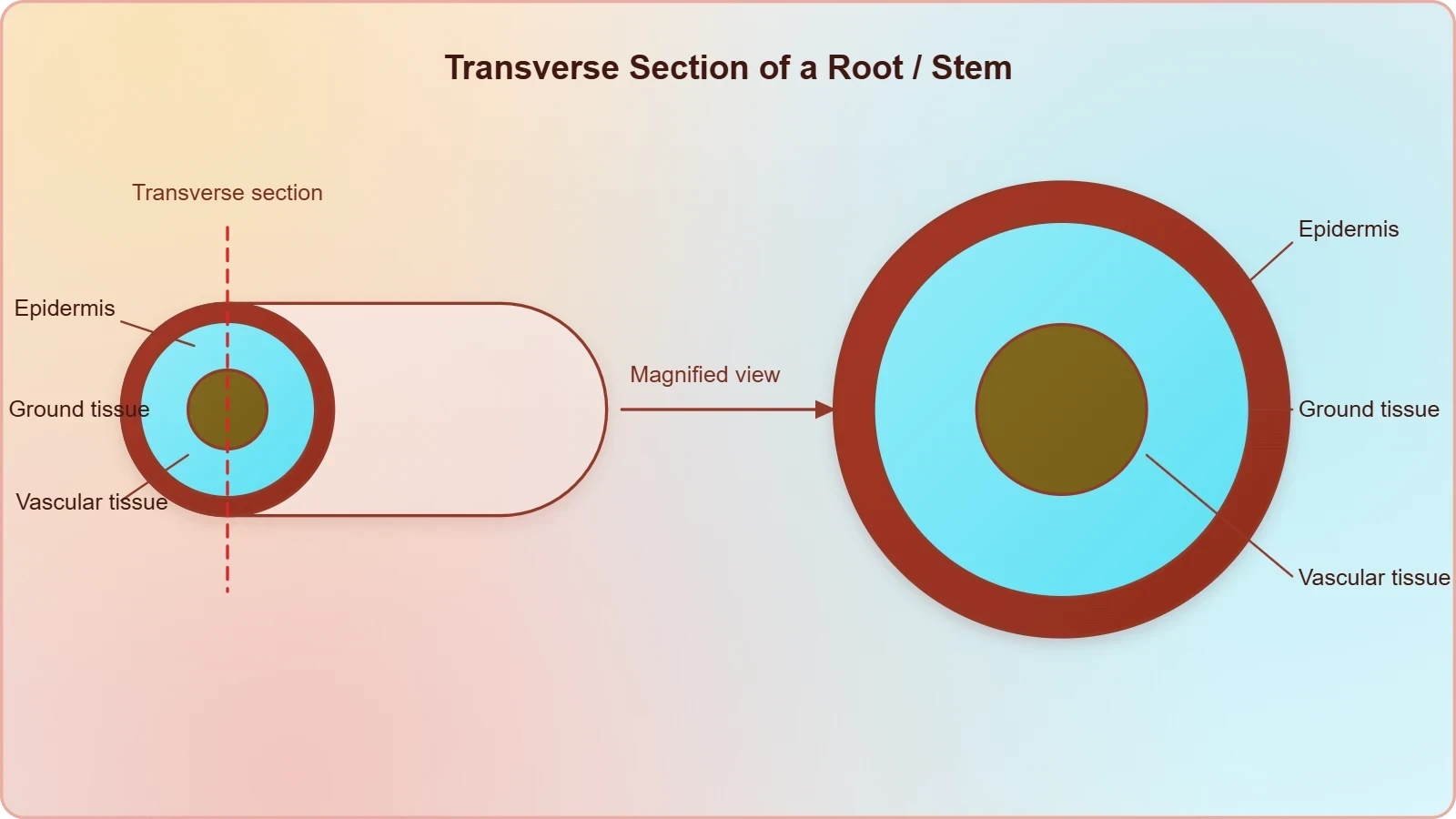
The transition from external to internal study is a change of scale and of instrument. External morphology can be observed with the naked eye; internal structure requires sectioning and magnification, and the transverse section becomes the standard view. A transverse section, cut across the long axis of an organ, reveals the concentric or scattered arrangement of tissues that defines that organ's identity. Learning to read such a section is the central practical skill of plant anatomy, and it depends on knowing in advance which tissues to expect and in what order they should appear.
This is why the course proceeds organ by organ and group by group. Once the three tissue systems are understood as categories, each organ can be examined as a particular arrangement of those categories. The dicot root, the monocot root, the dicot stem, the monocot stem, the dorsiventral leaf and the isobilateral leaf each present a recognisable pattern, and the differences between them are precisely what examination questions and practical identifications test. The vocabulary learned in this lesson is the toolkit for all of that later work.
Why Anatomy Compares Organs and Plant Groups
Comparison is built into the definition. Different organs in a plant show differences in their internal structure, and within angiosperms the monocots and dicots are also seen to be anatomically different. These two axes of comparison — organ against organ, and group against group — organise the entire subject. A stem is not simply a root seen from another angle; its vascular tissue is arranged on a different plan, and that plan differs again between a monocot and a dicot. Recognising these patterns is the substance of anatomical knowledge.
The third axis of comparison is environmental. Internal structures also show adaptations to diverse environments, which means anatomy carries ecological information. A plant in a dry habitat and a plant in a wet one may share the same organ types yet differ in the proportions and specialisations of their tissues. This is why the course keeps returning to specific structures such as the casparian strip, bulliform cells and the stomatal apparatus: each is a small anatomical feature with a large functional and environmental story attached to it.
The Ladder of Organisation: Cells, Tissues and Organs
The material states the hierarchy with deliberate clarity: plants have cells as the basic unit, cells are organised into tissues, and in turn the tissues are organised into organs. This three-rung ladder is the conceptual spine of the lesson. Each rung is a level of organisation with its own descriptive vocabulary, and each higher rung is built from the one below. A cell is not a tissue, and a tissue is not an organ; the distinctions matter because anatomical descriptions move constantly between these levels, naming a cell type in one sentence and an organ in the next.
Understanding the ladder also explains why the course begins with tissues rather than with cells. Cell types are introduced in the earlier study of tissues based on the types of cells present, and that groundwork is assumed here. What this chapter adds is a second way of classifying tissues: not by the cells they contain, but by where they sit in the plant body. Location, structure and function are treated as linked, so the same tissue concept can be recognised wherever it occurs and its role inferred from its position.
Cells as the Basic Unit
Every anatomical statement ultimately rests on cells. The plant body is cellular, and the properties of a tissue derive from the properties of its constituent cells: their walls, their shape, their living or non-living condition, and the way they are packed together. The material notes, for instance, that the epidermis is made up of elongated, compactly arranged cells, and that single phrase already tells the reader something about function, because compact arrangement limits the spaces through which water and gases might otherwise pass freely.
Keeping the cellular level in view prevents a common beginner's error, which is to treat tissues as abstract regions on a diagram. A tissue is a population of cells, and its appearance in section is the summed appearance of those cells. When later lessons describe open and closed vascular bundles, or the casparian strip in the root endodermis, the description is always traceable back to particular cells and particular features of their walls. The ladder of organisation is thus not a formality but a working method of explanation.
Tissues Grouped by Structure and Location
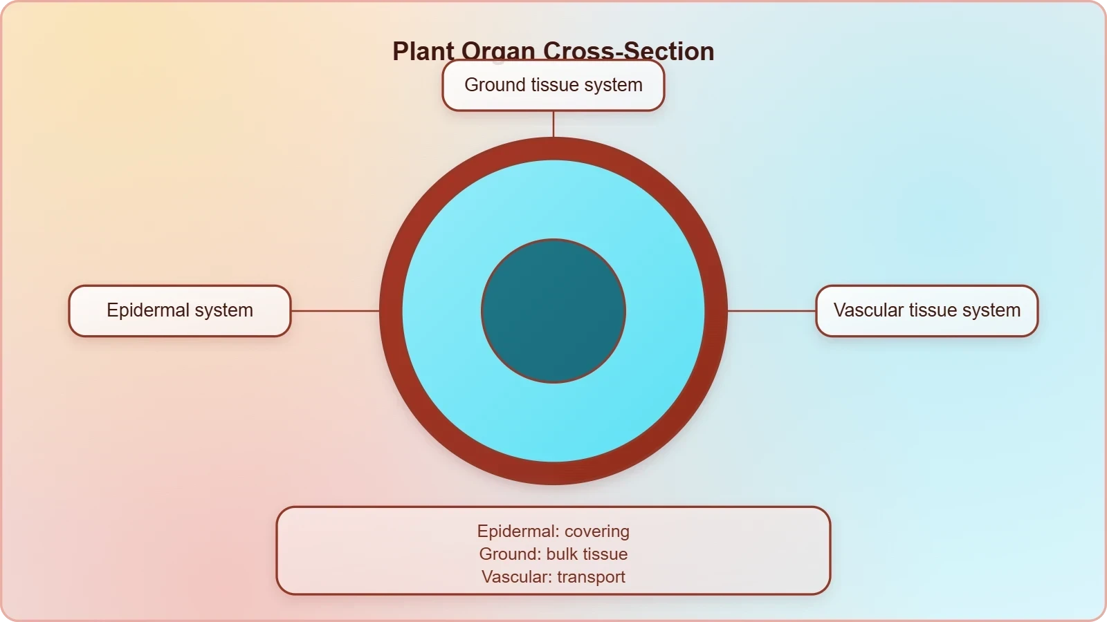
The decisive move in the chapter is the shift from classifying tissues by cell type to classifying them by location. Having discussed types of tissues based on the types of cells present, the material asks how tissues vary depending on their location in the plant body, noting that their structure and function would also be dependent on location. On the basis of their structure and location, there are three types of tissue systems: the epidermal tissue system, the ground or fundamental tissue system, and the vascular or conducting tissue system.
This threefold division is the single most useful framework in the whole course, because it applies to every organ. Whether the organ is a root, a stem or a leaf, its internal structure can be described as an arrangement of these three systems. The systems are not three separate tissues but three categories, each of which may be represented by more than one cell type. Learning to assign any observed structure to one of the three systems is the first competence a student of plant anatomy must acquire.
The Epidermal Tissue System: The Outer Covering
The epidermal tissue system forms the outer-most covering of the whole plant body and comprises epidermal cells, stomata and the epidermal appendages, namely the trichomes and hairs. The epidermis itself is the outermost layer of the primary plant body, and it is made up of elongated, compactly arranged cells. Even in this brief introduction, the material gives three distinct components, and the distinction is worth holding onto: the epidermal cells proper, the stomata that punctuate them, and the appendages that project from them are all parts of one system but are not the same thing.
Because this lesson is an overview, the epidermis is presented here only as the first of the three systems, in the order the material gives. Its detailed treatment — the stomatal apparatus, the structure of the guard cells, the variety of trichomes — belongs to the next lesson, which is devoted to it. What matters now is the principle the epidermis illustrates: a tissue system is defined by its location and structure together, and its position at the surface immediately suggests a protective and boundary role for the plant body.
Epidermal Cells, Stomata and Appendages
The three components named in the material each deserve a moment's attention at the level of definition. Epidermal cells form the continuous sheet; stomata are the structures associated with the epidermis that are treated in detail later; and the epidermal appendages are the trichomes and hairs. The word appendage is well chosen, because trichomes and hairs are outgrowths of the epidermis rather than separate layers. Recognising that all three belong to one system is the conceptual point, and it prepares the ground for the next lesson's closer inspection.
The description of epidermal cells as elongated and compactly arranged is a small but instructive example of anatomical reasoning. Elongation and compact packing are structural facts, but they carry functional implications about how the surface behaves. This is the pattern the whole course follows: observe the structure, then read its consequence. Students who practise this habit early will find the later lessons on stomatal apparatus and on leaf surfaces far easier, because they will already be asking what each arrangement achieves rather than merely memorising its name.
Why the Surface Layer Comes First
The material's order of ideas places the epidermal system first among the three, and there is a sound reason for that sequence. The epidermis is the outermost layer of the primary plant body, so it is the first tissue encountered when an organ is sectioned from the outside inward. Reading a transverse section therefore naturally begins at the epidermis and proceeds toward the centre. The order of the chapter mirrors the order of observation, which makes it easier to remember and easier to apply in the laboratory.
Beginning with the surface also establishes the habit of thinking in layers. Every organ examined later in the course will be described as a succession of regions from outside to inside, and the epidermal system is the fixed starting point of that succession. Once the student is comfortable with the idea that the outer covering is a system in its own right, the ground tissue and vascular tissue can be added beneath it without confusion. The framework is built from the outside in, exactly as the material presents it.
Organs Differ Internally: Roots, Stems and Leaves
A central claim of the material is that different organs in a plant show differences in their internal structure. This is not a trivial observation but the reason anatomy is worth studying organ by organ. A root, a stem and a leaf are all plant organs built from the same three tissue systems, yet they arrange those systems differently, and the differences are consistent enough to identify the organ from a section alone. The course is structured around exactly this fact, devoting successive lessons to roots, stems and leaves.
The differences are not random. Each organ's internal arrangement corresponds to what that organ does and where it sits in the plant body. The course description makes the plan explicit: after the tissue systems, it examines the transverse anatomy of dicotyledonous and monocotyledonous roots and stems, and the vertical anatomy of dorsiventral and isobilateral leaves. Note the change of plane as well as of organ: roots and stems are studied in transverse section, leaves in vertical section, because the informative arrangement in a leaf is best seen that way.
Transverse Anatomy of Roots and Stems
Roots and stems are both cylindrical organs, and both are conventionally studied in transverse section, cut across the axis. In such a section the arrangement of the vascular tissue is the decisive feature, and it differs between the two organs. The course later distinguishes open and closed vascular bundles, a distinction that belongs to the stem lessons, and examines the casparian strip, which belongs to the root. These are not isolated facts but markers of the different internal logic of the two organs.
The pairing of dicotyledonous and monocotyledonous within each organ type doubles the comparison. A dicot root and a monocot root are both roots, yet they differ anatomically; the same is true of stems. This is the second axis of comparison identified earlier, applied concretely. By the end of the relevant lessons, the student should be able to hold four patterns in mind at once — dicot root, monocot root, dicot stem, monocot stem — and to state how each differs from its partner.
Vertical Anatomy of Dorsiventral and Isobilateral Leaves
Leaves are studied in vertical section rather than transverse, and the course names two leaf types: dorsiventral and isobilateral. The distinction between them is anatomical as well as descriptive, and the vertical section is the view that makes the difference visible. The leaf lessons also bring in bulliform cells, which are among the specialised structures the course expects the student to recognise. As with roots and stems, the pattern is comparison: two leaf types, examined in the same plane, distinguished by their internal arrangement.
Placing leaves after roots and stems is pedagogically sound. By the time the leaf lessons arrive, the student will already know the three tissue systems and will have seen them arranged in two cylindrical organs. The leaf then appears as a third variation on a familiar theme, flattened rather than cylindrical, with its own characteristic distribution of tissues. The course thus builds a cumulative picture in which each organ is understood against the ones already studied, and no lesson stands alone.
Monocots and Dicots: Anatomical Difference Within Angiosperms
Within angiosperms, the monocots and dicots are seen to be anatomically different. This statement, brief in the material, carries a great deal of consequence for the course. It means that every organ examined must be examined twice, once in its dicotyledonous form and once in its monocotyledonous form. The course description reflects this directly, naming dicotyledonous and monocotyledonous roots and stems as separate subjects. The distinction is not a footnote to plant anatomy; it is one of its principal organising divisions.
The anatomical differences are consistent enough to be diagnostic, which is why they are taught as paired patterns rather than as isolated facts. A student who knows only one form of root will misread the other. The course therefore treats the pairs together, so that the contrast itself becomes the memorable unit. This is also why the material's opening paragraph mentions the monocot–dicot difference alongside the difference between organs: both are examples of the same principle, that internal structure varies in patterned ways.
Paired Patterns Across Organs
The pairing operates at every organ level. Roots come in a dicot form and a monocot form; stems likewise; and the leaf lessons present dorsiventral and isobilateral types, which are associated with the two groups in the standard treatment. The student's task is to learn each pair as a contrast, noting which features are shared because the organ is the same and which differ because the group is different. This double accounting is the discipline that makes anatomical identification reliable rather than guesswork.
Keeping the pairs distinct also prevents a common confusion, in which a feature belonging to one group is wrongly generalised to all flowering plants. The material is careful to say that monocots and dicots are anatomically different, not that one is normal and the other aberrant. Both are fully functional solutions to the same problems of support, transport and protection, and the differences between them are differences of arrangement rather than of adequacy. That framing keeps the comparison scientific and avoids implying a hierarchy between the two groups.
Reading the Difference in a Section
In practice, the monocot–dicot difference is read from the arrangement of the vascular tissue, which is why the course introduces open and closed vascular bundles in the stem lessons. The terms themselves signal the distinction: an open bundle retains the capacity for further growth, a closed bundle does not, and this bears directly on secondary growth, which the course also covers. The vocabulary is introduced where it is needed, in the context of the organ and group being studied, rather than as a list to be memorised in advance.
For the beginner, the practical advice is to learn each section as a pattern rather than as a collection of labels. A pattern can be recognised in an unfamiliar slide; a list of labels cannot. The three tissue systems provide the stable scaffolding, and the monocot–dicot contrast provides the variation that the pattern must accommodate. Once both are in place, the student can approach any new section with a method: locate the epidermis, identify the ground tissue, find the vascular tissue, and then ask which organ and which group the arrangement indicates.
Anatomy as Adaptation: Structure in Relation to Environment
Internal structures also show adaptations to diverse environments. This closing claim of the material's opening paragraph widens the subject from classification to explanation. Anatomy is not only a system for naming and distinguishing; it is also a record of how plants have adjusted their internal construction to the conditions they inhabit. The course keeps this ecological dimension alive through specific structures, and the phrase adaptations to diverse environments is the thread that connects the descriptive lessons to a larger biological argument.
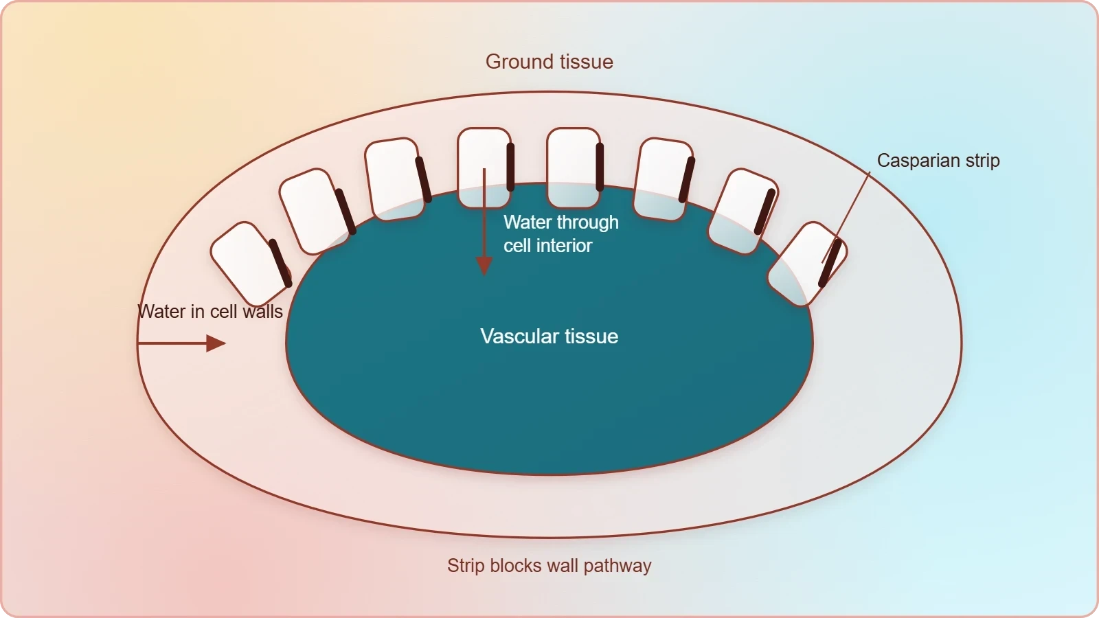
The structures named in the course description illustrate the point well. The casparian strip in the root is a feature of the endodermal cell walls that bears on the movement of water into the vascular tissue. Bulliform cells in the leaf are associated with the leaf's behaviour under conditions of water stress. The stomatal apparatus governs exchange between the interior of the plant and the air. Each is a small anatomical detail, but each is also a point where structure answers environment, and together they show why anatomy is studied functionally rather than as mere description.
Structural Features with Functional Meaning
The habit of linking structure to function is what turns a catalogue of terms into an understanding of plants. When the material describes the epidermis as made of elongated, compactly arranged cells, it is already inviting the reader to ask what such an arrangement achieves. The same invitation recurs with every specialised structure in the course. Students who accept it will find that the terminology becomes easier to retain, because each term is attached to a reason rather than to a diagram alone.
This functional reading also explains the course's insistence on staying close to the chapter's terminology and examples. The terms are not arbitrary labels but compressed descriptions of structure-function relationships. Casparian strip, bulliform cell, stomatal apparatus, open and closed vascular bundle: each name encodes a structural fact with a functional consequence. Learning the terminology precisely is therefore not pedantry but the necessary condition for thinking clearly about adaptation, since vague terms produce vague reasoning about how plants cope with their environments.
Preparing for the Lessons Ahead
This first lesson has established the frame within which all the others operate: anatomy as the study of internal structure; the ladder from cells to tissues to organs; the three tissue systems classified by structure and location; the internal differences between organs; the anatomical difference between monocots and dicots; and the adaptation of internal structure to environment. Each of these will be taken up in detail in the lessons that follow, and each will be assumed as known. The next lesson turns to the epidermal tissue system, examining the epidermis, its stomata and its appendages at close range.
The best preparation is to fix the framework firmly before adding detail. A student who can state the three tissue systems, explain the cell-to-organ hierarchy, and give one reason why organs and plant groups differ internally is ready for everything that follows. The course is cumulative by design, and the material's order of ideas — tissue systems first, then dicot and monocot anatomy — is the order in which the framework is best built. From here, each lesson adds a layer to a structure whose outline is already in place.
Going further
Two observations, offered as background rather than as part of the material above, may help the framework settle. The first is that plant anatomy is closely linked to physiology: the arrangement of tissues often reflects the functions they perform, such as transport or support, so a section can be read as a functional diagram as well as a structural one. The second is that the terms monocot and dicot refer to the number of cotyledons in the seed, a distinction that also shows up in internal structure. Neither point alters the material; both simply explain why the anatomical differences described here are so consistently patterned.
Also Read
- No links available.
From our network :
- Analyzing Trump Deportation Numbers Insights Into The 2026 Immigration Crackdown
- Vite 6/7 'Cold Start' Regression in Massive Module GraphsIn-depth analysis of Vite 6/7 cold start regressions in massive module graphs. Learn about barrel file issues, pre-bundling optimizations, and how to improve Vite dev server performance.
- 10 Physics Numerical Problems with Solutions for IIT JEEPrepare for IIT JEE with 10 physics numerical problems, complete with detailed solutions and conceptual explanations. Ideal for mastering kinematics, thermodynamics, electromagnetism, and optics.
- EV 2.0: The Solid-State Battery Breakthrough and Global Factory ExpansionDiscover the impact of the Solid-State Battery Breakthrough on the EV market. Learn about CATL and BYD stock growth, global factory expansion in Europe, and the future of EV 2.0 technology.
- Mastering DB2 12.1 Instance Design: A Technical Deep Dive into Modern Database ArchitectureComprehensive guide to DB2 12.1 Instance Design. Explore architectural components, HADR setup, buffer pool tuning, and security policies for IBM DB2 12.1. Includes 10 functional SQL and Shell samples.
- AI-Powered 'Precision Diagnostic' Replaces Standard GRE Score ReportsDiscover the new GRE Precision Diagnostic. Learn how AI-powered behavioral analytics and time-to-solve tracking are replacing standard GRE score reports to empower students.
- Trump Political Strategy How Geopolitical Stunts Serve As Media Diversions
- Mastering DB2 LUW v12 Tables: A Comprehensive Technical GuideComprehensive guide on DB2 LUW v12 tables. Explore CREATE TABLE syntax, BLU Acceleration, range partitioning, data types, and security features for IBM DB2 LUW v12.
- 98% of Global MBA Programs Now Prefer GRE Over GMAT Focus EditionExplore why 98% of global MBA programs now prefer the GRE over GMAT Focus. Learn about the shorter GRE format, verbal logic importance, and strategic versatility for students.
RESOURCES
- Plant Tissues and Organs | Biology for Majors II - Lumen Learningcourses.lumenlearning.comVascular plants have two distinct organ systems: a shoot system, and a root system. The shoot system consists of two portions: the vegetative (non-reproductive) ...
- Tissues, organs, & organ systems (article) - Khan Academykhanacademy.orgFrom left to right: single muscle cell, multiple muscle cells together forming muscle tissue ... tissues, organs, and organ systems—structure is closely related ...
- Plant tissues and organs | PPT - Slideshareslideshare.netThe document also outlines the three main tissue systems - vascular, dermal and ground tissues. Meristematic tissues contain stem cells that allow continuous ...
- 35.1.1: Plant Tissues and Organ Systems - Biology LibreTextsbio.libretexts.orgDec 17, 2021 ... Vascular plants have two distinct organ systems: a shoot system and a root system. The shoot system consists of two…
- Plant Cells, Tissues, and Organs Overview | PDF - Scribdscribd.comand carbon dioxide needed/produced during photosynthesis. Vascular Bundles: Xylem: water delivery to mesophyll tissue. Phloem: sugar delivery from mesophyll ...
- 30.1 The Plant Body - Biology 2e | OpenStaxopenstax.orgMar 28, 2018 ... Plant Tissues. Plants are multicellular eukaryotes with tissue systems made of various cell types that carry out specific functions. Plant ...
- Regeneration | National Institute of General Medical Sciences - NIHnigms.nih.govFeb 13, 2025 ... ... cells, tissues, organs, and even entire body parts to full function. ... Human organs and tissues have varied capacities…
- Traits, properties, and performance: how woody plants combine ...nph.onlinelibrary.wiley.comSep 23, 2014 ... ... plant unit (a cell, tissue, or organ/whole plant). We ... cells, cells in tissues, and tissues in organs and…
- Guidance on the Safety of Human Cells, Tissues and Organs for ...canada.caProfile page Guidance Document for Cell, Tissue and Organ Establishments.
- Tissue (biology) - Wikipediaen.wikipedia.orgIn plant anatomy, tissues are categorized broadly into three tissue systems ... The epithelial tissues are formed by cells that cover the organ ...
- Updating the Definition of Cancer - PMC - NIHpmc.ncbi.nlm.nih.govIgnoring the body's signal to stop, malignant cells multiply to form tumors in organs and tissues or, in the case of blood cancers, crowd…
- Introduction to Vascular Plant Structure - Digital Atlas of Ancient Lifedigitalatlasofancientlife.orgMar 30, 2020 ... Tissues & tissue systems. Tissues are regions of cells in the plant body that are functionally and physically connected to…
- Collective intelligence: A unifying concept for integrating biology ...nature.comMar 28, 2024 ... ... cells, tissues, organs, whole bodies, and swarms. Crucially however ... Toward uncovering an operating system in plant organs. Trends ...
- What Is Connective Tissue? Definition, Function, Typesmy.clevelandclinic.orgFeb 26, 2025 ... Some connective tissues carry and distribute immune cells. ... All of the organs and systems of your body are made…
- Inflammatory responses and inflammation-associated diseases in ...pmc.ncbi.nlm.nih.gov... tissue cells and inflammatory cells recruited from the blood [8]. ... Acute and chronic inflammation-mediated tissue injury is observed in many organ systems ...