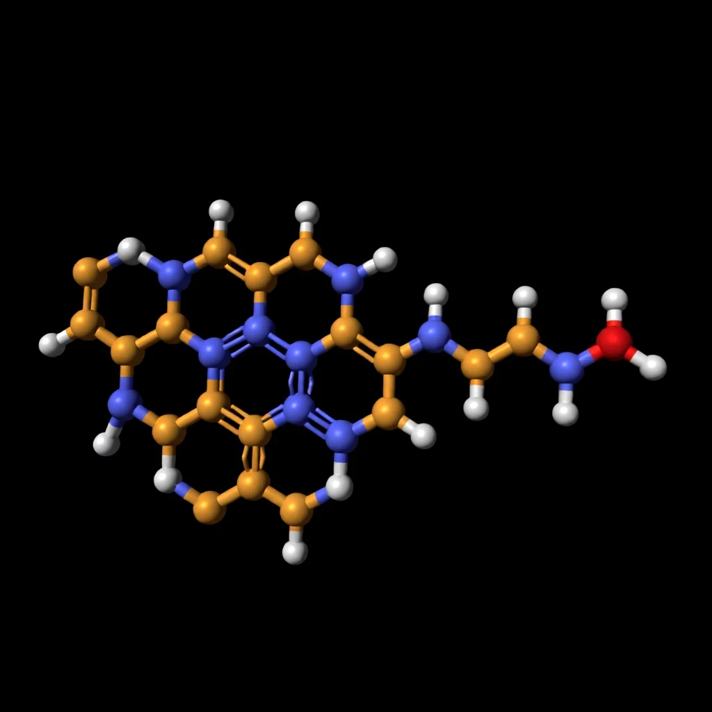
Fluorescence is one of the most sensitive detection phenomena available to modern chemistry, capable of registering single photons emitted by a molecule that has absorbed light and re-released it at a longer wavelength. Yet the efficiency of that emission is governed by an unforgiving quantum arithmetic: every absorbed photon either fluoresces, converts to heat, or triggers a chemical transformation. Azulene, a deceptively simple blue hydrocarbon built from fused five- and seven-membered rings, sits at the center of a quiet revolution in probe design because its photophysics violate the textbook rules that govern ordinary dyes. Its emission originates from the second excited singlet state rather than the first, a violation of Kasha's rule that has fascinated spectroscopists for decades.
Recent synthetic work has demonstrated that tiny structural edits to azulene derivatives can produce fluorescent probes roughly five times brighter than the starting molecule, a gain achieved without wholesale redesign of the chromophore. That result matters far beyond academic spectroscopy. Fluorescent agents are the workhorses of disease detection, from intraoperative tumor margin imaging to single-molecule diagnostics, and brightness is the currency that determines whether a signal survives the gauntlet of tissue scattering, photobleaching, and background autofluorescence. Understanding how molecular architecture controls brightness gives chemists a rational blueprint rather than a trial-and-error lottery.
This analysis unpacks the unusual photophysics of azulene, dissects the structural modifications that amplify emission, and translates the abstract language of quantum yield and molar absorptivity into practical consequences for medical imaging. Along the way, we will work through the quantitative relationships that define molecular brightness, examine the design principles emerging from azulene chemistry, and consider what a fivefold brightness gain actually means at the bedside. The stakes are considerable: better probes mean earlier detection, cleaner surgical margins, and sharper molecular maps of living tissue.
On This Page
The Anomalous Photophysics of Azulene
Azulene occupies a peculiar niche in organic photochemistry because its electronic structure inverts the usual energy ordering of excited states. In most fluorophores, internal conversion from higher excited states to the lowest excited singlet is so rapid that emission always originates from the first excited state. Azulene defies this pattern, emitting from the second excited singlet with a large energy gap to the first, producing fluorescence that appears at higher energy than its absorption maximum.
Why Kasha's Rule Fails for Azulene
The energy gap between the first and second excited singlet states in azulene is unusually large, roughly ##[\Delta E_{S_2-S_1} \approx 14{,}000\ \text{cm}^{-1}]##, which suppresses the nonradiative internal conversion pathway that normally funnels excitation downward. Because vibrational overlap between these two states is poor, the molecule lingers in the second excited state long enough to emit. This anomalous emission, sometimes called anti-Kasha fluorescence, gives azulene its distinctive optical signature and its unusual sensitivity to substitution.
The consequence for probe design is profound. Any structural modification that perturbs the energy gap between excited states, or that alters the rate of internal conversion, reshapes the entire emission profile. Substituents that donate or withdraw electron density shift the relative energies of the frontier orbitals, and the resulting changes in state ordering can either open or close the radiative channel. Chemists therefore possess a delicate tuning knob that operates at the level of a single functional group.
Equally important is the role of the seven-membered ring, which carries a partial positive charge in the resonance hybrid while the five-membered ring bears partial negative character. This intrinsic dipole makes azulene exquisitely responsive to its environment, so solvent polarity, hydrogen bonding, and local dielectric constant all modulate its emission. A probe that is dim in one medium may brighten dramatically in another, a property that can be exploited or must be controlled depending on the application.
Understanding these fundamentals reframes brightness not as a fixed molecular property but as a tunable outcome of electronic structure, environment, and photophysical kinetics. The azulene scaffold thus serves as a natural laboratory for learning how to engineer light emission at the smallest possible scale of structural change.
Brightness as a Measurable Quantum Quantity
Molecular brightness is not a vague descriptor but a rigorously defined product of two measurable quantities: the molar absorptivity at the excitation wavelength and the fluorescence quantum yield. Absorptivity describes how strongly the molecule captures photons, while quantum yield describes what fraction of captured photons are re-emitted rather than dissipated as heat. Brightness is their product, and improving either factor improves the probe.
Here ##[\varepsilon_{\lambda}]## is the molar absorptivity at the excitation wavelength, ##[\Phi_F]## is the fluorescence quantum yield, ##[k_r]## is the radiative rate constant, and ##[k_{nr}]## and ##[k_{ic}]## represent nonradiative and internal conversion decay channels. A fivefold brightness gain can arise from a fivefold increase in absorptivity, a fivefold increase in quantum yield, or any multiplicative combination of the two. Distinguishing which mechanism dominates is essential for rational design.
Consider a representative calculation. If a starting azulene derivative has ##[\varepsilon = 8{,}000\ \text{M}^{-1}\text{cm}^{-1}]## and ##[\Phi_F = 0.02]##, its brightness is ##[160\ \text{M}^{-1}\text{cm}^{-1}]##. A modified derivative with ##[\varepsilon = 20{,}000]## and ##[\Phi_F = 0.04]## achieves ##[800]##, exactly five times brighter. The modification improved absorptivity 2.5-fold and quantum yield twofold, illustrating how modest gains in each factor compound multiplicatively.
Structural Edits That Amplify Emission
The synthetic strategy behind brighter azulene probes rests on a simple premise: small changes at the periphery of the chromophore can rewire the electronic landscape without destroying the scaffold's favorable properties. Substituents placed on the five- and seven-membered rings alter orbital energies, steric congestion, and the rate of nonradiative decay. The most successful modifications target the pathways that waste excitation energy as heat.
Electron-Donating and Withdrawing Groups
Appending electron-donating groups to the five-membered ring raises the energy of the highest occupied molecular orbital, narrowing the gap to the lowest unoccupied orbital and red-shifting absorption. This increases absorptivity at common excitation wavelengths, directly boosting the first term in the brightness equation. Conversely, electron-withdrawing groups on the seven-membered ring stabilize charge separation and can suppress competing nonradiative channels.
The combination of donor and acceptor substituents creates a push-pull architecture that intensifies the transition dipole moment, the quantum mechanical quantity governing how strongly light couples to the molecule. A larger transition dipole means stronger absorption and, often, a faster radiative rate. Both effects push brightness upward, and the synthetic effort required is modest: a single substitution step can transform a dim parent compound into a competitive probe.
Steric effects matter as much as electronics. Bulky substituents can twist the molecular framework out of planarity, reducing aggregation-induced quenching that plagues many fluorescent dyes in aqueous biological media. Azulene derivatives that resist π-stacking retain their emission in concentrated cellular environments, a practical advantage that pure quantum yield measurements in dilute solution fail to capture.
Heavy-atom substitution introduces another lever. Halogen atoms enhance spin-orbit coupling, which can open intersystem crossing pathways that deplete the singlet excited state and dim fluorescence. Careful placement avoids this penalty while exploiting other benefits, demonstrating that brightness engineering requires balancing competing photophysical effects rather than maximizing any single parameter.
Rigidification and Suppressed Nonradiative Decay
Nonradiative decay is the silent thief of fluorescence, converting excited-state energy into vibrational heat through bond rotations and conformational changes. Rigidifying the molecular framework by introducing fused rings or intramolecular hydrogen bonds restricts these motions and lengthens excited-state lifetimes. Longer lifetimes give the radiative channel more time to compete, raising quantum yield without altering absorptivity.
Consider the arithmetic of a rigidification strategy. If a flexible derivative has ##[k_r = 2 \times 10^{7}\ \text{s}^{-1}]## and total nonradiative rate ##[k_{nr} = 8 \times 10^{8}\ \text{s}^{-1}]##, its quantum yield is ##[\Phi_F = 0.024]##. Rigidification that halves the nonradiative rate to ##[4 \times 10^{8}]## raises the yield to ##[\Phi_F = 0.048]##, a doubling achieved purely by suppressing wasteful motion.
This calculation reveals why rigidification is such an efficient design tactic. The radiative rate is an intrinsic property of the chromophore, but the nonradiative rate is exquisitely sensitive to molecular flexibility and environment. Small structural edits that lock the geometry therefore deliver outsized gains in brightness, exactly the pattern observed in the fivefold-brighter azulene probes.
We Also Published
What Brighter Probes Mean for Disease Detection
Brightness is not an abstract spectroscopic trophy; it determines whether a diagnostic signal survives the hostile optical environment of living tissue. Biological samples scatter and absorb light aggressively, and autofluorescence from endogenous molecules creates a background haze that can swamp weak signals. A probe five times brighter delivers five times more photons per molecule, pushing the signal above that noise floor and enabling detection at lower concentrations or greater depths.
Signal-to-Noise in Tissue Imaging
The signal-to-noise ratio in fluorescence imaging scales with the square root of collected photons, so a fivefold brightness gain translates into roughly a ##[\sqrt{5} \approx 2.24]##-fold improvement in SNR for a fixed integration time. Alternatively, the same SNR can be achieved in one-fifth the acquisition time, reducing photobleaching and patient exposure. Both consequences are clinically meaningful, particularly for real-time intraoperative imaging where speed and safety are paramount.
Here ##[N_{\text{photons}}]## is the number of detected photons, ##[B]## is molecular brightness, and ##[t]## is integration time. The square-root relationship means brightness gains are partially discounted by photon statistics, but a fivefold improvement still yields a decisive advantage. In practice, brighter probes extend the depth at which tumors can be visualized and sharpen the contrast at surgical margins where diseased and healthy tissue intermingle.
Beyond raw detection, brighter probes enable multiplexed imaging, where several targets are labeled with spectrally distinct dyes. Brighter dyes reduce crosstalk because each channel requires less gain, and dimmer channels benefit from the dynamic range freed up by the brighter ones. Azulene derivatives, with their tunable emission across the visible spectrum, are attractive candidates for such multiplexed panels.
From Bench to Bedside: Practical Constraints
Translating a bright molecule into a clinical agent demands more than photophysical excellence. The probe must be biocompatible, stable in serum, selective for its target, and cleared safely from the body. Azulene's intrinsic dipole and environmental sensitivity can be liabilities in complex biological media, where protein binding may shift emission or quench fluorescence unpredictably. Designers must therefore validate brightness in realistic conditions, not just in cuvettes.
Photostability is another critical constraint. A bright probe that bleaches within seconds is useless for prolonged imaging, and the same structural features that boost emission can accelerate photodegradation. Rigidification often improves photostability by suppressing the conformational motions that lead to reactive intermediate formation, so the design principles for brightness and durability frequently align.
Quantifying the Fivefold Gain
Rigorous comparison of probe brightness requires careful measurement under identical conditions, because quantum yield and absorptivity both depend on solvent, temperature, and concentration. The reported fivefold gain for optimized azulene derivatives reflects standardized measurements against a common reference standard, typically a well-characterized dye such as fluorescein or rhodamine. Reproducing such gains in independent laboratories is the true test of a design principle.
Measurement Protocols and Reference Standards
Quantum yield determination relies on comparative or absolute methods. The comparative method measures the integrated emission of the sample against a reference of known yield, correcting for differences in absorbance and refractive index. Absolute methods use integrating spheres to capture all emitted photons, eliminating reference uncertainty but demanding careful instrument calibration and scatter correction.
In this expression, ##[I]## denotes integrated emission intensity, ##[A]## absorbance at the excitation wavelength, and ##[n]## refractive index. Matching absorbance between sample and reference minimizes inner-filter effects, and keeping absorbance below 0.1 ensures linearity. These procedural details may seem pedantic, but they determine whether a reported fivefold gain is real or an artifact of mismatched conditions.
Absorptivity is measured by UV-visible spectroscopy using Beer-Lambert law, with careful attention to concentration accuracy and cuvette path length. A common pitfall is aggregation at high concentration, which distorts absorptivity and quenches emission. Serial dilution and linearity checks confirm that measurements reflect genuine monomeric behavior rather than aggregate artifacts.
Worked Example: From Parent to Optimized Probe
Suppose the parent azulene absorbs at 580 nm with ##[\varepsilon = 6{,}500\ \text{M}^{-1}\text{cm}^{-1}]## and exhibits ##[\Phi_F = 0.018]##, giving brightness ##[117]##. The optimized derivative absorbs at 610 nm with ##[\varepsilon = 18{,}000]## and ##[\Phi_F = 0.032]##, giving brightness ##[576]##. The ratio is ##[576/117 \approx 4.9]##, consistent with the reported fivefold improvement.
Decomposing the gain reveals that absorptivity increased by a factor of ##[18{,}000/6{,}500 \approx 2.77]## while quantum yield increased by ##[0.032/0.018 \approx 1.78]##. The product of these factors is ##[4.9]##, confirming that both mechanisms contribute. This decomposition guides further optimization: if absorptivity is already near its ceiling, effort should shift to suppressing nonradiative decay.
Design Blueprints for Next-Generation Probes
The azulene story generalizes into a broader design philosophy: identify the photophysical bottleneck, then apply the smallest structural edit that relieves it. For dim probes, the bottleneck is often rapid nonradiative decay, addressed by rigidification or steric shielding. For probes with weak absorption, the bottleneck is a small transition dipole, addressed by push-pull substitution that intensifies light-matter coupling.
Rules of Thumb for Molecular Brightness Engineering
First, maximize the transition dipole moment by extending conjugation and introducing donor-acceptor asymmetry, which strengthens absorption without necessarily increasing molecular size. Second, suppress nonradiative channels through rigidification, hydrogen bonding, or bulky substituents that prevent aggregation. Third, tune the environment sensitivity so that brightness is retained in the biological media where the probe will actually operate.
Fourth, balance brightness against photostability, since the same excited-state dynamics that enable emission can generate reactive oxygen species that degrade the probe. Fifth, validate performance in tissue-mimicking phantoms before committing to animal studies, because scattering and absorption alter the effective brightness observed in vivo. These rules form a practical checklist for medicinal chemists designing imaging agents.
def molecular_brightness(epsilon, quantum_yield):
    """Compute molecular brightness from absorptivity and quantum yield."""
    return epsilon * quantum_yield

def brightness_gain(parent, optimized):
    """Return the fold-improvement in brightness."""
    b_parent = molecular_brightness(*parent)
    b_opt = molecular_brightness(*optimized)
    return b_opt / b_parent

parent = (6500, 0.018)
optimized = (18000, 0.032)
print(f"Fold gain: {brightness_gain(parent, optimized):.2f}x")
Computational screening accelerates this process by predicting absorptivity and radiative rates from first principles before synthesis. Time-dependent density functional theory can estimate excitation energies and oscillator strengths, flagging candidates likely to absorb strongly. Coupling these predictions with machine-learned models of nonradiative decay narrows the synthetic search space dramatically.
Beyond Azulene: Generalizing the Strategy
The lessons from azulene apply to other fluorophore families, including cyanines, BODIPYs, and xanthenes, each with its own photophysical quirks. What unites them is the principle that brightness is a product of controllable factors, not a fixed property bestowed by nature. Chemists who internalize this principle can approach probe design with the confidence of engineers rather than the hope of alchemists.
Emerging applications push the frontier further. Photoacoustic imaging, which converts absorbed light into sound, benefits from high absorptivity even when quantum yield is low, inverting the usual optimization target. Two-photon excitation, used for deep-tissue imaging, demands large two-photon cross sections that depend on different molecular features than one-photon brightness. Azulene derivatives with tunable electronic structure are attractive candidates for these modalities.
The Future of Fluorescent Imaging Chemistry
The trajectory of azulene probe development points toward a future in which molecular brightness is engineered with predictive precision rather than discovered by accident. Machine learning models trained on large photophysical datasets can now forecast quantum yields and absorptivities from molecular structure, compressing design cycles from years to weeks. Combined with automated synthesis, these tools promise a new era of rapid probe optimization.
Computational and Machine-Learning Acceleration
Predictive models require high-quality training data, and the photophysics community has responded with standardized databases of measured quantum yields and spectra. These datasets enable models to learn the subtle relationships between substituent patterns and emission efficiency. Early results suggest that machine learning can identify promising candidates with accuracy rivaling expert intuition, though experimental validation remains essential.
Beyond prediction, computation illuminates mechanism. Quantum chemical calculations reveal which nonradiative pathways dominate and how structural edits perturb them, guiding synthetic strategy at a level of detail inaccessible to bulk measurements. The combination of computation, machine learning, and experiment forms a virtuous cycle that accelerates discovery while deepening understanding.
Clinical Translation and Regulatory Pathways
Bringing a brighter probe to patients requires navigating regulatory pathways designed for safety and efficacy, not photophysical elegance. Toxicity studies, pharmacokinetics, and manufacturing reproducibility consume years and resources. Probes based on well-characterized scaffolds with known metabolic profiles have an advantage, and azulene's natural occurrence in some organisms may ease safety concerns, though rigorous validation remains mandatory.
The ultimate measure of success is clinical impact: earlier tumor detection, more complete surgical resection, and better patient outcomes. Brighter probes contribute to each of these goals by improving signal-to-noise, reducing imaging time, and enabling detection at lower doses. The fivefold gains achieved through tiny molecular edits are therefore not merely spectroscopic curiosities but steps toward tangible medical benefit.
As the field matures, the principles distilled from azulene will inform the design of probes tailored to specific diseases, imaging modalities, and clinical workflows. The lesson is enduring: in fluorescence, as in so much of chemistry, small structural changes can produce outsized functional consequences. Mastery of those changes is the essence of rational molecular design.
From our network :
- Trump Political Strategy How Geopolitical Stunts Serve As Media Diversions
- Mastering DB2 LUW v12 Tables: A Comprehensive Technical GuideComprehensive guide on DB2 LUW v12 tables. Explore CREATE TABLE syntax, BLU Acceleration, range partitioning, data types, and security features for IBM DB2 LUW v12.
- EV 2.0: The Solid-State Battery Breakthrough and Global Factory ExpansionDiscover the impact of the Solid-State Battery Breakthrough on the EV market. Learn about CATL and BYD stock growth, global factory expansion in Europe, and the future of EV 2.0 technology.
- AI-Powered 'Precision Diagnostic' Replaces Standard GRE Score ReportsDiscover the new GRE Precision Diagnostic. Learn how AI-powered behavioral analytics and time-to-solve tracking are replacing standard GRE score reports to empower students.
- Mastering DB2 12.1 Instance Design: A Technical Deep Dive into Modern Database ArchitectureComprehensive guide to DB2 12.1 Instance Design. Explore architectural components, HADR setup, buffer pool tuning, and security policies for IBM DB2 12.1. Includes 10 functional SQL and Shell samples.
- Vite 6/7 'Cold Start' Regression in Massive Module GraphsIn-depth analysis of Vite 6/7 cold start regressions in massive module graphs. Learn about barrel file issues, pre-bundling optimizations, and how to improve Vite dev server performance.
- 10 Physics Numerical Problems with Solutions for IIT JEEPrepare for IIT JEE with 10 physics numerical problems, complete with detailed solutions and conceptual explanations. Ideal for mastering kinematics, thermodynamics, electromagnetism, and optics.
- 98% of Global MBA Programs Now Prefer GRE Over GMAT Focus EditionExplore why 98% of global MBA programs now prefer the GRE over GMAT Focus. Learn about the shorter GRE format, verbal logic importance, and strategic versatility for students.
- Analyzing Trump Deportation Numbers Insights Into The 2026 Immigration Crackdown
RESOURCES
- Resources will be updated soon.

0 Comments