
Organoids in drug discovery have emerged as the cornerstone of a transformative era in biomedical research, signaling the potential conclusion of the century-long reliance on animal models. As of December 2025, the U.S. Food and Drug Administration (FDA) has significantly accelerated its roadmap to transition from traditional in vivo mammalian testing to human-relevant New Approach Methodologies (NAMs). This shift is not merely a policy adjustment but a fundamental scientific evolution necessitated by the high attrition rates in clinical trials. Statistics from the past decade indicate that approximately 90% of drug candidates that pass animal safety and efficacy tests ultimately fail in human clinical phases, often due to unforeseen toxicity or lack of efficacy arising from interspecies biological divergence. The biological complexity of the human system, characterized by unique genetic expressions, metabolic pathways, and cellular architectures, cannot be fully recapitulated in murine or canine models. The introduction of the FDA Modernization Act 2.0 in late 2022 provided the initial legal framework for this transition, and the subsequent efforts throughout 2025 have solidified the role of microphysiological systems (MPS) and organoids as the primary tools for IND (Investigational New Drug) applications. By utilizing human-derived stem cells to grow three-dimensional tissue structures, researchers can now simulate human physiology with a degree of fidelity previously deemed impossible.
Biogenesis: Engineering Human Tissues from Stem Cells
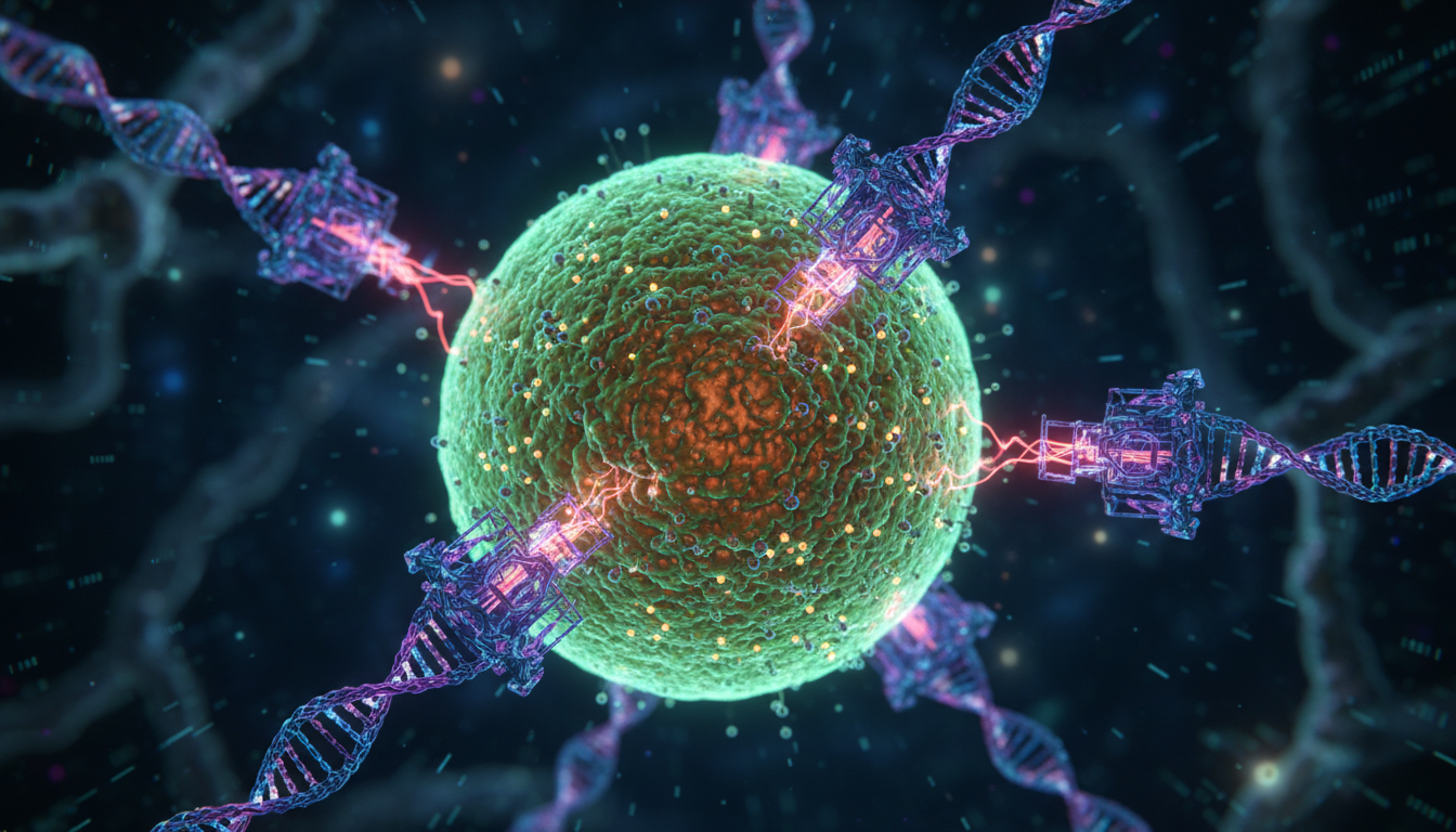
The development of organoids begins with the manipulation of human pluripotent stem cells (hPSCs), which include both embryonic stem cells (ESCs) and induced pluripotent stem cells (iPSCs). The process of differentiation is governed by precise biochemical signaling that mimics embryonic development. Researchers employ specific growth factors and small molecules to activate or inhibit various signaling pathways, such as the Wnt/β-catenin, Notch, and TGF-β/BMP pathways.
For instance, the formation of intestinal organoids requires the sequential addition of Activin A to induce definitive endoderm, followed by FGF4 and Wnt3a to promote posteriorization and the emergence of hindgut spheroids. The maturation of these spheroids into functional organoids involves embedding them in a three-dimensional extracellular matrix (ECM) surrogate, such as Matrigel, which provides the necessary structural support and biochemical cues. The growth kinetics of these cell populations can be modeled using the Monod equation or similar saturable growth models:
###\mu = \mu_{max} \frac{[S]}{K_s + [S]}###
Where ##\mu## is the specific growth rate, ##\mu_{max}## is the maximum growth rate, ##[S]## is the concentration of the limiting substrate, and ##K_s## is the half-velocity constant. In the context of organoids in drug discovery, maintaining the stability of ##[S]## within the 3D matrix is a critical engineering challenge, as nutrient diffusion is often limited by the radius of the organoid structure.
Physics of Microphysiological Systems: Fluid Dynamics and Shear Stress
While organoids provide the 3D cellular architecture, microphysiological systems (MPS) provide the dynamic environment. Often referred to as human-on-a-chip technology, these devices integrate microfluidics to simulate the circulatory system and mechanical forces. The movement of fluid through microchannels is typically laminar, characterized by a low Reynolds number (##Re##):
##Re = \frac{\rho v D_h}{\mu}##
In this equation, ##\rho## represents fluid density, ##v## is the velocity, ##D_h## is the hydraulic diameter of the channel, and ##\mu## is the dynamic viscosity. For most MPS applications, ##Re < 1##, ensuring that transport is dominated by diffusion rather than convection at the cellular interface. However, the shear stress (##\tau##) exerted by the fluid on the cell surface is a vital physiological cue, particularly for endothelial and renal tissues. For a Newtonian fluid in a rectangular channel, the wall shear stress can be approximated by:
###\tau = \frac{6 \mu Q}{w h^2}###
Where ##Q## is the volumetric flow rate, ##w## is the channel width, and ##h## is the channel height. Researchers must precisely calibrate these parameters to ensure that the mechanical environment of the chip matches the in vivo conditions of the target organ. This physical fidelity allows organoids in drug discovery to exhibit mature phenotypes that are often absent in static 2D cultures.
Pharmacokinetic and Pharmacodynamic Modeling in Human-on-a-Chip
The integration of organoids into MPS allows for the derivation of sophisticated pharmacokinetic (PK) and pharmacodynamic (PD) data. Unlike animal models, which provide a whole-organism readout that may not translate to humans, MPS platforms allow for the isolation of organ-specific metabolic contributions. This is often modeled using Physiologically Based Pharmacokinetic (PBPK) frameworks. A single organ compartment can be described by the following mass balance differential equation:
###V_i \frac{dC_i}{dt} = Q_i (C_{art} - \frac{C_i}{K_{p,i}}) - R_{met,i}###
In this model, ##V_i## is the volume of the organoid compartment, ##C_i## is the drug concentration within that compartment, ##Q_i## is the blood flow rate to the organ, ##C_{art}## is the arterial drug concentration, ##K_{p,i## is the tissue-to-blood partition coefficient, and ##R_{met,i}## is the rate of metabolism or elimination within the tissue.
By interconnecting multiple organ modules—such as a liver organoid for metabolism, a gut organoid for absorption, and a kidney organoid for excretion—researchers can create a body-on-a-chip. This multi-organ integration enables the study of first-pass metabolism and the generation of toxic metabolites that might affect distant organs. The accuracy of these models is quantified by comparing the area under the curve (AUC) and maximum concentration (##C_{max}##) values derived from the chip to known human clinical data.
The Role of Artificial Intelligence in Mechanistic Modeling
Artificial intelligence (AI) has become an indispensable partner for organoids in drug discovery, particularly in processing the massive datasets generated by high-content imaging and multi-omics analysis. Machine learning algorithms, specifically deep convolutional neural networks (CNNs), are employed to monitor organoid morphology and health in real-time. These systems can detect subtle phenotypic shifts caused by drug toxicity that might be invisible to the human eye.
Beyond image analysis, AI-driven mechanistic modeling uses Bayesian inference and neural ODEs (Ordinary Differential Equations) to predict how a drug will interact with biological pathways. If we consider a drug's effect on a protein network, the state of the system ##x(t)## can be modeled as:
###\frac{dx}{dt} = f(x(t), u(t), \theta) + \epsilon###
Where ##u(t)## is the drug input, ##\theta## represents the biological parameters (such as binding affinities), and ##\epsilon## accounts for stochastic noise. AI models are trained on vast libraries of chemical structures and historical biological data to optimize the parameters ##\theta##, allowing for the simulation of drug interactions across thousands of virtual patient profiles. This computational approach reduces the reliance on iterative trial-and-error testing and focuses experimental efforts on the most promising candidates.
The FDA Modernization Act and Regulatory Transformation
The regulatory landscape underwent a seismic shift with the passage of the FDA Modernization Act 2.0, which removed the absolute requirement for animal testing for new drug approvals. Throughout 2025, the FDA has issued several guidance documents outlining the validation criteria for NAMs. To be accepted as a primary safety tool, organoid-based platforms must demonstrate reproducibility, robustness, and sensitivity.
The agency's roadmap emphasizes the use of
cell lines and standardized microfluidic architectures. This December, the announcement that major pharmaceutical firms have adopted these platforms as their primary safety testing tool marks a critical milestone. The regulatory acceptance of data from organoids in drug discovery is predicated on the ability of these models to predict human-specific adverse drug reactions (ADRs), such as drug-induced liver injury (DILI) or cardiotoxicity, which are frequently missed in animal trials due to species-specific metabolic differences.
Comparison: Mammalian Models versus Human-Relevant Platforms
A technical comparison between traditional animal models and human-relevant organoid systems reveals stark differences in predictive power. For example, in the study of neurodevelopmental disorders, murine models often fail to replicate the complex folding of the human cerebral cortex. Human brain organoids, however, naturally develop organized layers of neurons and glia that reflect human neuroanatomy.
In toxicology, the cytochrome P450 (CYP) enzyme profiles in mice differ significantly from those in humans. The metabolic rate of a drug in a mouse might be described by different Michaelis-Menten constants:
###v = \frac{V_{max} [S]}{K_m + [S]}###
Where the human ##K_m## and ##V_{max}## values are fundamentally different from the murine counterparts. This leads to inaccurate predictions of drug half-life and effective dosage. By using human liver organoids, researchers can measure human-specific metabolic clearance (##CL_{int}##), leading to much safer Phase 1 clinical trial designs.
High-Throughput Screening and Industrial Scalability
The industrialization of organoids in drug discovery has required significant advancements in automation and bioprocessing. Scaling from a single lab-grown organoid to thousands of standardized units for high-throughput screening (HTS) involves the use of robotic liquid handling systems and advanced bioreactors. These bioreactors maintain precise control over dissolved oxygen (##dO_2##), pH, and temperature.
The oxygen transfer rate (OTR) in a bioreactor is governed by the following relationship:
###OTR = k_L a (C^* - C_L)###
Where ##k_L a## is the volumetric mass transfer coefficient, ##C^*## is the saturated oxygen concentration, and ##C_L## is the actual oxygen concentration in the medium. Ensuring an adequate OTR is essential for the survival of the dense cellular cores of large organoids. Furthermore, the use of AI in HTS allows for automated dose-response curve fitting and the identification of
with high statistical confidence, significantly shortening the early stages of the drug development lifecycle.
Challenges: Vascularization, Complexity, and the Path Forward
Despite the rapid progress of organoids in drug discovery, several technical challenges remain. One of the primary hurdles is the lack of a functional vasculature within most organoid models. Without blood vessels, organoids are limited in size by the diffusion limit of oxygen, which is approximately 200 micrometers. Researchers are currently exploring co-culture techniques that integrate endothelial cells to form primitive vascular networks, often guided by microfluidic patterning.
Another challenge is the inclusion of the immune system. Many drug responses, particularly for biologics and immunotherapies, are mediated by T-cells and macrophages. Developing
organoids requires the integration of peripheral blood mononuclear cells (PBMCs) into the MPS platform. Solving these complexities involves interdisciplinary collaboration between biologists, physicists, and data scientists to create increasingly holistic models of human biology.
The Future of Personalized Medicine and Clinical Standards
The ultimate goal of using organoids in drug discovery is the realization of truly personalized medicine. By creating patient-derived organoids (PDOs), clinicians can test the efficacy of different treatments on a patient's own cells before administering them. This is particularly valuable in oncology, where the heterogeneity of tumors often makes standard treatments ineffective.
As we look beyond 2025, the integration of AI, MPS, and organoid technology is set to become the global clinical standard. This synergy not only addresses the ethical concerns surrounding animal welfare but also provides a more rigorous, data-driven approach to medical science. The end of the lab rat era does not signify the end of preclinical testing; rather, it marks the beginning of a more accurate and human-centric methodology that promises to bring safer, life-saving therapies to patients with unprecedented speed.
From our network :
- EV 2.0: The Solid-State Battery Breakthrough and Global Factory Expansion
- 98% of Global MBA Programs Now Prefer GRE Over GMAT Focus Edition
- Mastering DB2 12.1 Instance Design: A Technical Deep Dive into Modern Database Architecture
- https://www.themagpost.com/post/trump-political-strategy-how-geopolitical-stunts-serve-as-media-diversions
- 10 Physics Numerical Problems with Solutions for IIT JEE
- Vite 6/7 'Cold Start' Regression in Massive Module Graphs
- https://www.themagpost.com/post/analyzing-trump-deportation-numbers-insights-into-the-2026-immigration-crackdown
- Mastering DB2 LUW v12 Tables: A Comprehensive Technical Guide
- AI-Powered 'Precision Diagnostic' Replaces Standard GRE Score Reports
RESOURCES
- Alternatives to Animal Testing | National Institute of Environmental ...
- Alternatives to Animal Research | Harvard Medical School
- In Vitro Methods and More Animal Testing Alternatives | PETA
- Alternatives to animal testing: A review - PMC
- ALTBIB - Alternatives to Animal Testing
- When Are Alternatives to Animals Used in Research? | Grants ...
- Reimagining alternatives to animal testing | Hub
- The U.S. wants to phase out animal research. Are the alternatives ...
- Alternatives to animal testing under REACH - ECHA
- Alternatives to animal testing are the future — it's time that journals ...
- FDA Announces Plan to Phase Out Animal Testing Requirement for ...
- Alternatives to animal testing: A review
- Welcome Alternatives to Animal Testing
- ALTBIB - Alternatives to Animal Testing | Rutgers University Libraries
- Public views of animal testing and alternatives in chemical risk ...


1 Comment